Ruptured Uterine Leiomyosarcoma With Heterologous Components Including Osteosarcoma and Chondrosarcoma
DOI:
https://doi.org/10.14740/jmc5266Keywords:
Leiomyosarcoma, Osteosarcoma, Chondrosarcoma, Ruptured uterine leiomyosarcomaAbstract
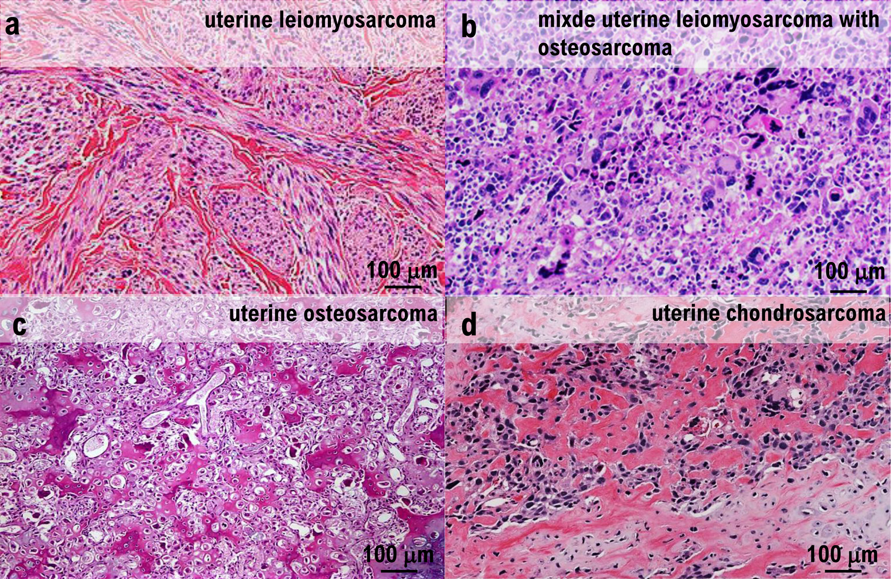
Tumor rupture is a rare complication of uterine leiomyosarcoma. We report a case of ruptured uterine leiomyosarcoma diagnosed after the onset of abdominal pain following endoscopic examination of the lower gastrointestinal tract. The patient was a 56-year-old woman who was diagnosed with anemia at 48 years of age when she was first referred to our medical team. Contrast-enhanced magnetic resonance imaging (MRI) revealed a 48 × 50 mm mass in the anterior wall of the uterine body, which was diagnosed as a uterine fibroid. After 8 years of regular follow-up once or twice a year, the patient developed abdominal pain after undergoing lower gastrointestinal endoscopy, which was prompted by a positive fecal occult blood test result. Contrast-enhanced computed tomography (CT) showed that the uterine mass had enlarged to 90 × 69 mm. T2-weighted contrast-enhanced MRI demonstrated moderate signal intensity and restricted diffusion, whereas contrast-enhanced T1-weighted MRI revealed high signal intensity, suggestive of hemorrhage. The outlines of the tumor and uterus were interrupted cephalad to the lesion, raising the suspicion of rupture of a malignant uterine mesenchymal tumor. Therefore, total hysterectomy, bilateral salpingo-oophorectomy, and partial omentectomy were performed. Intraoperatively, tumor rupture and adhesion of the ruptured tissue to the ileum were observed, necessitating partial ileectomy. Pathological examination of the resected specimen revealed irregularly proliferating spindle cells with marked nuclear atypia, 12 mitotic figures per 10 high-power fields, coagulative necrosis, and multinucleated giant cell infiltration. Another notable finding was the presence of ectopic osteosarcoma and chondrosarcoma.
Published
Issue
Section
License
Copyright (c) 2026 The authors

This work is licensed under a Creative Commons Attribution 4.0 International License.