An Unexpected Intraoperative Discovery: A Parasitic Leiomyoma in a Perimenopausal Patient With Complex Abnormal Uterine Bleeding
DOI:
https://doi.org/10.14740/jmc5254Keywords:
Parasitic leiomyoma, Abnormal uterine bleeding, Robotic-assisted hysterectomy, Minimally invasive, Invasive gynecologic surgeryAbstract
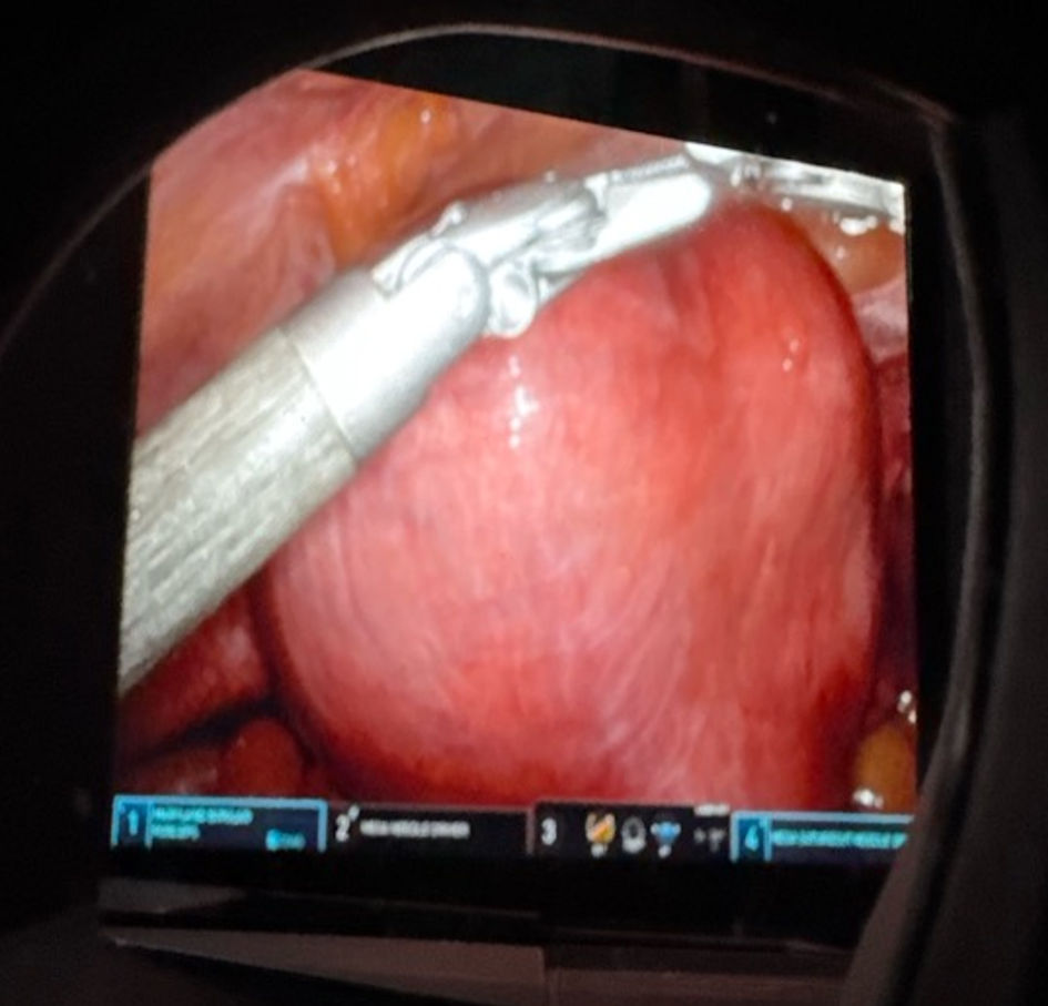
Parasitic leiomyomas are an uncommon subtype of extra-uterine smooth muscle tumors characterized by complete detachment from the uterus and subsequent development of an autonomous vascular supply. Their atypical anatomical locations, independent blood flow, and frequent coexistence with large fibroid uteri make preoperative diagnosis difficult. Abnormal uterine bleeding (AUB) is one of the leading indications for hysterectomy worldwide, and leiomyomas remain the most common structural cause within the International Federation of Gynecology and Obstetrics (FIGO) PALM–COEIN classification. Although hysterectomy is considered curative for symptomatic fibroid disease, unexpected intraoperative findings, such as parasitic leiomyomas, can significantly alter operative planning, increase surgical complexity, and necessitate careful dissection to prevent bowel or vascular injury. We report the case of a patient who underwent robotic-assisted total laparoscopic hysterectomy with bilateral salpingectomy for symptomatic AUB and a markedly enlarged multi-fibroid uterus. During surgery, a distinct parasitic leiomyoma was discovered, densely adherent to the large intestine with no connection to the uterus. The mass had mesenteric-derived vascularity and required meticulous adhesiolysis for safe mobilization. The uterus and parasitic mass were delivered vaginally. Pathology confirmed benign intramural leiomyomas and separately submitted benign smooth muscle fragments consistent with parasitic leiomyoma. No atypia, necrosis, or abnormal mitotic activity was present. The patient recovered well and reported significant symptom relief at the 1-week postoperative visit. This case shows how difficult it can be to diagnose parasitic leiomyomas and why careful assessment is needed during hysterectomy for large fibroid uteri. We also discuss their causes, imaging challenges, surgical management, and follow-up to help inform clinicians.
Published
Issue
Section
License
Copyright (c) 2026 The authors

This work is licensed under a Creative Commons Attribution 4.0 International License.