

A Silent Mimic: Biopsy-Proven Focal Nodular Hyperplasia Masquerading as Chronic Intrahepatic Biliary Obstruction
DOI:
https://doi.org/10.14740/jmc5364Keywords:
Focal nodular hyperplasia, Biliary obstruction, Liver mass, Cholestatic liver injury, Liver biopsy, Hepatic adenomaAbstract
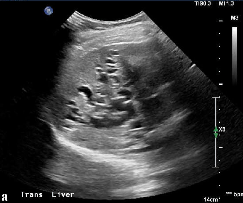
Focal nodular hyperplasia (FNH) is a benign hepatic lesion that is typically asymptomatic and most often found incidentally. Biliary obstruction caused by FNH is rare and can create diagnostic uncertainty, particularly when imaging can mimic vascular lesions, hepatocellular adenoma, bile duct adenoma, or congenital hepatobiliary abnormalities. We present the case of a 38-year-old woman with hypertension who was noted to have persistently abnormal liver enzymes on routine outpatient blood tests. She denied symptoms such as jaundice, pruritus, pale stools, dark urine, weight loss, nausea, vomiting, or abdominal pain. Laboratory tests showed a chronic cholestatic-predominant pattern with elevated alkaline phosphatase, mildly elevated aspartate aminotransferase (AST) and alanine aminotransferase (ALT), fluctuating hyperbilirubinemia, and markedly elevated gamma-glutamyl transferase (GGT). Ultrasound revealed intrahepatic biliary ductal dilatation and indeterminate cystic areas within the liver. Computed tomography revealed chronic biliary ductal dilatation, hepatic parenchymal deformity, a mass-like hypertrophied hepatic segment, and a central arterial-enhancing structure initially concerning for aneurysm or pseudoaneurysm. Magnetic resonance imaging (MRI) and magnetic resonance cholangiopancreatography (MRCP) demonstrated marked intrahepatic biliary dilatation, an arterially enhancing lesion suspicious for atypical FNH, and a second exophytic lesion causing mass effect on the biliary tree. Differentials included atypical adenoma, intrahepatic bile duct adenoma, vascular lesion, or congenital anomaly. Because serial imaging remained indeterminate, percutaneous liver biopsy was performed. Histopathology showed preserved lobular architecture, intermittent thin fibrous septa, focal sinusoidal dilatation, ductular reaction, and map-like glutamine synthetase staining with membranous beta-catenin staining, supporting the diagnosis of FNH. This case highlights an atypical presentation of FNH causing chronic intrahepatic biliary obstruction and emphasizes the importance of integrating imaging, multidisciplinary review, and histopathology when benign hepatic lesions present atypically and influence management decisions.
Published
Issue
Section
License
Copyright (c) 2026 The authors

This work is licensed under a Creative Commons Attribution 4.0 International License.