Fetal Ovarian Cysts in Prenatal Imaging: Diagnostic Challenges and Management Options
DOI:
https://doi.org/10.14740/jmc5173Keywords:
Fetal ovarian cysts, Ultrasound, Prenatal diagnosisAbstract
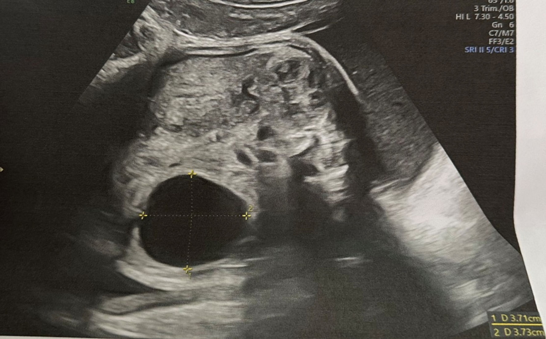
Fetal ovarian cysts (FOCs) are a rare prenatal finding that may be associated with maternal, fetal, or neonatal complications. They are classified by various features - small or large, simple or complex, unilateral or bilateral - which determine whether active treatment or simple observation is required. Prenatal ultrasound enables diagnosis as early as the first trimester, though most cases are detected in the second or third trimester. We present a case of a simple, small FOC diagnosed at 27 weeks of gestation in primigravida without accompanying diseases. The cyst remained uncomplicated throughout pregnancy and after birth, with spontaneous regression observed within the first year of life. We also conducted a brief literature review on the management of different types of FOCs. Small, asymptomatic FOCs detected in the second or third trimester usually require only ultrasound monitoring, as most regress spontaneously within the first year after birth. Symptomatic neonatal ovarian cysts, as well as those that enlarge during follow-up in pregnancy, carry a risk of ovarian torsion and generally require surgical intervention. Complex cysts and large cysts may be monitored conservatively unless they cause symptoms or show growth on serial ultrasounds.
Published
Issue
Section
License
Copyright (c) 2025 The authors

This work is licensed under a Creative Commons Attribution-NonCommercial 4.0 International License.