


A Cadaveric Case of Lusoria Artery With Calcification
DOI:
https://doi.org/10.14740/jmc5426Keywords:
Developmental anatomy, Aberrant right subclavian artery, Thyroglossal duct, Embryology, Kommerell diverticulum, DysphagiaAbstract
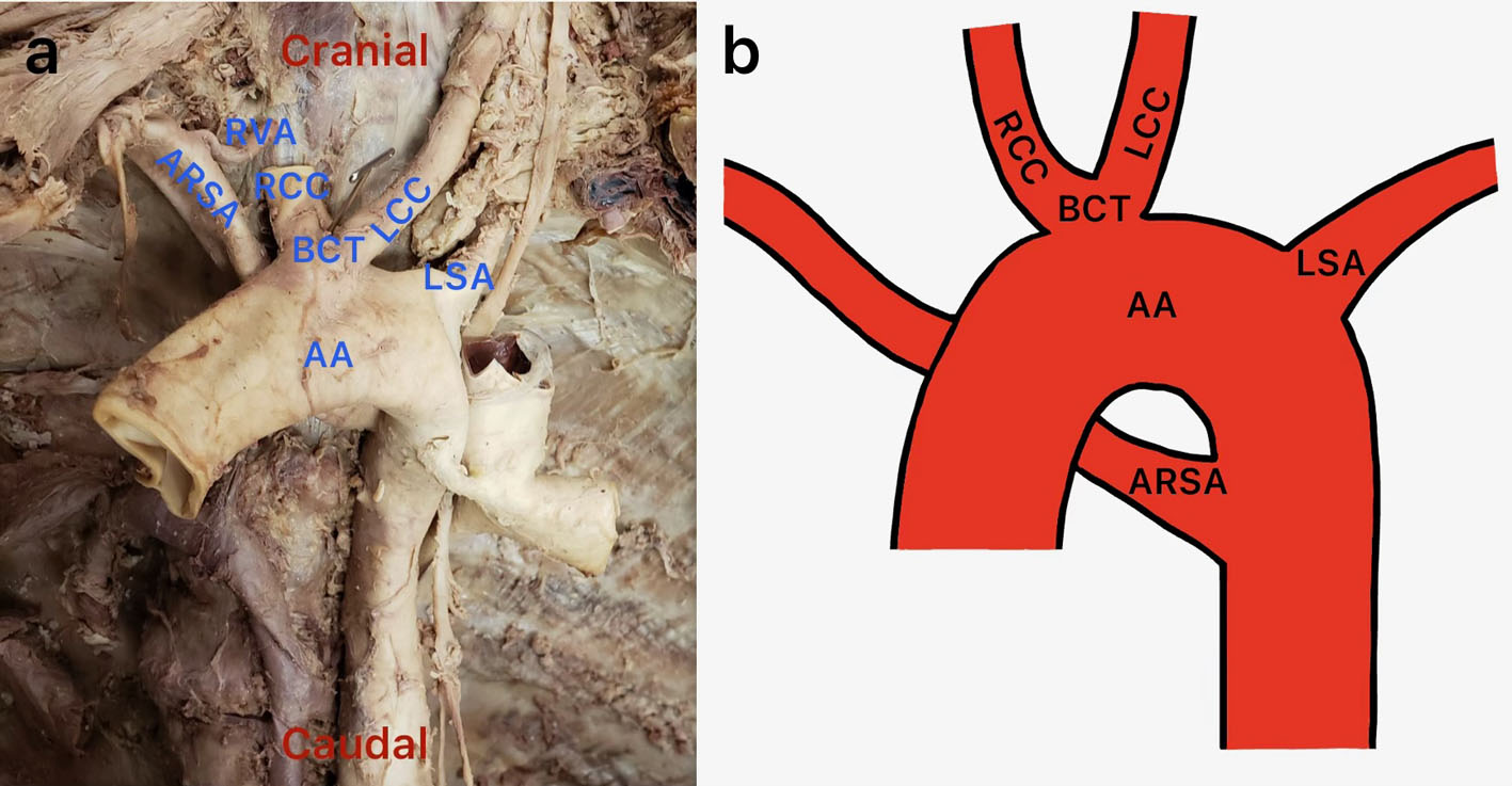
The right subclavian artery is commonly derived from the first branch of the arch of the aorta, the brachiocephalic trunk. However, there have been reported cases where the right subclavian artery did not follow this typical anatomy. Several anatomical variants of the right subclavian artery point of origin have been described in literature; one uncommon variation is the lusoria artery, which is a right subclavian artery that emerges from the proximal descending aorta. This case describes a lusoria artery that originated distal to the arch of the aorta, at the level of T5, then coursed to the right across the posterior mediastinum deep to the esophagus. A bicarotid trunk was also observed originating from the aortic arch at its most superior portion. The origin of the lusoria artery exhibited areas of extensive calcification and the posterior wall was markedly attenuated, exhibiting folding with loss of structural integrity and the presence of several full-thickness perforations. Although the embryologic relationship remains unclear, this case was also notable for the presence of a persistent thyroglossal duct. Presence of the lusoria artery is rare and 2/3 of these cases are asymptomatic. The presence of symptoms likely depends on other features. Patients with a lusoria artery may present with mild respiratory distress, dysphagia, and other less frequently observed symptoms. Diminished vascular wall thickness at the lusoria artery origin can lead to a Kommerell diverticulum, which can compound the lusoria artery symptoms and present with additional symptoms due to the mass compressing adjacent tissues. This anomaly is adequately common to demand the attention of vascular surgeons confronted with a mediastinal mass accompanied by symptoms of lusoria artery.
Published
Issue
Section
License
Copyright (c) 2026 The authors

This work is licensed under a Creative Commons Attribution 4.0 International License.