An Atypical Case of Superior Vena Cava Syndrome Due to Sarcomatoid Lung Cancer With Pericardial Metastasis
DOI:
https://doi.org/10.14740/jmc5293Keywords:
Superior vena cava syndrome, Lung cancer, NSCLCAbstract
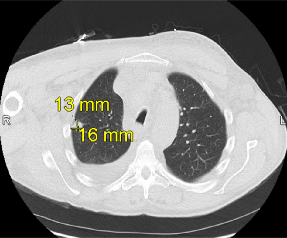
Superior vena cava (SVC) syndrome is commonly caused by malignancies from lung cancers, thrombus, and indwelling intravascular devices. Specifically, SVC syndrome is mostly associated with malignancies such as small cell lung cancer (SCLC) and non-small cell lung cancer (NSCLC). Pulmonary sarcomatoid carcinoma (PSC), as a rare type of NSCLC, accounts for 0.1% to 0.4% of pulmonary tumors. PSC’s rapid progression, aggressive growth, and complexity to diagnose help differentiate from other thoracic malignancies, yet due to its rarity, there is limited literature documenting an association between this entity and SVC syndrome. Here we present an atypical case of SVC syndrome with a history of tobacco use and chronic right upper extremity lymphedema who developed facial swelling, dyspnea, and worsening right upper extremity swelling. Imaging revealed a large mediastinal mass compressing both the SVC and pulmonary artery. Further workup and endobronchial ultrasound (EBUS) were performed, which led to a diagnosis of poorly differentiated sarcomatoid carcinoma. Additionally, pericardiocentesis revealed features consistent with adenocarcinoma. The case was further complicated with hypoxic respiratory failure and new-onset atrial fibrillation. With extensive intensive care unit (ICU) care, the patient was discharged with a medication regiment, and referred to oncology, cardiology, and palliative care. Patient declined chemotherapy and immunotherapy, opting for hospice care. Patient stay was complicated by hypoxia respiratory failure, pleural effusions, pericardial effusion, and atrial fibrillation with rapid ventricular response (RVR). This case emphasizes that SVC syndrome can be associated with poorly differentiated sarcomatoid carcinoma.
Published
Issue
Section
License
Copyright (c) 2026 The authors

This work is licensed under a Creative Commons Attribution 4.0 International License.