


Asymptomatic Intracardiac Migration of a Cane-Shaped Polymethylmethacrylate Cement Embolus Following Percutaneous Vertebroplasty
DOI:
https://doi.org/10.14740/jmc5403Keywords:
Percutaneous vertebroplasty, Polymethylmethacrylate, Bone cement embolism, Intracardiac embolismAbstract
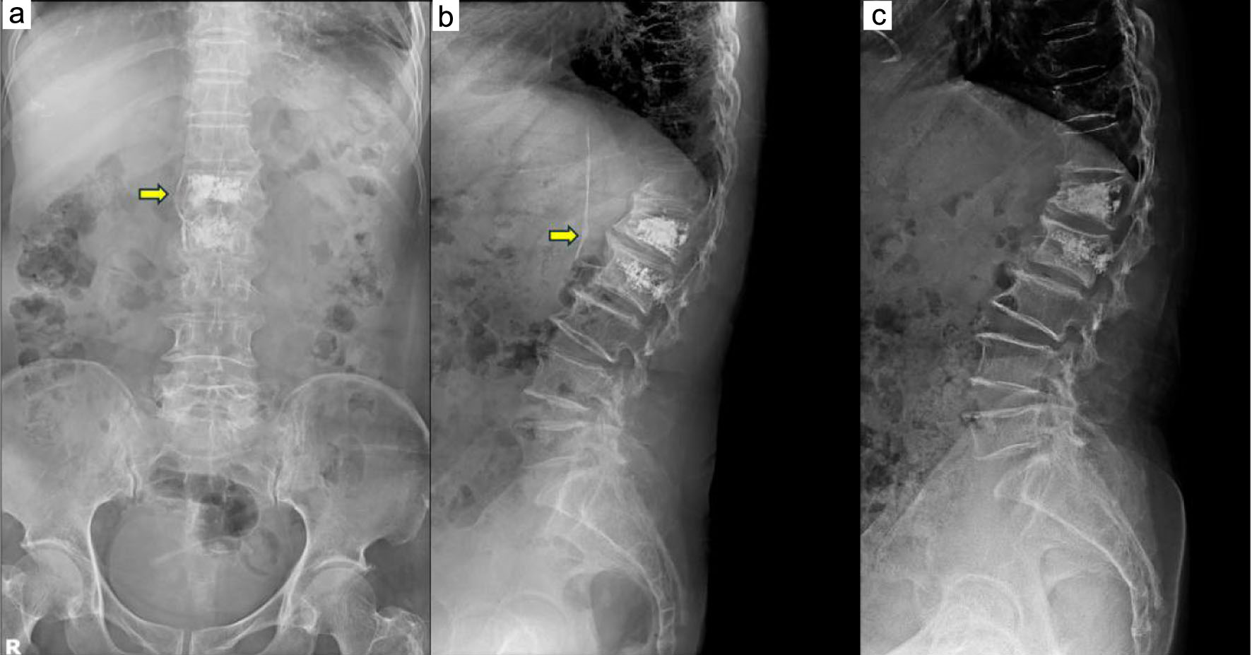
Percutaneous vertebroplasty (PVP) is a minimally invasive and effective treatment for osteoporotic vertebral compression fractures. Although generally safe, cement leakage is a relatively common complication and may rarely result in intracardiac embolism. We report the case of a 75-year-old woman who underwent PVP for an L2 compression fracture after failed conservative treatment. Postoperative imaging revealed venous cement leakage, but she remained asymptomatic. Six months later, during a routine health examination, echocardiography incidentally detected an abnormal structure in the right atrium. Cardiac computed tomography demonstrated a linear, cane-shaped polymethylmethacrylate embolus extending from the right atrium to the right ventricle. Despite the absence of cardiopulmonary symptoms, surgical removal was performed because of the elongated and sharp morphology of the embolus and the associated risk of cardiac perforation. Open-heart surgery was successfully completed without major complications. Although many intracardiac cement embolisms are asymptomatic and can be managed conservatively, embolus morphology and location should guide treatment decisions. Early recognition and individualized management are essential to prevent life-threatening complications.
Published
Issue
Section
License
Copyright (c) 2026 The authors

This work is licensed under a Creative Commons Attribution 4.0 International License.