Multi-Modality Imaging for Accurate Valvular Lesion Diagnosis: A Case Report of Catastrophic Outcomes From Unrecognized Severe Aortic Regurgitation
DOI:
https://doi.org/10.14740/jmc5177Keywords:
Aortic regurgitation, Cardiac MRI, Cardiac arrest, Aortic valve replacementAbstract
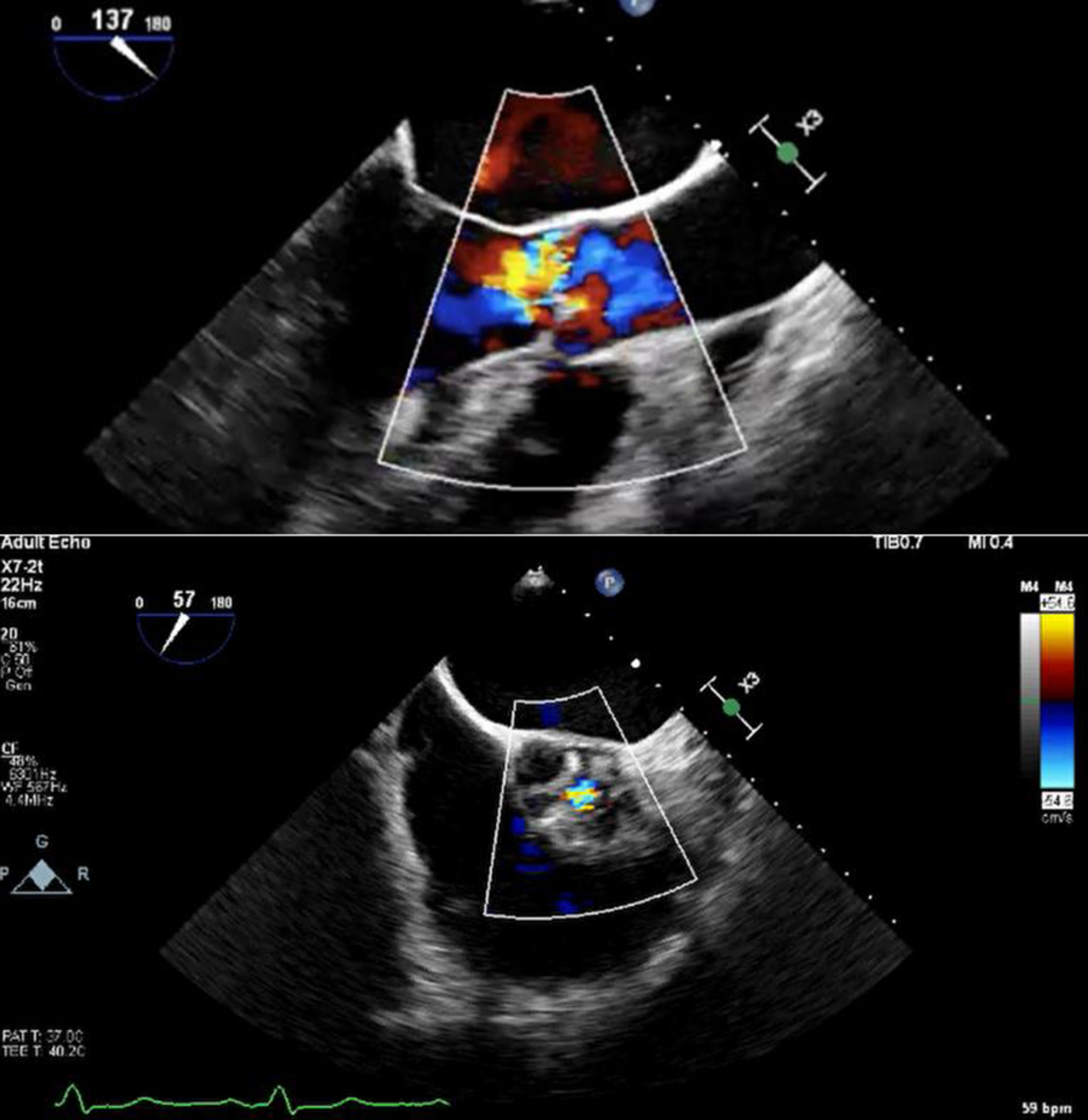
Aortic regurgitation (AR) can be difficult to accurately quantify on echocardiography alone, potentially leading to erroneous grading. A 52-year-old male presented following resuscitation after an out-of-hospital cardiac arrest. He had been followed in the cardiology outpatient clinic for a number of years for monitoring of bicuspid aortic valve and associated AR. Regular transthoracic echocardiography and one transesophageal echocardiogram had shown moderate range AR. Cardiac magnetic resonance imaging reported the AR as severe with associated severely dilated left ventricle. Echocardiography grading and the patient’s lack of symptoms supported a strategy of active surveillance. The presentation with cardiac arrest prompted re-evaluation of the severity of this patient’s AR. Repeat cardiac magnetic resonance imaging re-affirmed severe AR, and the patient proceeded to surgical aortic valve replacement with a bioprosthetic valve. Post-operatively, the patient had heart failure with severely reduced ejection fraction. During hospital stay, he developed thyrotoxicosis secondary to amiodarone. This case demonstrates the discrepancy in assessing severity between different imaging techniques and highlights the potential complications in delayed intervention in AR.
Published
Issue
Section
License
Copyright (c) 2025 The authors

This work is licensed under a Creative Commons Attribution-NonCommercial 4.0 International License.