

Cardiac Magnetic Resonance-Supported Diagnosis of Acute Myocarditis Mimicking Acute Coronary Syndrome in a Patient With Sepsis Due to a Massive Perianal Abscess
DOI:
https://doi.org/10.14740/jmc5410Keywords:
Acute myocarditis, Perianal abscess, Sepsis, Emergency surgery, Myocardial injury, Mitral regurgitation, Myocardial infarction with non-obstructive coronary arteriesAbstract
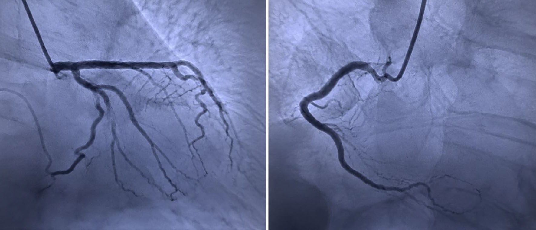
Acute myocarditis is a rare but potentially life-threatening inflammatory disease that may mimic acute myocardial infarction, particularly in septic patients where myocardial injury may have other causes. We report the case of a 46-year-old man with sepsis secondary to a massive perianal abscess who underwent emergency surgical drainage under general anesthesia. Postoperatively, persistent respiratory deterioration and elevated cardiac biomarkers prompted further cardiac evaluation. Echocardiography showed newly reduced left ventricular (LV) systolic function, with an ejection fraction (EF) of 35%, and moderate-to-severe mitral regurgitation (MR). Although acute myocardial infarction was initially suspected, coronary angiography showed non-obstructive coronary arteries (MINOCA), with only a 40% right coronary artery stenosis. Cardiac magnetic resonance (CMR) demonstrated elevated T2 values and late gadolinium enhancement (LGE) of the interventricular septum, with additional enhancement of the LV lateral wall, consistent with myocardial edema and inflammation and supportive of acute myocarditis. The patient received source control, broad-spectrum antibiotics, intravenous diuretics, norepinephrine and dobutamine, oxygen therapy, and heart failure-directed therapy. EF improved to 45% at 10 days, with complete recovery of cardiac function and resolution of MR at the 3-month follow-up. This case highlights the diagnostic challenge of acute myocarditis in septic surgical patients and suggests that early multidisciplinary assessment, coronary evaluation, and CMR may help clarify the diagnosis and guide management.
Published
Issue
Section
License
Copyright (c) 2026 The authors

This work is licensed under a Creative Commons Attribution 4.0 International License.