Paraspinal Intramuscular Hemangioma at L5-S1 With Concurrent Disc Herniation
DOI:
https://doi.org/10.14740/jmc5132Keywords:
Intramuscular hemangioma, Lumbar paraspinal muscles, Erector spinae, Surgical excision, Histopathology, Vascular tumor, Literature reviewAbstract
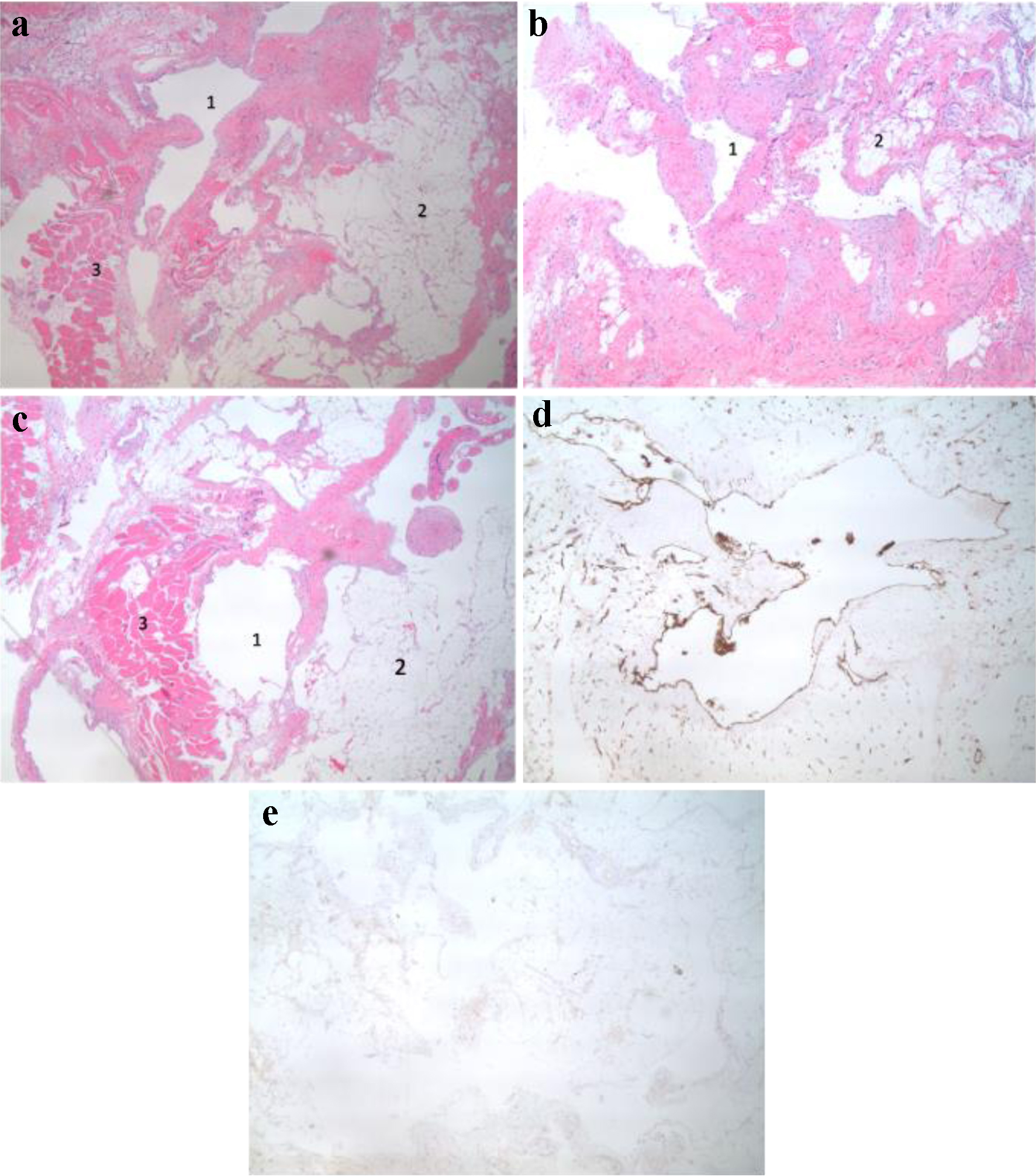
Intramuscular hemangiomas are rare, benign vascular tumors, with very few reported cases arising in lumbar paraspinal muscles. We describe the seventh documented adult case involving a 39-year-old male presenting with acute severe right-sided S1 radiculopathy. Magnetic resonance imaging (MRI) identified an 8.0 × 3.0 × 3.3 cm lesion within the erector spinae muscles at the L5-S1 level, accompanied by concurrent right-sided L5-S1 disc herniation compressing the S1 nerve root. Conservative treatment initially alleviated radicular pain, but persistent back pain and diagnostic uncertainty necessitated further evaluation. Negative metastatic screening and an inconclusive computed tomography (CT)-guided biopsy led to surgical excision, revealing an intramuscular hemangioma with significant adipocytic stromal components. This case highlights diagnostic challenges and underscores the importance of including vascular lesions in the differential diagnosis of persistent back pain, particularly when coexisting spinal pathologies complicate clinical presentation.
Published
Issue
Section
License
Copyright (c) 2025 The authors

This work is licensed under a Creative Commons Attribution-NonCommercial 4.0 International License.