| Journal of Medical Cases, ISSN 1923-4155 print, 1923-4163 online, Open Access |
| Article copyright, the authors; Journal compilation copyright, J Med Cases and Elmer Press Inc |
| Journal website https://jmc.elmerpub.com |
Case Report
Volume 18, 2027, e5440

Serial Custom 3D-Printed Nostril Retainers After Unilateral Cleft Lip Repair in Infants: A Comparative Report of Two Cases
Dieu Hong Dinha, b, Dinh Hai Trinha, Trieu Hung Dangb, Thi Minh Huyen Nguyenb, Hoang Hiep Danga, Viet Hoangc, d, e
aSchool of Dentistry, Hanoi Medical University, Vietnam
bFaculty of Dentistry, VNU - University of Medicine and Pharmacy, Hanoi, Vietnam
cDigital Industrial Design and Manufacturing Research Unit, Faculty of Engineering at Sriracha, Kasetsart University, Sriracha, Chonburi 20110, Thailand
dFaculty of Dentistry, Thammasat University, Pathum Thani 12120, Thailand
eCorresponding Author: Viet Hoang, Faculty of Dentistry, Thammasat University, Pathum Thani 12120, Thailand
Manuscript submitted September 19, 2026, accepted November 13, 2026, published online November 20, 2026
Short title: 3D-Printed Nostril Retainer After Cleft Lip Repair
doi: https://doi.org/10.14740/jmc5440
| Abstract | ▴Top |
Postoperative nasal support may help maintain the contour established by cleft lip repair, but the contribution of an individual appliance protocol requires careful evaluation. This report describes sequential use of custom bilateral nostril retainers and compares the clinical course with that of an infant treated without retention. Two 3-month-old infants, one boy and one girl, with complete left unilateral cleft lip and palate underwent Fisher lip and nasal repair by the same surgeon. Neither had received presurgical molding. Random allocation determined postoperative management with or without retention. For the intervention infant, an immediate postoperative nasal impression was digitized to design six sequential bilateral retainers incorporating staged enlargement and later-stage mirroring of the non-cleft side. The appliances were fabricated using TFDH resin and a UNIZ UBEE printer. Retention began after suture removal at postoperative week 1 and continued for approximately 6 months. Clinical and photographic assessments were performed at 1 week, 3 months, and 6 months. At the later reviews, cleft-side alar collapse appeared more pronounced in the infant without retention. Initial columellar blanching in the intervention infant resolved within approximately 20–30 s; no local allergic reaction or appliance loosening was reported. Outcomes were assessed qualitatively, without standardized nasal measurements. This two-case report illustrates the clinical implementation of serial custom bilateral nostril retainers following unilateral cleft lip repair. The observed difference in postoperative alar contour provides a basis for further investigation but cannot establish a causal benefit of retention. Larger studies with objective measurements, standardized photography, and follow-up after appliance discontinuation are needed to evaluate effectiveness and longer-term safety.
Keywords: Cleft lip and palate; Nostril retainer; Postoperative nasal retention; Three-dimensional printing; Digital fabrication
| Introduction | ▴Top |
Management of cleft lip and palate extends across multiple stages of childhood and requires coordination of surgical, dental, feeding, and speech-related care [1]. Differences in occurrence across populations and the potential psychological effects of the condition further emphasize the need for treatment that considers the child beyond the initial operation [2, 3]. Within this broader pathway, preserving the nasal correction achieved during primary repair is a specific clinical challenge.
Primary lip repair may follow anatomical subunit approximation or a rotation-advancement approach [4, 5]. Contemporary descriptions of unilateral cleft repair relate lip reconstruction to the underlying nasal deformity and the goals of primary rhinoplasty [6]. Because nasal shape reflects cartilage position, soft-tissue relationships, healing, and growth, an acceptable early appearance does not by itself establish durable correction [7]. Postoperative appliances are intended to support the reconstructed tissues during this evolving period.
Literature provides a rationale for nasal retention, although devices and clinical protocols vary. Chimruang et al synthesized outcomes of nasal conformers after primary cheiloplasty [8]. Nguyen et al identified substantial heterogeneity in postoperative stenting studies, including differences in device design and reported complications [9]. Clinical investigations have also evaluated retention after presurgical nasoalveolar molding and compared support delivered at different treatment stages [10, 11]. These findings make the timing and context of an appliance relevant to interpretation of its outcomes.
Patient-specific fabrication has already been described. Carter et al presented a digital pathway encompassing nasoalveolar molding and a postoperative stent, while Chaikangwan et al investigated image-based development of custom retainers [12, 13]. Khodir et al subsequently assessed digitally fabricated nasal conformers in a pilot randomized trial [14]. Digital manufacture therefore represents an established approach rather than, on its own, a new methodology.
The present report focuses on a serial bilateral appliance protocol whose initial geometry was obtained from the surgically reconstructed nasal cavity. It documents the design, manufacture, insertion, and successive use of six removable retainers, together with the postoperative course of a second infant who did not receive retention. The purpose is to describe this clinical implementation and the observed difference between two individual courses, providing a basis for subsequent objective investigation.
| Case Report | ▴Top |
Clinical characteristics and treatment allocation
Both patients were 3-month-old infants, one boy and one girl, with complete left unilateral cleft lip and complete cleft palate. Examination identified loss of continuity of the lip and vermilion, collapse of the left ala, widening of the left nostril, and rightward columellar deviation. Feeding difficulties and nasal regurgitation were reported before treatment. Neither infant had undergone nasoalveolar molding or other presurgical orthopedic treatment.
Primary lip and nasal reconstruction was undertaken at Ha Dong General Hospital in Hanoi, Vietnam. One surgeon treated both infants using Fisher anatomical subunit approximation [4]. Random allocation assigned one child to postoperative retainer use and the other to care without a retainer. They are described below as the intervention case and the control case, respectively. Each condition included only one infant; the comparison is descriptive.
Intervention case and digital appliance workflow
An impression was recorded after the lip and nasal repair had been completed, before emergence from the operative general anesthesia. Elite HD silicone was used to register the nasal cavity while respiratory control was maintained. Acquisition took approximately 3 min, after which the impression was retrieved and the operative area cleaned. This step captured the anatomy that the first retainer was intended to support, rather than the untreated cleft configuration.
The impression was digitized with a Medit i900 intraoral scanner (Medit Corp., South Korea) in approximately 1 min. An STL file was transferred to Meshmixer (Autodesk), where excess mesh regions were trimmed and the surfaces refined. The appliance included two hollow intranasal components with a planned wall thickness of 0.5 mm, openings intended to allow airflow, and an external T-shaped handling extension. The digital geometry is illustrated in Figure 1a. Mesh inspection preceded export for printing.
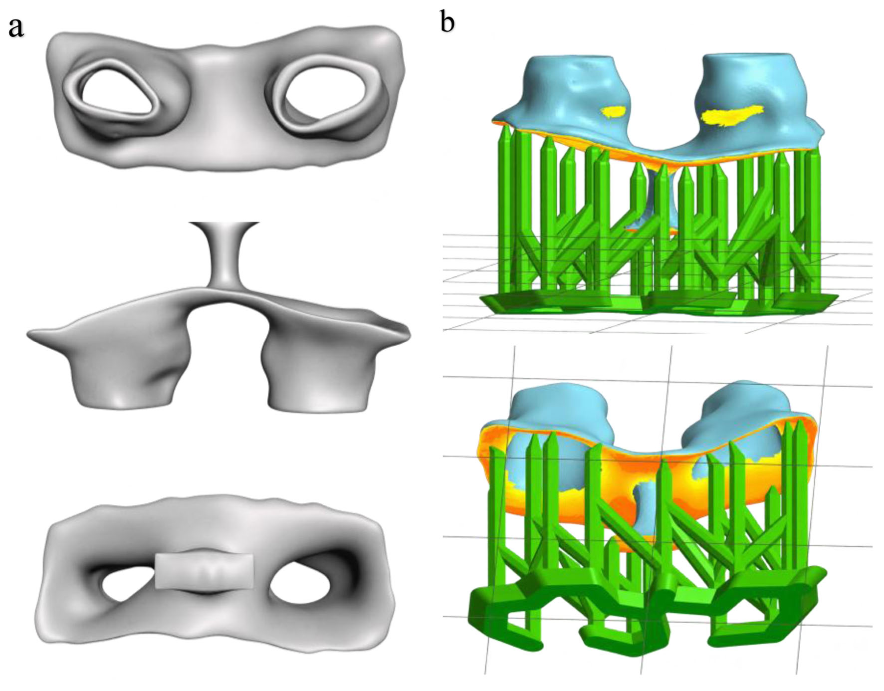 Click for large image | Figure 1. Digital design and preparation of the custom nostril retainer. (a) Computer-aided design of the appliance. (b) Appliance positioned in UNIZ Slicer for 3D printing. |
Six designs were prepared for sequential delivery. The first reproduced the immediate postoperative cavity; subsequent versions introduced approximately 5% increments in planned size and gradual enlargement on the cleft side. In the fifth and sixth designs, a mirrored representation of the non-cleft component guided the cleft-side shape and size. These increments refer to the appliance designs and do not quantify tissue movement or improvement in symmetry. Depending on the extent of modification, digital modeling took approximately 10–15 min.
UNIZ Slicer (China) was used to prepare the printing file. The appliance was arranged horizontally, with supports restricted to surfaces that would not contact nasal tissues (Fig. 1b). Manufacture used TFDH resin (Graphy, South Korea) in a UNIZ UBEE printer (UNIZ Dental, China), with layers of 0.05 mm. The printing stage lasted approximately 30 min. Figure 2 shows the printed appliance.
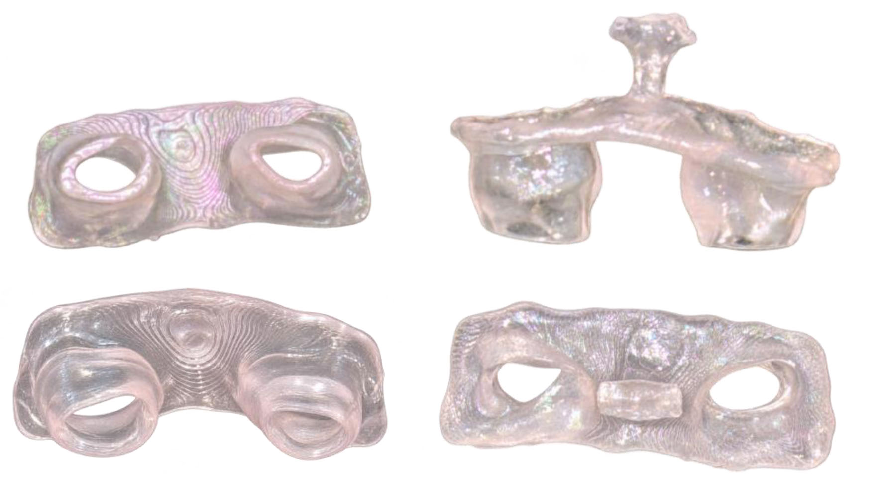 Click for large image | Figure 2. Custom nostril retainer after 3D printing. |
Centrifugation removed excess unpolymerized resin (6 min); no solvent-washing step was used. A Terahertz unit provided nitrogen-atmosphere post-curing. After support removal, fine abrasives were used to finish and polish the tissue-facing surfaces, followed by sodium hypochlorite disinfection before clinical insertion. The reported cleaning and post-processing estimate were approximately 15 min, bringing fabrication to approximately 55–60 min for one retainer. The equipment used is specified above. The reasons for brand selection and the team’s prior experience with comparable nasal appliances were not documented.
Appliance delivery and clinical monitoring
During the 1-week postoperative hospital stay, caregivers were instructed in insertion, removal, and hygiene. The first appliance was delivered after suture removal at the week-1 review. This interval allowed fabrication from the postoperative impression before fitting. Continuous wear was prescribed, interrupted twice daily for saline cleaning of the device and nasal cavity.
The six appliances were used during an approximately 6-month retention course. Replacement was described as occurring at roughly 3–4-week intervals according to growth and adaptation, although individual insertion dates and the duration of each stage were not recorded. These approximate intervals should not be interpreted as six identical wear periods. Retention relied on anatomical engagement. The handle also offered a potential attachment site for adhesive fixation, but routine taping was not reported.
Initial fitting produced temporary blanching where the appliance contacted the columella; normal coloration returned within approximately 20–30 s. The device did not touch the repaired lip. Follow-up included assessment of local tissue appearance, appliance stability, and feeding-related concerns. No local allergic reaction or loosening was reported. Caregivers reported no nasal regurgitation during retainer use, although feeding was not evaluated with a standardized instrument and this observation cannot establish an effect on the unrepaired palate.
Control case and perioperative comparison
The second infant underwent the same surgical approach and postoperative care without a nostril retainer, in accordance with the allocation. Age, cleft side, diagnosis, surgeon, and absence of presurgical molding were shared with the intervention case. These characteristics describe clinical similarity, rather than quantitatively established equivalence in baseline deformity.
Figure 3 presents the perioperative appearances: the intervention infant before and after surgery in panels a and b, and the control infant in panels c and d. Both children were subsequently reviewed at postoperative week 1 and months 3 and 6. At later reviews, the cleft-side ala appeared more collapsed in the child without retention than in the child wearing the serial appliances.
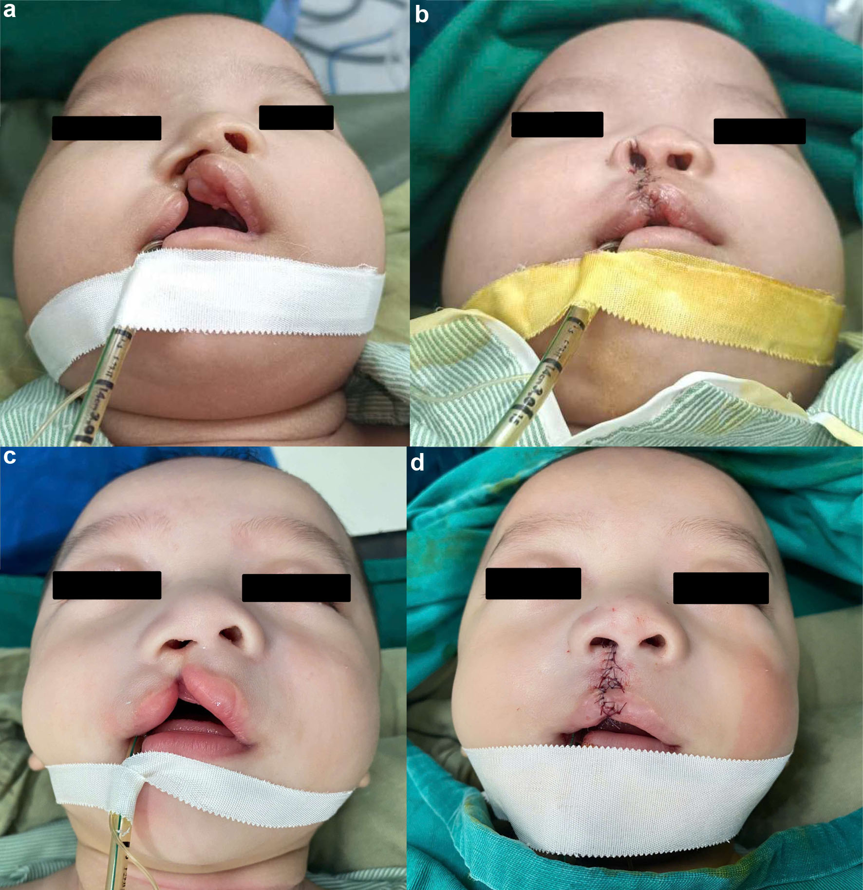 Click for large image | Figure 3. Preoperative and postoperative photographs of the two infants. (a, b) Intervention infant before and after surgery, respectively. (c, d) Control infant before and after surgery, respectively. Preoperative views appear on the left and postoperative views on the right. |
Outcomes and comparative assessment
Figure 4 documents the follow-up sequence. Panels a, b, and c correspond to the intervention infant at 1 week, 3 months, and 6 months, respectively; panels d, e, and f show the control infant at the same intervals. Assessment concerned the visible alar contour, nostril configuration, and overall nasal asymmetry. The photographs support descriptive comparison, but were not analyzed as calibrated anthropometric records.
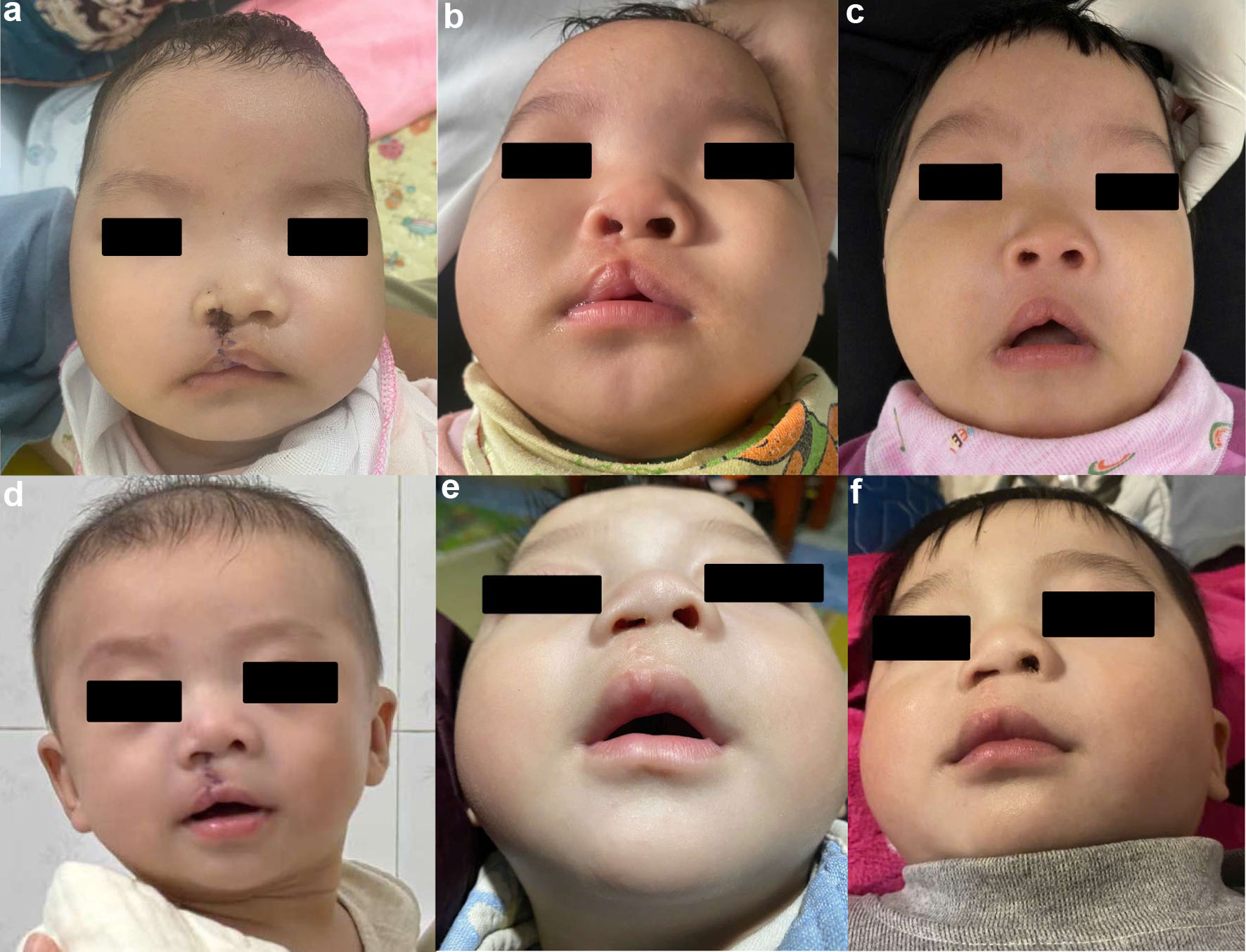 Click for large image | Figure 4. Postoperative follow-up photographs of the two infants. (a–c) Intervention infant at 1 week, 3 months, and 6 months after surgery, respectively. (d–f) Control infant at the corresponding intervals. Photographs are presented for qualitative assessment and were not used for calibrated anthropometric measurements. |
At the 3- and 6-month reviews, cleft-side alar collapse was clinically more pronounced in the control case. The direction of this observation was consistent with the photographic comparison, but its magnitude could not be calculated. No standardized nostril dimensions, alar-width measurements, columellar angles, or serial three-dimensional facial measurements were obtained. Consequently, neither the extent of change within each patient nor a numerical difference between patients can be reported.
Tolerance in the intervention case was characterized by the transient pressure-related blanching at first insertion and the absence of reported local allergy or loosening during use. Wear adherence was based on caregiver accounts and clinical review, without an objective wear-time record or a validated satisfaction scale. Table 1 summarizes the shared characteristics, treatment difference, and available outcomes.
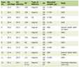 Click to view | Table 1. Clinical Characteristics, Treatment, and Descriptive Outcomes of the Two Infants |
Primary repair occurred at age 3 months, with retainer insertion at postoperative week 1 in the intervention case and subsequent staged use over approximately 6 months. Follow-up photography was obtained at week 1 and months 3 and 6. Palatal closure was planned for age 9–12 months; completed palate surgery is not an outcome of this report.
| Discussion | ▴Top |
The clinical observation was a more collapsed cleft-side alar contour in the infant managed without postoperative retention at subsequent reviews. Adding a control case gives context to the course of the infant wearing the appliance, but does not isolate the effect of retention. The infants shared several recorded characteristics and the same surgeon, yet the severity of the initial deformity and the immediate surgical correction were not measured. Differences in these factors could contribute to the later appearances.
The comparison should be considered within a heterogeneous retention literature. Systematic reviews support continued investigation of postoperative conformers, while indicating variation in devices, outcomes, and study quality [8, 9]. Al-Qatami et al examined postsurgical retention within a treatment pathway that included presurgical nasoalveolar molding [10]; this differs from the present infants, neither of whom received molding. Funayama et al compared presurgical nasal stenting with postsurgical retainer placement [11]. Such differences prevent direct transfer of an effect estimate from one protocol to another.
Timing also distinguishes protocols. Zhang et al evaluated a nasal clip and conventional retainers introduced 7 days after primary repair, with support continued for 6–12 months [15]. This provides clinical context for initiating retention at the week-1 visit, but does not establish that this timing is optimal for the present design. Here, impression-taking occurred immediately after surgery, whereas insertion followed suture removal. The intervening week was therefore a period without appliance support. The report cannot determine whether earlier insertion would have altered the outcome.
Surgical correction remains an important alternative explanation for postoperative nasal form. Fisher repair and contemporary anatomical descriptions address both lip reconstruction and nasal relationships [4, 6]. Murali et al investigated primary rhinoplasty with overcorrection using outcomes assessed after skeletal maturity [16]. Their study illustrates the importance of evaluating the surgical contribution and following patients beyond the early healing period. The present 6-month comparison cannot establish persistence of the observed contour through growth.
Khodir et al provide the closest methodological comparison: their pilot randomized study included 14 infants, used Fisher repair, and assessed postoperative outcomes objectively at 6 months [14]. Their unilateral conformer was based on mirroring the unaffected side, placed during surgery, and secured with sutures for 3 months. In contrast, the present protocol used a bilateral removable series based initially on postoperative anatomy, with enlargement across stages and mirroring introduced only in the fifth and sixth designs. Delivery began at week 1 and use extended to approximately 6 months.
The practical contribution is the description of serial appliance delivery from one postoperative anatomical record. Existing digital pathways already include custom postoperative stents and image-derived retainers [12, 13]. In the present infant, the initial record supported repeated modifications without taking a new impression for each described stage. The bilateral configuration, hollow components, handling extension, and staged geometry were intended to facilitate support and everyday use. Neither airflow nor pressure distribution was measured, and the chosen 0.5-mm wall thickness and approximately 5% increments should be understood as design parameters rather than validated therapeutic thresholds.
Daily use of the removable retainer required caregiver involvement in insertion, removal, and cleaning. Hennocq et al prospectively assessed parental information and tolerance of nasal splints, demonstrating that family experience is a distinct outcome worth evaluating [17]. Here, instruction and twice-daily cleaning formed part of delivery, but adherence and burden were captured only descriptively. The reported absence of loosening and local allergy is relevant to this child’s course; it cannot establish general tolerance or exclude uncommon adverse events. Transient columellar blanching also warrants reporting as a pressure-related finding.
Material selection requires an equally specific interpretation. The FDA 510(k) summary for Graphy’s TERA HARZ DENTURE concerns denture and baseplate indications [18]. That document does not establish an infant intranasal indication or validate the particular printer, layer thickness, centrifugation, nitrogen-curing, and disinfection sequence used here. The present report makes no claim of infant-specific intranasal clearance. Clinical use in one child is insufficient to characterize biological safety under prolonged nasal contact, and the finished appliance requires evaluation for its intended application.
The fabrication estimate describes the time reported for this workflow, rather than a demonstrated advantage in accuracy, cost, or efficiency. Exact centrifugation settings, post-curing duration, and hypochlorite concentration and exposure time were not documented, limiting reproducibility. Home care involved saline cleaning; repeated hypochlorite disinfection by caregivers was not part of the reported regimen. Future work should document the manufacturing conditions and each appliance’s delivery and replacement dates alongside clinical outcomes.
Related dental studies illustrate the wider use of digital fabrication, including AI-assisted design of lingual indirect bonding trays and AI-assisted segmentation with printed models for impacted-canine surgery in a patient with cleft lip and palate [19, 20]. These examples support the relevance of digital tools across dental workflows, but do not validate this nasal appliance. The workflow described here used scanning, manual digital modeling, slicing, and printing; no AI-assisted step was documented.
This report includes only one infant in each treatment condition. Random allocation was reported, but sequence generation and allocation-concealment details were unavailable. Regardless of allocation, this sample supports descriptive reporting rather than a reliable estimate of treatment effect. Qualitative assessment remains vulnerable to differences in head position, camera perspective, lighting, and baseline anatomy. Photographs at 1 week, 3 months, and 6 months document the course but do not substitute for standardized measurements. The interval between any retainer removal and photography was not established, and there was no documented assessment of stability after retention ended.
We plan to evaluate this protocol in a larger clinical study using standardized nasal measurements. A prospective protocol should record the immediate postoperative baseline, standardized serial images, nostril dimensions, columellar alignment, appliance-processing conditions, wear adherence, and replacement dates. Follow-up after discontinuation would help distinguish contour maintained during active support from subsequent stability. These are proposed research objectives; the present contribution remains the description of two clinical courses and the implementation of the serial retainer protocol.
Conclusion
Serial bilateral nostril retainers were fabricated from a postoperative nasal impression and used in one infant following Fisher repair. A second infant treated without retention showed more pronounced cleft-side alar collapse on qualitative follow-up. This comparison provides clinical context for the appliance protocol but cannot establish a causal benefit. Objective measurements, reproducible processing records, and larger comparative studies are needed to evaluate effectiveness and longer-term safety.
Acknowledgments
None to declare.
Financial Disclosure
Dieu Hong Dinh was funded by the PhD Scholarship Programme of the Vingroup Innovation Foundation (VINIF), VinUniversity, grant code VINIF.2025.TS51.
Conflict of Interest
The authors declare no conflict of interest related to this work.
Informed Consent
Written informed consent for treatment and publication of clinical information and photographs was obtained from the parents or legal guardians of both infants.
Author Contributions
Dieu Hong Dinh: conceptualization, digital workflow design, manuscript writing, supervision. Dinh Hai Trinh: clinical treatment, patient management, data collection. Trieu Hung Dang: digital design, 3D printing workflow, technical support. Thi Minh Huyen Nguyen: literature review, manuscript editing. Hoang Hiep Dang: digital workflow design. Viet Hoang: digital workflow design, manuscript writing, supervision, manuscript revision, academic consultation. All authors read and approved of the final manuscript.
Data Availability
The clinical data supporting this report are included in the article. Additional de-identified information may be made available by the corresponding author upon reasonable request, subject to patient consent and applicable privacy restrictions.
| References | ▴Top |
- Mink van der Molen AB, van Breugel JMM, Janssen NG, Admiraal RJC, van Adrichem LNA, Bierenbroodspot F, Bittermann D, et al. Clinical practice guidelines on the treatment of patients with cleft lip, alveolus, and palate: an executive summary. J Clin Med. 2021;10(21):4813.
doi pubmed - Putri FA, Pattamatta M, Anita SES, Maulina T. The global occurrences of cleft lip and palate in pediatric patients and their association with demographic factors: a narrative review. Children (Basel). 2024;11(3):322.
doi pubmed - Al-Namankany A, Alhubaishi A. Effects of cleft lip and palate on children's psychological health: a systematic review. J Taibah Univ Med Sci. 2018;13(4):311-318.
doi pubmed - Fisher DM. Unilateral cleft lip repair: an anatomical subunit approximation technique. Plast Reconstr Surg. 2005;116(1):61-71.
doi pubmed - Romero R. The Millard rotation-advancement lip repair using accurate measurements. Oral Surg Oral Med Oral Pathol Oral Radiol Endod. 1997;84(4):335-338.
doi pubmed - Tse RW, Fisher DM. State of the art: The unilateral cleft lip and nose deformity and anatomic subunit approximation. Plast Surg (Oakv). 2024;32(1):138-147.
doi pubmed - Ye J, Wang X, Xiao M. Current treatment progress of unilateral cleft lip nasal deformities. Aesthetic Plast Surg. 2024;48(23):4895-4901.
doi pubmed - Chimruang J, Worasakwutiphong S, Tansalarak R. The effectiveness of nasal conformers in patients with unilateral cleft lip and palate following primary cheiloplasty: a systematic review and meta-analysis. Arch Plast Surg. 2025;52(4):225-233.
doi pubmed - Nguyen DC, Myint JA, Lin AY. The role of postoperative nasal stents in cleft rhinoplasty: a systematic review. Cleft Palate Craniofac J. 2024;61(12):1941-1950.
doi pubmed - Al-Qatami F, Avinoam SP, Cutting CB, Grayson BH, Shetye PR. Efficacy of postsurgical nostril retainer in patients with unilateral cleft lip and palate treated with presurgical nasoalveolar molding and primary cheiloplasty-rhinoplasty. Plast Reconstr Surg. 2022;150(3):623-629.
doi pubmed - Funayama E, Yamamoto Y, Oyama A, Furukawa H, Murao N, Hayashi T, Maeda T, et al. Comparison of nasal symmetry between presurgical nasal stenting and postsurgical nasal retainer placement in unilateral clefts. J Craniofac Surg. 2019;30(1):133-136.
doi pubmed - Carter CB, Gallardo FF, Jr., Colburn HE, Schlieder DW. Novel digital workflow for nasoalveolar molding and postoperative nasal stent for infants with cleft lip and palate. Cleft Palate Craniofac J. 2023;60(9):1176-1181.
doi pubmed - Chaikangwan I, Yodrabum N, Kusakunniran W, Tachavijijaru R, Aojanepong C. Utilization of images and three-dimensional custom-made nostril retainer fabricate for patients with cleft lip and cleft lip nose deformities at Siriraj Hospital: preliminary phase. Sci Rep. 2023;13(1):19109.
doi pubmed - Khodir MA, Osman SM, Ragab HR, AboulHassan MA, Oraby MS. Digital fabrication of customized nasal conformers for unilateral cleft lip repair: a pilot randomized controlled trial. BMC Oral Health. 2026;26(1):735.
doi pubmed - Zhang S, Yang C, Wu M, Yin J, Zeng N, Shi B, Huang H. Application of a novel nasal clip for nostril retention after primary unilateral cleft rhinoplasty. Cleft Palate Craniofac J. 2023;60(7):851-857.
doi pubmed - Murali SP, Denadai R, Sato N, Lin HH, Hsiao J, Pai BCJ, Chou PY, et al. Long-term outcome of primary rhinoplasty with overcorrection in patients with unilateral cleft lip: avoiding intermediate rhinoplasty. Plast Reconstr Surg. 2023;151(3):441e-451e.
doi pubmed - Hennocq Q, Person H, Hachani M, Bertin H, Corre P, Gorbonosov V, Ivanov A, et al. Quality of life and nasal splints after primary cleft lip and nose repair: Prospective assessment of information and tolerance. J Craniomaxillofac Surg. 2018;46(10):1783-1789.
doi pubmed - U.S. Food and Drug Administration. TERA HARZ DENTURE: 510(k) summary, K222414. 2022. Accessed November 11, 2026. https://www.accessdata.fda.gov/cdrh_docs/pdf22/K222414.pdf.
- Hoang V, Vuong TQT, Nguyen PH, Pham NH, Hoang KL, Hoang TTT, Nguyen TK, et al. AI-assisted design of 3D-printed lingual indirect bonding trays: a comparative evaluation of bracket transfer accuracy. J Clin Med. 2025;14(12):4303.
doi pubmed - Hoang V, Khoi BH, Chantarapanich N, Phuoc TH, Marya A. AI and 3D printing-assisted surgical exposure of impacted canines and tooth extraction in complex cleft lip and palate orthodontic treatment. J Surg Case Rep. 2026;2026(4):rjag323.
doi pubmed
This article is distributed under the terms of the Creative Commons Attribution 4.0 International License (CC BY 4.0), which permits unrestricted use, distribution, and reproduction in any medium, including commercial use, provided the original work is properly cited.
Journal of Medical Cases is published by Elmer Press Inc.