| Journal of Medical Cases, ISSN 1923-4155 print, 1923-4163 online, Open Access |
| Article copyright, the authors; Journal compilation copyright, J Med Cases and Elmer Press Inc |
| Journal website https://jmc.elmerpub.com |
Case Report
Volume 18, 2027, e5433

From a Cutaneous Nodule to Presumed Multisystem Sarcoidosis Amid Persistent Tuberculosis Uncertainty
Gan Lin Qiaoa, Wen Qing Chenga, Wen Hua Zhanga, Yi Fan Songa, Xiang Yi Zanb, c
aThe Second Clinical Hospital of Lanzhou University, Lanzhou 730000, Gansu, China
bSpecial Needs Department of Internal Medicine, The Second Clinical Hospital of Lanzhou University, Lanzhou 730000, Gansu, China
cCorresponding Author: Xiang Yi Zan, Special Needs Department of Internal Medicine, The Second Clinical Hospital of Lanzhou University, Lanzhou 730000, Gansu, China
Manuscript submitted September 21, 2026, accepted November 4, 2026, published online November 20, 2026
Short title: Cutaneous Clues in Presumed Sarcoidosis
doi: https://doi.org/10.14740/jmc5433
| Abstract | ▴Top |
Sarcoidosis requires a compatible phenotype, granulomatous inflammation, and reasonable exclusion of alternative causes. That framework becomes difficult when tissue samples are discordant and tuberculosis remains plausible. A 60-year-old woman had a right-eyebrow cutaneous nodule removed in 2024. Histology showed chronic inflammation and granuloma formation with an intradermal nevus, but it was interpreted as chronic dermatitis and did not establish cutaneous sarcoidosis. In 2025, she was evaluated for respiratory symptoms and found to have diffuse pulmonary micronodules, extensive thoracic and extrathoracic lymphadenopathy, bilateral pleural effusions, hypercalcemia, kidney dysfunction, parotid abnormalities, dry eye, and multiple subcutaneous nodules. A bronchial biopsy showed severe acute and chronic inflammation with necrosis and was non-diagnostic. A later right supraclavicular-node biopsy showed multiple epithelioid granulomas with multinucleated giant cells and no definite necrosis. Five respiratory acid-fast smears, bronchoalveolar-lavage Xpert MTB/RIF and metagenomic sequencing, and tissue Mycobacterium tuberculosis polymerase-chain-reaction testing were negative. No respiratory or lymph-node specimen was submitted for mycobacterial culture, and special-stain results were unavailable. Multidisciplinary assessment supported a working diagnosis of presumed multisystem sarcoidosis. Prednisone was administered with preventive isoniazid and rifampicin because immunosuppression was considered necessary despite residual tuberculosis uncertainty. Symptoms, subcutaneous nodules, hypercalcemia, kidney dysfunction, and thoracic imaging improved during concurrent treatment, but residual supraclavicular lymph nodes were interpreted as possibly tuberculous. This case demonstrates how an initially nonspecific cutaneous granulomatous lesion may become an important longitudinal clue while also showing why negative molecular tests and improvement during overlapping therapies cannot fully exclude tuberculosis or confirm treatment attribution.
Keywords: Sarcoidosis; Tuberculosis; Cutaneous nodule; Granuloma; Hypercalcemia; Lymph node biopsy
| Introduction | ▴Top |
Sarcoidosis is a multisystem granulomatous disorder with no single diagnostic test. Current diagnostic frameworks require a compatible clinical and radiological presentation, histological evidence of non-necrotizing granulomatous inflammation when tissue confirmation is needed, and reasonable exclusion of competing diagnoses [1, 2]. Each component is probabilistic. Granulomas are not specific to sarcoidosis, characteristic thoracic imaging can occur in other diseases, and negative tests for infection do not always exclude a paucibacillary process. Diagnostic confidence therefore depends on integration of organ findings, repeated sampling, microbiology, epidemiology, and longitudinal follow-up rather than on a single biopsy result.
Tuberculosis is a particularly important alternative diagnosis in regions where it is prevalent. Both diseases can produce pulmonary nodules, lymphadenopathy, constitutional or respiratory symptoms, and epithelioid granulomas. Although caseating necrosis and microbiological confirmation favor tuberculosis, absence of necrosis and negative acid-fast stains or molecular tests do not completely exclude it [3]. This distinction matters because glucocorticoids used for symptomatic sarcoidosis may worsen unrecognized infection, while empirical or preventive antituberculosis treatment can confound interpretation of a subsequent clinical response.
Cutaneous involvement may provide an accessible diagnostic window into systemic sarcoidosis. Its morphology and histology are variable, however, and a skin specimen showing granulomatous inflammation can remain nonspecific without the later systemic context [4]. We describe a woman whose earliest right-eyebrow nodule preceded recognition of a multisystem phenotype, but whose bronchial and lymph-node samples, absence of mycobacterial culture, and persistent lymph-node abnormalities sustained uncertainty about tuberculosis. The educational focus is the longitudinal reconciliation of discordant evidence and the limits of causal inference during concurrent prednisone and preventive antituberculosis therapy. The Ethics Committee of Lanzhou University Second Hospital approved this case report (Approval No. 2026A-750).
| Case Report | ▴Top |
A 60-year-old woman with a history of hypertension and hypertensive nephropathy for more than 10 years had experienced intermittent dyspnea, cough, and sputum production for over 1 year by 2025. The longstanding renal disease predated the current granulomatous illness and was not considered evidence of renal sarcoidosis. The earliest documented skin manifestation was a solitary right-eyebrow cutaneous nodule that had been surgically removed. The pathology report dated October 14, 2024, described a fragmented skin specimen with dermal collagen-fiber proliferation and degeneration, chronic inflammatory-cell infiltration, and granuloma formation, together with an intradermal nevus (Fig. 1a). The final report favored chronic dermatitis with an intradermal nevus and did not diagnose cutaneous sarcoidosis. No dermatology consultation or systemic sarcoidosis evaluation was documented at that time.
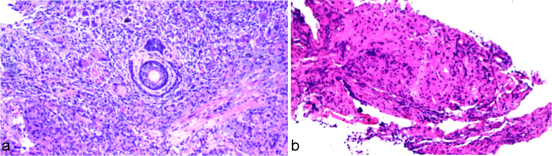 Click for large image | Figure 1. Representative deidentified pathology screenshots. (a) Right-eyebrow skin specimen, report dated October 14, 2024; the report described chronic inflammation and granuloma formation with an associated intradermal nevus. (b) Left-main-bronchial biopsy, report dated May 27, 2025; the report described severe acute and chronic mucosal inflammation with necrosis. The pathology department could not provide staining methods, objective magnifications, image calibration, or validated scale bars; none has been inferred or added. |
During evaluation in May 2025, chest computed tomography (CT) demonstrated diffuse bilateral pulmonary micronodules and patchy opacities, bilateral hilar and mediastinal lymphadenopathy, additional extrathoracic lymph-node enlargement, bronchial narrowing, bilateral pleural effusions, and a small pericardial effusion (Fig. 2a, d). Pulmonary function testing showed preserved spirometry and lung volumes with mildly reduced diffusing capacity. The combination of nodular parenchymal disease and extensive lymphadenopathy raised concern for a granulomatous disorder, but infection and malignancy remained in the differential diagnosis.
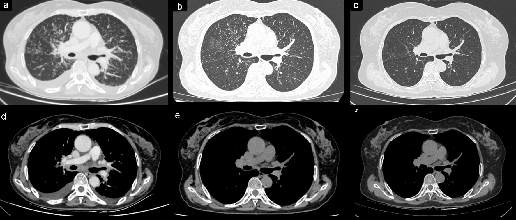 Click for large image | Figure 2. Serial chest computed tomography images at three time points. Lung-window images were obtained on May 28, 2025 (a), January 7, 2026 (b), and June 10, 2026 (c); mediastinal-window images obtained at the same time points are shown in (d–f), respectively. The baseline images (a, d) show diffuse bilateral micronodular and peribronchovascular abnormalities, thoracic lymphadenopathy, and bilateral pleural effusions. The images obtained on January 7, 2026 (b, e) show marked interval improvement, and the latest images obtained on June 10, 2026 (c, f) show sustained radiographic improvement. |
Bronchoscopy on May 26, 2025, showed diffuse nodular mucosal elevations along the trachea and left main bronchus (Supplementary Material 1, jmc.elmerpub.com). A left-main-bronchial biopsy reported on May 27 showed severe acute and chronic mucosal inflammation with necrosis (Fig. 1b). It did not demonstrate the well-formed non-necrotizing granulomas required to provide positive histological support for sarcoidosis. The sample was therefore treated as non-diagnostic rather than as evidence against sarcoidosis. This discordance between the radiological pattern and the first respiratory tissue sample prompted continued assessment.
The available infection evaluation included five respiratory specimens for acid-fast staining: one sputum specimen collected on May 22, 2025, three tracheal-secretion specimens collected on May 26, and one sputum specimen collected on August 18; all were negative for acid-fast bacilli. The records did not specify whether the sputum was spontaneous or induced, and cough with sputum production was not a leading symptom at the May or August presentations. Bronchoscopy on May 26 documented bronchoalveolar-lavage sampling from the upper and lower lobes of both lungs, but did not identify a single target segment or state whether sampling was directed to the most radiologically involved area. Xpert MTB/RIF testing of bronchoalveolar-lavage fluid on May 26 did not detect Mycobacterium tuberculosis (M. tuberculosis) complex. The laboratory report separately displayed a negative rifampicin-resistance entry and a passed specimen-processing-control probe; no inference regarding rifampicin susceptibility was made because M. tuberculosis complex was not detected. DNA-based metagenomic next-generation sequencing of bronchoalveolar-lavage fluid reported no suspected pathogen; organisms listed as likely colonizing flora were not interpreted as causative infection. Mycobacterial cultures were not submitted from the respiratory specimens.
Routine bacterial and fungal culture of sputum on May 22 showed normal flora and no fungal growth; bronchoalveolar-lavage culture on May 26 showed no pathogenic bacterial or fungal growth. A sputum bacterial and fungal smear on August 18 detected gram-positive cocci and gram-negative bacilli but no fungal hyphae or spores. No fungal polymerase-chain-reaction result was available. Neither tuberculin skin testing nor a conventional interferon-gamma release assay was performed. A hospital-designated “M. tuberculosis dual-factor” whole-blood assay on May 22 was reported negative (M. tuberculosis-specific interferon-gamma 0.33 pg/mL (reference interval 0.0–7.0) and interleukin-2 0.31 pg/mL (reference interval 0.0–20.0)). Because the report did not identify this platform as a conventional interferon-gamma release assay, the result was reported separately and was not treated as equivalent to tuberculin skin testing or an interferon-gamma release assay. Metagenomic sequencing was not regarded as a substitute for mycobacterial culture.
On readmission in August 2025, the systemic nature of the illness became more evident. The patient had dry eye, parotid abnormalities, supraclavicular lymphadenopathy, and multiple small subcutaneous nodules in addition to the thoracic findings. Serum calcium was 3.24 mmol/L, creatinine was 187.3 µmol/L, and parathyroid hormone was suppressed at 8.76 pg/mL. The combination of hypercalcemia with suppressed parathyroid hormone was compatible with active granulomatous disease. However, the pre-existing hypertensive nephropathy and the absence of renal histology prevented attribution of the full creatinine rise to sarcoidosis.
Because the earlier skin and bronchial specimens were not definitive, an accessible right supraclavicular lymph node was sampled. The biopsy reported on August 21, 2025, showed multiple epithelioid granulomas composed predominantly of epithelioid cells with fewer multinucleated giant cells and no definite necrosis (Fig. 3). A paraffin-tissue M. tuberculosis nucleic-acid test reported on August 25 was negative. The lymph-node specimen was not submitted for mycobacterial or fungal culture, and results for mycobacterial and fungal special stains could not be retrieved. Multidisciplinary review integrated the supraclavicular-node histology with the pulmonary, lymphatic, cutaneous, lacrimal/parotid, calcium, and kidney findings and favored presumed multisystem sarcoidosis as the most coherent working diagnosis while acknowledging that active tuberculosis had not been definitively excluded.
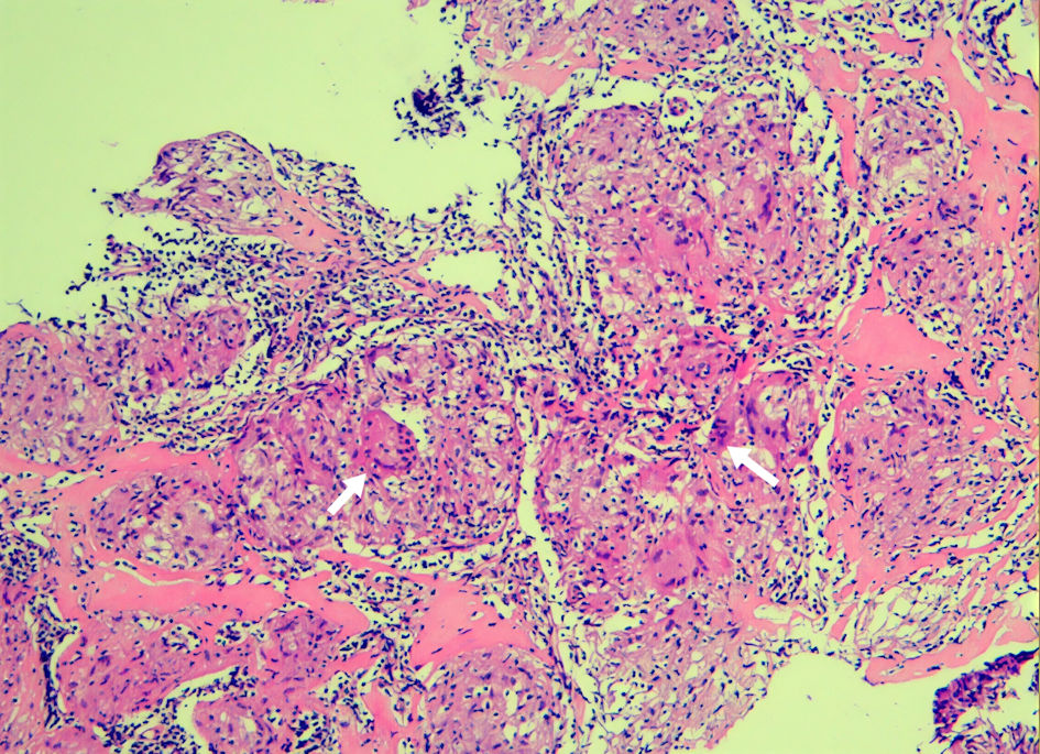 Click for large image | Figure 3. Right supraclavicular-node biopsy reported on August 21, 2025. The report described multiple epithelioid granulomas with multinucleated giant cells and no definite necrosis. White arrows were present in the source image. The pathology department could not provide the staining method, objective magnification, image calibration, or a validated scale bar; none has been inferred or added. |
Cardiac evaluation was reviewed because dyspnea and a small pericardial effusion could not be assumed to be exclusively pulmonary. On May 22, 2025, electrocardiography showed sinus rhythm and left-axis deviation without documented arrhythmia or conduction block. Transthoracic echocardiography showed normal cardiac structure, left-ventricular ejection fraction (LVEF) of 76%, and grade I diastolic dysfunction, with no pericardial abnormality at that examination. On August 18–19, electrocardiography was normal and echocardiography showed LVEF of 64%, grade I diastolic dysfunction, and a small pericardial effusion without another structural abnormality. During the August admission, high-sensitivity troponin T was 30.10 ng/L and N-terminal pro-B-type natriuretic peptide was 4,556 pg/mL. Concurrent kidney dysfunction and uncertain volume status limited interpretation of these biomarkers. Whole-body fluorodeoxyglucose positron-emission tomography, cardiac magnetic resonance imaging, and ambulatory electrocardiographic monitoring were not performed; the record did not state why positron-emission tomography was omitted.
After discharge on August 26, 2025, prednisone was administered at a dose of 20 mg once daily for 1 month, 15 mg once daily for 2 months, 10 mg once daily for 3 months, and 5 mg once daily for 3 months and 15 days, with the daily dose continued through June 10, 2026. The 5-mg dose was then taken every other day from June 11 to 18, followed by a regimen with two drug-free days between doses from June 19 to 26 and three drug-free days between doses from June 27 through July 3, after which prednisone was stopped. The later outpatient taper was reconstructed from the patient’s confirmed account because prescriptions and dispensing records were unavailable. Preventive antituberculosis treatment was administered concurrently: isoniazid 300 mg once daily for 6 months and rifampicin 450 mg once daily for 3 months, both commencing after the August 26 discharge. The records described this regimen as a precaution because immunosuppression was considered necessary despite residual concern for occult tuberculosis. It was not based on a documented tuberculin skin test or interferon-gamma release assay, and no specific institutional or national latent-tuberculosis protocol was identified in the available record.
Follow-up chest CT on January 7, 2026, showed interval improvement in the pulmonary and mediastinal abnormalities compared with the May 28, 2025 baseline, while residual thoracic findings remained (Fig. 2b, e; Supplementary Material 2B, E, jmc.elmerpub.com). An additional chest CT obtained on June 10, 2026, while the patient was receiving the final day of prednisone 5 mg daily before intermittent tapering, showed sustained radiographic improvement compared with baseline (Fig. 2c, f; Supplementary Material 2C, F, jmc.elmerpub.com). On July 22, 2026, serum calcium was 2.33 mmol/L, creatinine 69.5 µmol/L, C-reactive protein 1.54 mg/L, and white blood cell count 4.30 × 10⁹/L. Liver enzymes and thyroid indices were within the reported reference intervals. The 25-hydroxyvitamin D concentration was low at 16.38 ng/mL. Ultrasound nevertheless continued to show abnormal bilateral supraclavicular lymph nodes, measuring up to 0.7 × 0.4 cm on the left and 0.9 × 0.5 cm on the right. The radiology impression, after considering the clinical history, favored lymph-node tuberculosis; this was an imaging interpretation rather than microbiological proof. Treatment-related adverse-event information was not documented in the available records.
At telephone follow-up on September 20, 2026, the patient reported no recurrence of chest tightness, dyspnea, or cough. The multiple subcutaneous nodules had resolved without recurrence, and no medication had been restarted. The clinical and biochemical course was therefore favorable after treatment withdrawal, but persistent sonographic lymph-node abnormalities prevented equating symptomatic remission with complete radiological resolution. The principal events are summarized in Table 1.
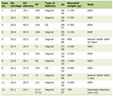 Click to view | Table 1. Clinical Timeline |
| Discussion | ▴Top |
This case was managed under a working diagnosis of presumed multisystem sarcoidosis reached through convergence rather than through a single decisive test. The patient had a compatible thoracic pattern, granulomatous inflammation in a supraclavicular node, and manifestations involving several systems, including skin and subcutaneous tissue, lymph nodes, lacrimal/parotid structures, calcium metabolism, and possibly pleural/pericardial compartments. The World Association of Sarcoidosis and Other Granulomatous Disorders (WASOG) organ-assessment approach similarly emphasizes that organ involvement is assigned after granulomatous inflammation has been established and reasonable alternatives have been considered [5]. In this patient, the longitudinal sequence increased diagnostic coherence, but the absence of mycobacterial culture and unavailable special-stain results appropriately limited certainty and precluded describing sarcoidosis as microbiologically exclusive or definitively confirmed.
The right-eyebrow nodule is important because it was the first observable event, not because it was pathognomonic. Cutaneous sarcoidosis can present with papules, plaques, nodules, scar infiltration, or other morphologies, and histology may be subtle or nonspecific [4]. The original report in this case described granuloma formation but interpreted the specimen as chronic dermatitis with an intradermal nevus. Retrospectively relabeling it as histologically proven cutaneous sarcoidosis would exceed the evidence. A more defensible conclusion is that it represented an early cutaneous clue whose significance became apparent only after systemic disease evolved. For clinical practice, a granulomatous skin lesion of uncertain cause should prompt review of exposures, infection risk, respiratory symptoms, lymph nodes, and other organ findings, with dermatology and pathology input when available.
Published periocular cases provide a useful contrast. An eyebrow-tattoo-associated case had bilateral eyebrow plaques with biopsy-proven non-caseating granulomas, whereas a periocular-scar case had non-necrotizing granulomatous inflammation that directly prompted systemic assessment [6, 7]. In the present patient, no tattoo or scar trigger was documented, the lesion was solitary, and the original specimen was interpreted as chronic dermatitis with an intradermal nevus. Its value was therefore retrospective and longitudinal rather than diagnostic at presentation.
The sequence of tissue sampling also illustrates why a non-diagnostic biopsy should not be treated as a negative disease test. The bronchial biopsy showed severe inflammation and necrosis but did not provide a coherent explanation for the radiological and systemic pattern. Sampling error, lesion heterogeneity, and the small size of endobronchial specimens may all reduce diagnostic yield. When the patient later developed accessible supraclavicular lymphadenopathy, biopsy of that site demonstrated multiple epithelioid granulomas without definite necrosis. This second specimen did not erase the first; instead, the discordance required synthesis with the entire phenotype. Selecting the safest active and accessible lesion for repeat sampling can be more informative than repeatedly sampling the same organ.
Because necrosis and granuloma morphology overlap across infectious and non-infectious disorders, the main histopathological alternatives and their relationship to the available specimens are summarized in Table 2 [3, 8]. Typical patterns are not absolute and must be interpreted with microbiology, immunophenotype, clinical features, and specimen adequacy.
Click to view | Table 2. Histopathological Differential Diagnosis Relevant to This Case |
Tuberculosis remained the most consequential competing diagnosis. Histological overlap is substantial: non-necrotizing granulomas can occur in tuberculosis, and absence of acid-fast organisms or caseation does not exclude paucibacillary disease [3, 9]. In this case, several negative results reduced the likelihood of active tuberculosis: five acid-fast smears from respiratory samples, bronchoalveolar-lavage Xpert MTB/RIF, bronchoalveolar-lavage metagenomic sequencing, and tissue M. tuberculosis nucleic-acid testing. Neither a tuberculin skin test nor a conventional interferon-gamma release assay was performed. A hospital-designated “M. tuberculosis dual-factor” whole-blood assay on May 22, 2025, was reported negative (M. tuberculosis-specific interferon-gamma 0.33 pg/mL (reference interval 0.0–7.0) and interleukin-2 0.31 pg/mL (reference interval 0.0–20.0)). Because the available report did not identify this platform as a conventional interferon-gamma release assay, it was reported separately rather than treated as an equivalent. None of the negative results alone has perfect negative predictive value across specimen types and disease burdens, and no respiratory or lymph-node specimen was submitted for mycobacterial culture. The appropriate wording is therefore that active tuberculosis was not microbiologically demonstrated and was judged less likely, not that it was definitively excluded.
Treatment further complicated attribution. The patient received a prolonged prednisone taper and concurrent preventive isoniazid and rifampicin. Glucocorticoids are recommended first-line for clinically significant symptomatic sarcoidosis, while steroid-sparing therapy is considered for relapse, refractory disease, or unacceptable toxicity [10]. The observed course is best described as clinical improvement following treatment for presumed sarcoidosis while receiving concurrent antituberculosis therapy. Response to treatment is not a diagnostic criterion, and the simultaneous regimens prevent causal attribution of the improvement in respiratory symptoms, subcutaneous nodules, hypercalcemia, kidney function, and CT abnormalities to prednisone alone. The later ultrasound impression favoring lymph-node tuberculosis was an imaging interpretation rather than microbiological proof and reinforces the need to separate confirmed evidence, radiological interpretation, and therapeutic precaution.
A practical diagnostic sequence emerges from this case. First, clinicians should map all organ manifestations and identify the safest lesion that is most likely to yield diagnostic tissue. Second, pathology should be interpreted together with specimen adequacy and the exact microbiological methods performed, rather than reduced to a binary label of granuloma present or absent. Third, tuberculosis testing should be matched to the sampled compartment and should include culture whenever feasible, because nucleic-acid amplification and metagenomic sequencing answer related but non-identical questions. Finally, follow-up should test the working diagnosis: expected improvement increases coherence, whereas persistent or discordant findings should reopen the differential. This approach does not eliminate uncertainty, but it makes the residual uncertainty visible and clinically manageable [1, 3].
The calcium and kidney findings added biological coherence but also required restraint. Sarcoidosis can increase extrarenal conversion of 25-hydroxyvitamin D to active 1,25-dihydroxyvitamin D, resulting in hypercalcemia with suppressed parathyroid hormone. The observed calcium of 3.24 mmol/L and parathyroid hormone of 8.76 pg/mL supported granulomatous activity. However, the documented renal history was hypertensive nephropathy for more than 10 years, which predated the granulomatous illness and was not considered sarcoidosis-related. The creatinine rise at readmission could therefore have represented an acute, multifactorial deterioration superimposed on chronic hypertensive kidney disease. Recovery of creatinine and calcium during concurrent treatment supports reversibility but does not establish renal sarcoidosis in the absence of renal histology or other organ-specific evidence. Similarly, the low 25-hydroxyvitamin D level at follow-up should not be confused with a measured 1,25-dihydroxyvitamin D concentration.
From a rheumatology perspective, sarcoidosis can include acute or chronic arthralgia and arthritis, periarticular ankle inflammation, enthesitis, myopathy, and osseous disease. A rheumatology-clinic series emphasized frequent ankle, metacarpophalangeal, wrist, and knee involvement, while a systematic review found that sarcoid arthritis most often presents as acute oligoarthritis or polyarthritis affecting lower-extremity large joints [11, 12]. No inflammatory joint or other musculoskeletal manifestation was documented in this patient, but explicit screening is relevant because musculoskeletal symptoms may precede or accompany pulmonary and cutaneous disease.
Cardiac involvement deserved explicit evaluation because it can be clinically silent and carries disproportionate risk. Serial electrocardiograms did not show conduction block or arrhythmia, and echocardiograms showed preserved LVEF without a focal structural lesion. These findings argued against clinically overt cardiac sarcoidosis. They did not, however, establish its absence. Grade I diastolic dysfunction, a small pericardial effusion, and elevated cardiac biomarkers were nonspecific, particularly during kidney dysfunction and uncertain volume status. Contemporary guidance conceptualizes cardiac sarcoidosis probability as a continuum and uses cardiac magnetic resonance or fluorodeoxyglucose positron-emission tomography when clinical suspicion persists [13]. Because neither modality nor ambulatory rhythm monitoring was performed here, subclinical cardiac involvement could not be fully excluded. The reason for not performing positron-emission tomography was not documented and should not be inferred.
Two follow-up observations are clinically useful. First, symptomatic and biochemical remission persisted after prednisone was stopped and no treatment was restarted. Second, abnormal supraclavicular nodes persisted on ultrasound. Together, these findings show why follow-up outcomes should be reported by domain. Resolution of respiratory symptoms and subcutaneous nodules, normalization of calcium and creatinine, and improvement on chest CT are not equivalent to complete anatomical remission. Continued surveillance is reasonable when imaging abnormalities remain or when a competing infection has not been fully excluded.
The principal limitations are the retrospective reconstruction of parts of the outpatient prednisone taper, absence of dispensing records for the late taper, lack of mycobacterial cultures and tuberculin skin or interferon-gamma release testing, unavailable tissue special-stain results, lack of whole-body and advanced cardiac imaging, and the use of pathology screenshots without retrievable staining methods, objective magnifications, or validated scale bars. A preoperative clinical photograph of the right-eyebrow lesion was also unavailable. These limitations reduce the ability to prove organ-specific involvement and to distinguish presumed sarcoidosis from coexisting or occult tuberculosis with absolute certainty. They are disclosed because transparent boundaries are more informative than filling missing evidence by assumption. The case follows the reporting principle that the clinical timeline, diagnostic alternatives, treatment details, outcomes, and limitations should be explicit [14].
Conclusions
An initially nonspecific cutaneous granulomatous nodule may become an important clue when later multisystem findings emerge. In patients with a phenotype compatible with presumed sarcoidosis and continuing tuberculosis plausibility, diagnostic reasoning should integrate serial tissue samples, specimen-specific microbiology, organ assessment, treatment overlap, and longitudinal outcomes. Negative molecular tests and improvement during concurrent therapy should narrow, but not prematurely close, the differential diagnosis.
| Supplementary Material | ▴Top |
Suppl 1. Bronchoscopic findings on May 26, 2025.
Suppl 2. Serial chest computed tomography images at a second representative axial level.
Acknowledgments
None to declare.
Financial Disclosure
No specific funding was received for the clinical care, data collection, analysis, or preparation of this case report. The article processing charge for this publication was supported by the Gansu Provincial Department of Science and Technology Joint Research Fund, General Program (Grant No. 25JRRA1281), the Natural Science Foundation of Gansu Province (Grant No. 22JR5RA993), and the Gansu Provincial Drug Regulatory Science Research Program (Grant No. 2023GSMPA019), all awarded to Xiang Yi Zan. The funders had no role in the clinical management of the patient, data collection or interpretation, preparation of the manuscript, or the decision to publish.
Conflict of Interest
The authors declare no conflict of interest.
Informed Consent
Written informed consent was obtained from the patient for publication of the case details and accompanying images. All identifying marks were removed from the submitted figures.
Author Contributions
Gan Lin Qiao: guarantor, data curation, case analysis, and writing – original draft. Wen Qing Cheng: data curation, figure preparation, and writing – review and editing. Wen Hua Zhang: data curation, literature review, and writing – review and editing. Yi Fan Song: data curation, figure preparation, and writing – review and editing. Xiang Yi Zan: conceptualization, supervision, project administration, and critical review. All authors reviewed and approved the final submitted version and accept responsibility for the work.
Data Availability
All data supporting the findings of this case report are included within the article. Additional deidentified information may be available from the corresponding author on reasonable request and subject to institutional and patient-privacy requirements.
AI Use Declaration
DeepSeek and ChatGPT (versions and dates not retained) and OpenAI Codex (GPT-5; used on September 17, 20, 21, and 22, October 28–29 and 31, and November 2, 2026) were used under author direction to organize source material, assist English translation and substantive language editing, check journal formatting and references, prepare responses to peer review, and assemble deterministic figure layouts from author-supplied images. No generative system was used to create or alter clinical data or diagnostic image content. The authors reviewed and verified the scientific content and accept full responsibility for the accuracy, originality, and integrity of the manuscript.
Abbreviations
AFB: acid-fast bacilli; BALF: bronchoalveolar-lavage fluid; CT: computed tomography; CRP: C-reactive protein; LVEF: left-ventricular ejection fraction; mNGS: metagenomic next-generation sequencing; PCR: polymerase chain reaction; WASOG: World Association of Sarcoidosis and Other Granulomatous Disorders
| References | ▴Top |
- Crouser ED, Maier LA, Wilson KC, Bonham CA, Morgenthau AS, Patterson KC, Abston E, et al. Diagnosis and detection of sarcoidosis. An official American thoracic society clinical practice guideline. Am J Respir Crit Care Med. 2020;201(8):e26-e51.
doi pubmed - Belperio JA, Shaikh F, Abtin FG, Fishbein MC, Weigt SS, Saggar R, Lynch JP, 3rd. Diagnosis and treatment of pulmonary sarcoidosis: a review. JAMA. 2022;327(9):856-867.
doi pubmed - Valeyre D, Brauner M, Bernaudin JF, Carbonnelle E, Duchemann B, Rotenberg C, Berger I, et al. Differential diagnosis of pulmonary sarcoidosis: a review. Front Med (Lausanne). 2023;10:1150751.
doi pubmed - Caplan A, Rosenbach M, Imadojemu S. Cutaneous sarcoidosis. Semin Respir Crit Care Med. 2020;41(5):689-699.
doi pubmed - Judson MA, Costabel U, Drent M, Wells A, Maier L, Koth L, Shigemitsu H, et al. The WASOG Sarcoidosis Organ Assessment Instrument: an update of a previous clinical tool. Sarcoidosis Vasc Diffuse Lung Dis. 2014;31(1):19-27.
pubmed - Nie S, Li K, Gao C, Yin N, Chen Z, Wu Z. Eyebrow tattoo-associated sarcoidosis: A case report. Front Med (Lausanne). 2022;9:1009135.
doi pubmed - Diaz-Cabanas L, Belani-Raju M, Gonzalez-Marquez PI, Artioli-Schellini S, Guerenu-Panero I, Galindo-Ferreiro A. Sarcoidosis in the periocular scar as the first finding of systemic sarcoidosis: Clinical-radiological characteristics. Arch Soc Esp Oftalmol (Engl Ed). 2019;94(9):453-459.
doi pubmed - Ohshimo S, Guzman J, Costabel U, Bonella F. Differential diagnosis of granulomatous lung disease: clues and pitfalls: Number 4 in the Series "Pathology for the clinician" Edited by Peter Dorfmuller and Alberto Cavazza. Eur Respir Rev. 2017;26(145):170012.
doi pubmed - Weeratunga P, Moller DR, Ho LP. Immune mechanisms of granuloma formation in sarcoidosis and tuberculosis. J Clin Invest. 2024;134(1):e175264.
doi pubmed - Baughman RP, Valeyre D, Korsten P, Mathioudakis AG, Wuyts WA, Wells A, Rottoli P, et al. ERS clinical practice guidelines on treatment of sarcoidosis. Eur Respir J. 2021;58(6):2004079.
doi pubmed - Bulut Gokten D, Mercan R. Sarcoidosis from the perspective of rheumatology: three years of experience. Nam Kem Med J. 2024;12(1):1-7.
doi - Yeung T, Grebowicz A, Nevskaya T, Zahid S, Pope JE. Joint involvement in sarcoidosis: systematic review and meta-analysis of prevalence, clinical pattern and outcome. Rheumatology (Oxford). 2024;63(7):1803-1814.
doi pubmed - Cheng RK, Kittleson MM, Beavers CJ, Birnie DH, Blankstein R, Bravo PE, Gilotra NA, et al. Diagnosis and management of cardiac sarcoidosis: a scientific statement from the American Heart Association. Circulation. 2024;149(21):e1197-e1216.
doi pubmed - Gagnier JJ, Kienle G, Altman DG, Moher D, Sox H, Riley D, CARE Group. The CARE guidelines: consensus-based clinical case reporting guideline development. BMJ Case Rep. 2013;2013:bcr2013201554.
doi pubmed
This article is distributed under the terms of the Creative Commons Attribution 4.0 International License (CC BY 4.0), which permits unrestricted use, distribution, and reproduction in any medium, including commercial use, provided the original work is properly cited.
Journal of Medical Cases is published by Elmer Press Inc.