Journal of Medical Cases, ISSN 1923-4155 print, 1923-4163 online, Open Access
Article copyright, the authors; Journal compilation copyright, J Med Cases and Elmer Press Inc
Journal website https://jmc.elmerpub.com

Case Report

Volume 18, 2027, e5433


From a Cutaneous Nodule to Presumed Multisystem Sarcoidosis Amid Persistent Tuberculosis Uncertainty

Figures

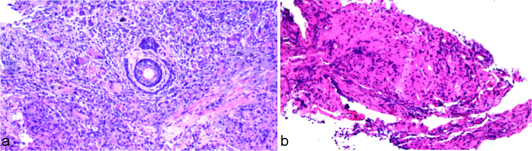
↓  Figure 1. Representative deidentified pathology screenshots. (a) Right-eyebrow skin specimen, report dated October 14, 2024; the report described chronic inflammation and granuloma formation with an associated intradermal nevus. (b) Left-main-bronchial biopsy, report dated May 27, 2025; the report described severe acute and chronic mucosal inflammation with necrosis. The pathology department could not provide staining methods, objective magnifications, image calibration, or validated scale bars; none has been inferred or added.
Figure 1.
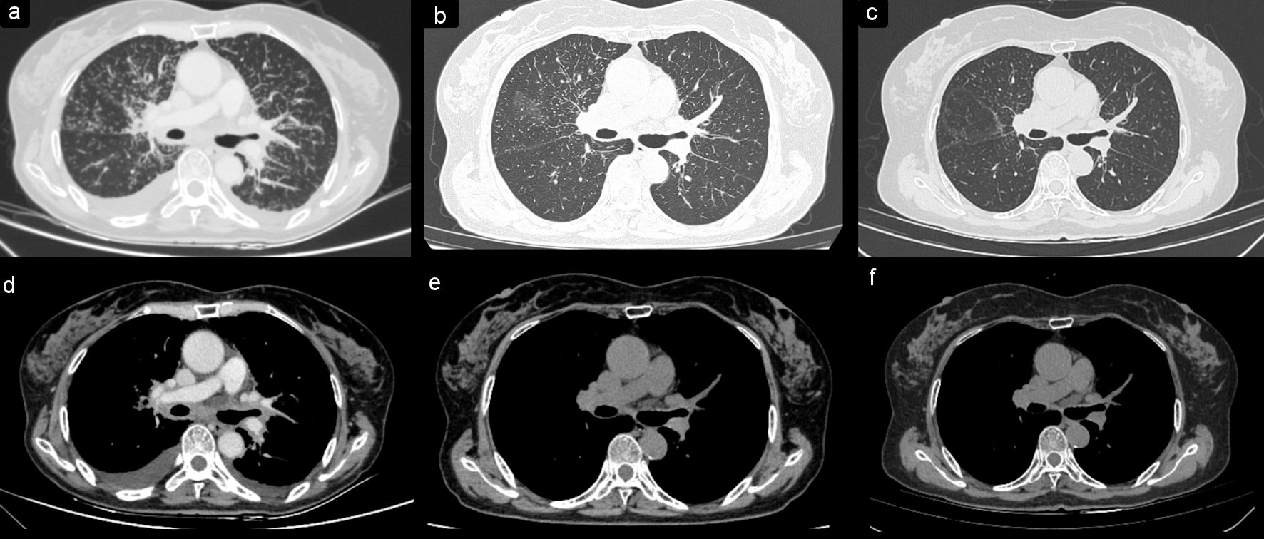
↓  Figure 2. Serial chest computed tomography images at three time points. Lung-window images were obtained on May 28, 2025 (a), January 7, 2026 (b), and June 10, 2026 (c); mediastinal-window images obtained at the same time points are shown in (d–f), respectively. The baseline images (a, d) show diffuse bilateral micronodular and peribronchovascular abnormalities, thoracic lymphadenopathy, and bilateral pleural effusions. The images obtained on January 7, 2026 (b, e) show marked interval improvement, and the latest images obtained on June 10, 2026 (c, f) show sustained radiographic improvement.
Figure 2.
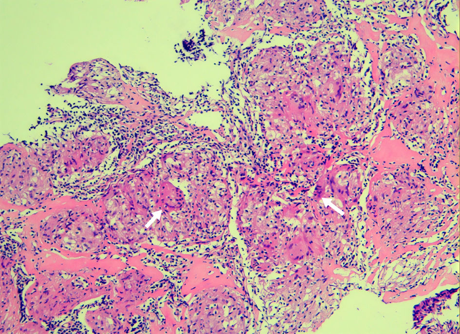
↓  Figure 3. Right supraclavicular-node biopsy reported on August 21, 2025. The report described multiple epithelioid granulomas with multinucleated giant cells and no definite necrosis. White arrows were present in the source image. The pathology department could not provide the staining method, objective magnification, image calibration, or a validated scale bar; none has been inferred or added.
Figure 3.

Tables

↓  Table 1. Clinical Timeline
 
DateEventKey finding
AFB: acid-fast bacilli; BALF: bronchoalveolar-lavage fluid; CT: computed tomography; CRP: C-reactive protein; LVEF: left-ventricular ejection fraction; mNGS: metagenomic next-generation sequencing; PCR: polymerase chain reaction; WBC: white blood cell.
October 14, 2024Right-eyebrow nodule pathologyChronic inflammation, granuloma formation, and intradermal nevus; not diagnostic of sarcoidosis
May 22–28, 2025Thoracic and cardiac evaluationDiffuse thoracic disease; preserved LVEF; grade I diastolic dysfunction; small effusions on CT
May 26–27, 2025Bronchoscopy and biopsyNodular mucosal elevations; severe acute and chronic inflammation with necrosis; non-diagnostic
May 22–26, and August 18, 2025MicrobiologyFive respiratory AFB smears negative; whole-blood M. tuberculosis dual-factor assay negative; BALF Xpert MTB/RIF and mNGS negative; routine bacterial/fungal cultures showed no pathogenic bacteria or fungi; no mycobacterial cultures submitted
August 18–25, 2025Systemic assessment and lymph-node biopsyHypercalcemia, kidney dysfunction, multiple-organ findings, non-necrotizing granulomas; tissue tuberculosis PCR negative
August 26, 2025Treatment after dischargePrednisone plus preventive isoniazid/rifampicin because immunosuppression was required despite residual tuberculosis uncertainty
January 7, 2026Follow-up chest CTInterval improvement with residual thoracic abnormalities
June 10, 2026Additional follow-up chest CTSustained radiographic improvement in pulmonary and thoracic abnormalities compared with baseline
July 3, 2026Prednisone stoppedEnd of patient-confirmed extended taper
July 22, 2026Laboratory and ultrasound follow-upCalcium, creatinine, CRP, and WBC improved; abnormal supraclavicular nodes persisted
September 20, 2026Clinical follow-upNo recurrent respiratory symptoms; subcutaneous nodules resolved; no treatment restarted


↓  Table 2. Histopathological Differential Diagnosis Relevant to This Case
 
ConditionTypical histopathological patternNecrosisRelevance to this case
AFB: acid-fast bacilli; BALF: bronchoalveolar-lavage fluid; mNGS: metagenomic next-generation sequencing; PCR: polymerase chain reaction.
SarcoidosisWell-formed epithelioid granulomas, usually with sparse surrounding inflammationUsually absent; focal fibrinoid necrosis may occurSupraclavicular-node granulomas without definite necrosis supported, but did not prove, the working diagnosis
TuberculosisEpithelioid granulomas, often caseating; non-necrotizing granulomas may occur in paucibacillary diseaseCommon but not obligatoryRespiratory AFB smears, BALF Xpert/mNGS, and tissue PCR were negative; no mycobacterial culture was submitted
Histiocytosis, particularly Langerhans cell histiocytosisLangerhans-cell aggregates with grooved nuclei, often accompanied by eosinophils; immunophenotyping is diagnosticNot a defining featureNo characteristic histiocytic infiltrate was reported; confirmatory immunostains were not available
Granulomatosis with polyangiitisNecrotizing granulomatous inflammation with necrotizing vasculitisUsually presentNo vasculitis was described in the available skin, bronchial, or node specimens
Bronchocentric granulomatosisDestructive necrotizing granulomatous inflammation centered on bronchioles, often with eosinophilsUsually presentBronchial necrosis was reported, but bronchiolocentric granulomatous destruction and an eosinophil-rich pattern were not described
HistoplasmosisNecrotizing granulomas containing small intracellular yeasts demonstrable with fungal stainsOften presentRespiratory cultures showed no fungal growth, and the sputum smear showed no hyphae or spores; tissue fungal stains and fungal PCR results were unavailable