| Journal of Medical Cases, ISSN 1923-4155 print, 1923-4163 online, Open Access |
| Article copyright, the authors; Journal compilation copyright, J Med Cases and Elmer Press Inc |
| Journal website https://jmc.elmerpub.com |
Case Report
Volume 17, Number 12, December 2026, pages 759-764

Emergency Physician-Performed Point-of-Care Ultrasound Identified Left Atrial Myxoma in Acute Cerebellar Infarction
Takanobu Satoa, Ryo Ichibayashia, c  , Sota Sadamotob
aDivision of Emergency Medicine, Department of Internal Medicine, Toho University Sakura Medical Center, Sakura, Chiba, Japan
bDepartment of Surgical Pathology, Toho University Omori Medical Center, Tokyo, Japan
cCorresponding Author: Ryo Ichibayashi, Division of Emergency Medicine, Department of Internal Medicine, Toho University Sakura Medical Center, 564-1 Shimoshizu Sakura-shi, Chiba 285-8741, Japan
Manuscript submitted September 20, 2026, accepted October 27, 2026, published online November 9, 2026
Short title: POCUS Diagnosis of Cardiac Myxoma
doi: https://doi.org/10.14740/jmc5432
| Abstract | ▴Top |
Cardiac myxoma is a rare but potentially curable cause of cardioembolic stroke. Early identification of a potential cardiac embolic source may be challenging when neurological symptoms predominate during the initial emergency department evaluation. A 72-year-old woman presented to the emergency department with acute-onset dizziness and vomiting. Although her National Institutes of Health Stroke Scale score was 0, she was unable to walk independently. Electrocardiography demonstrated newly diagnosed atrial fibrillation. Because a cardioembolic mechanism was considered, the emergency physician performed bedside cardiac point-of-care ultrasound (POCUS), which revealed a highly mobile left atrial mass attached to the interatrial septum. POCUS was performed 28 min after arrival, before brain magnetic resonance imaging, which demonstrated multiple acute cerebellar infarctions without large-vessel occlusion. Intravenous alteplase was not administered despite presentation within the therapeutic time window. Anticoagulation with intravenous unfractionated heparin was initiated. Surgical resection was postponed after hemorrhagic transformation of the cerebellar infarction was detected on hospital day 14. The patient underwent complete tumor resection on hospital day 41, and histopathological examination confirmed cardiac myxoma. She was discharged on hospital day 54 with a favorable outcome. This case illustrates the potential value of emergency physician-performed cardiac POCUS for early identification of structural cardiac abnormalities during the initial evaluation of selected patients with suspected cardioembolic stroke.
Keywords: Left atrial myxoma; Cardioembolic stroke; Cerebellar infarction; Emergency medicine; Transthoracic echocardiography; Point-of-care ultrasound
| Introduction | ▴Top |
Cardiac myxoma is the most common primary cardiac tumor but remains rare [1, 2]. Most cardiac myxomas arise in the left atrium, typically near the fossa ovalis of the interatrial septum [3–5].
Although cardiac myxoma is histologically benign, it can cause systemic embolization, which may be its initial clinical manifestation [4, 6, 7]. Ischemic stroke is a major neurological complication, although cardiac myxoma is an uncommon cause of stroke [7]. Because neurological symptoms may precede cardiac manifestations, recognition of the underlying cardiac lesion can be delayed [7, 8]. Cardiac point-of-care ultrasound (POCUS) may facilitate early identification of structural cardiac abnormalities during emergency department evaluation. Previous case reports have described the identification of left atrial myxoma by POCUS during emergency department evaluation of patients presenting with acute neurological symptoms [9–11].
We report a patient with acute cerebellar infarction who had a National Institutes of Health Stroke Scale (NIHSS) score of 0 despite being unable to walk independently and in whom emergency physician-performed cardiac POCUS identified a left atrial mass before brain magnetic resonance imaging (MRI). This case highlights the potential role of bedside cardiac ultrasound in the early identification of structural cardiac abnormalities in selected patients with suspected cardioembolic stroke.
| Case Report | ▴Top |
A 72-year-old woman with no significant medical history or regular medications was transported to the emergency department because of sudden-onset dizziness and repeated vomiting. She arrived at the emergency department approximately 110 min after symptom onset. She had no history of smoking. On arrival, she was alert, with a Glasgow Coma Scale score of 15. Her vital signs were as follows: body temperature, 36.7 °C; blood pressure, 131/97 mm Hg; heart rate, 110 beats/min with an irregular rhythm; respiratory rate, 18 breaths/min; and oxygen saturation, 98% on room air.
Neurological examination revealed no obvious focal deficits, including nystagmus, limb weakness, sensory disturbance, dysarthria, or facial palsy. Finger-to-nose testing was normal, and the NIHSS score was 0. Although she could sit independently, movement-provoked dizziness prevented independent walking and required full assistance for ambulation. Formal truncal ataxia assessment and the Head Impulse, Nystagmus, Test of Skew (HINTS) examination were not performed. Laboratory tests showed no clinically significant abnormalities. C-reactive protein was 0.03 mg/dL, prothrombin time-international normalized ratio was 0.90, activated partial thromboplastin time was 24.8 s, and D-dimer was 0.88 µg/mL. Liver and renal function tests were also unremarkable.
A 12-lead electrocardiogram demonstrated newly diagnosed atrial fibrillation with a heart rate of approximately 110 beats/min. Because a cardioembolic mechanism was considered, bedside cardiac POCUS was performed by the emergency physician, who had 4 years of experience performing cardiac ultrasonography, 28 min after emergency department arrival. Parasternal long-axis and apical four-chamber views revealed a highly mobile mass measuring approximately 35 × 20 mm attached to the left atrial side of the interatrial septum. The mass prolapsed through the mitral valve orifice during diastole. Left ventricular systolic function appeared preserved (Fig. 1a, b and Videos 1, 2, jmc.elmerpub.com).
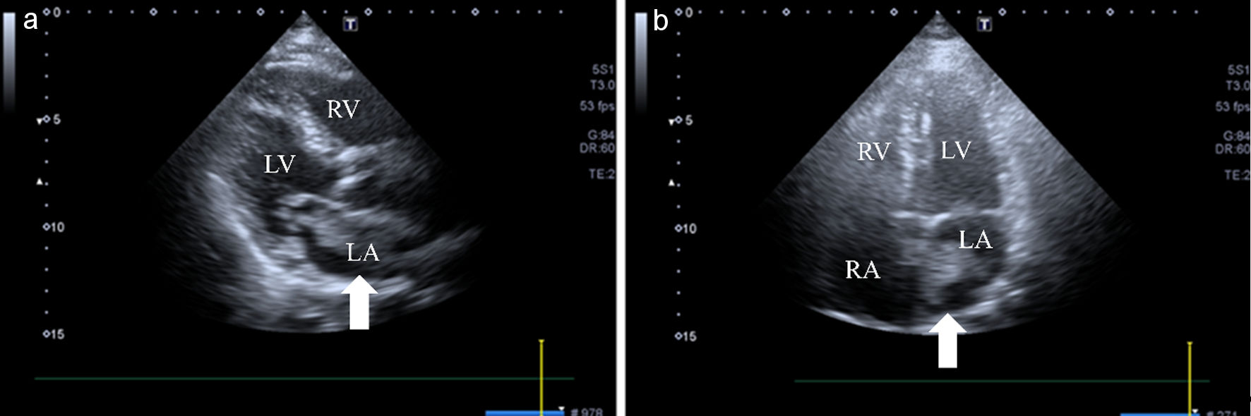 Click for large image | Figure 1. Cardiac point-of-care ultrasound findings. (a) Parasternal long-axis view demonstrating a highly mobile left atrial mass attached to the interatrial septum. The arrow indicates the mass. Left ventricular systolic function appeared preserved. (b) Apical four-chamber view demonstrating a highly mobile left atrial mass measuring approximately 35 × 20 mm. The arrow indicates the mass prolapsing through the mitral valve orifice during diastole. LA: left atrium; LV: left ventricle; RA: right atrium; RV: right ventricle. |
Based on these findings, a cardioembolic mechanism was suspected, with both the left atrial mass and newly diagnosed atrial fibrillation considered potential embolic sources. Brain MRI was subsequently performed 62 min after emergency department arrival and revealed multiple hyperintense lesions scattered in both cerebellar hemispheres on diffusion-weighted imaging, consistent with acute cerebellar infarctions. No corresponding signal changes were evident on fluid-attenuated inversion recovery imaging. Magnetic resonance angiography showed no significant stenosis, aneurysmal lesion, or large-vessel occlusion in the major intracranial arteries (Fig. 2a, b). Cardiology consultation was obtained 85 min after emergency department arrival for further evaluation of the left atrial mass.
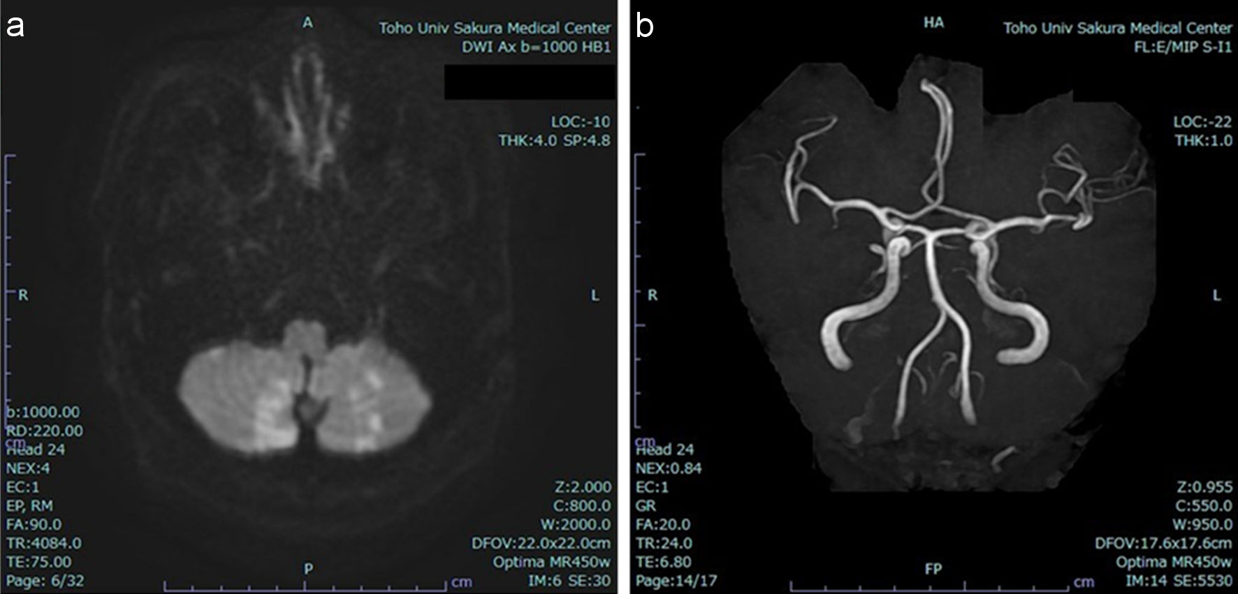 Click for large image | Figure 2. Brain magnetic resonance imaging and magnetic resonance angiography findings. (a) Diffusion-weighted imaging shows multiple hyperintense lesions scattered in both cerebellar hemispheres, consistent with acute cerebellar infarctions. (b) Magnetic resonance angiography shows no significant stenosis, aneurysmal lesion, or large-vessel occlusion in the major intracranial arteries. |
Although the patient presented within the therapeutic time window for intravenous thrombolysis, alteplase was not administered. The clinical assessment emphasized the absence of obvious focal neurological deficits and an NIHSS score of 0, although her inability to walk represented a potentially disabling symptom. Intravenous unfractionated heparin was initiated on the day of admission for presumed atrial fibrillation-related cardioembolic stroke, at 560 units/h without an initial bolus, targeting an activated partial thromboplastin time of 60–80 s. Intravenous glycerol was also administered to prevent or reduce cerebral edema. The patient was admitted to the high-care unit for close neurological and cardiovascular monitoring.
Contrast-enhanced computed tomography performed for further evaluation demonstrated a low-attenuation mass within the left atrium during the arterial phase. The mass was less clearly delineated on non-contrast and equilibrium-phase images. No evidence of embolic lesions in the major thoracic vessels was identified (Fig. 3a–c).
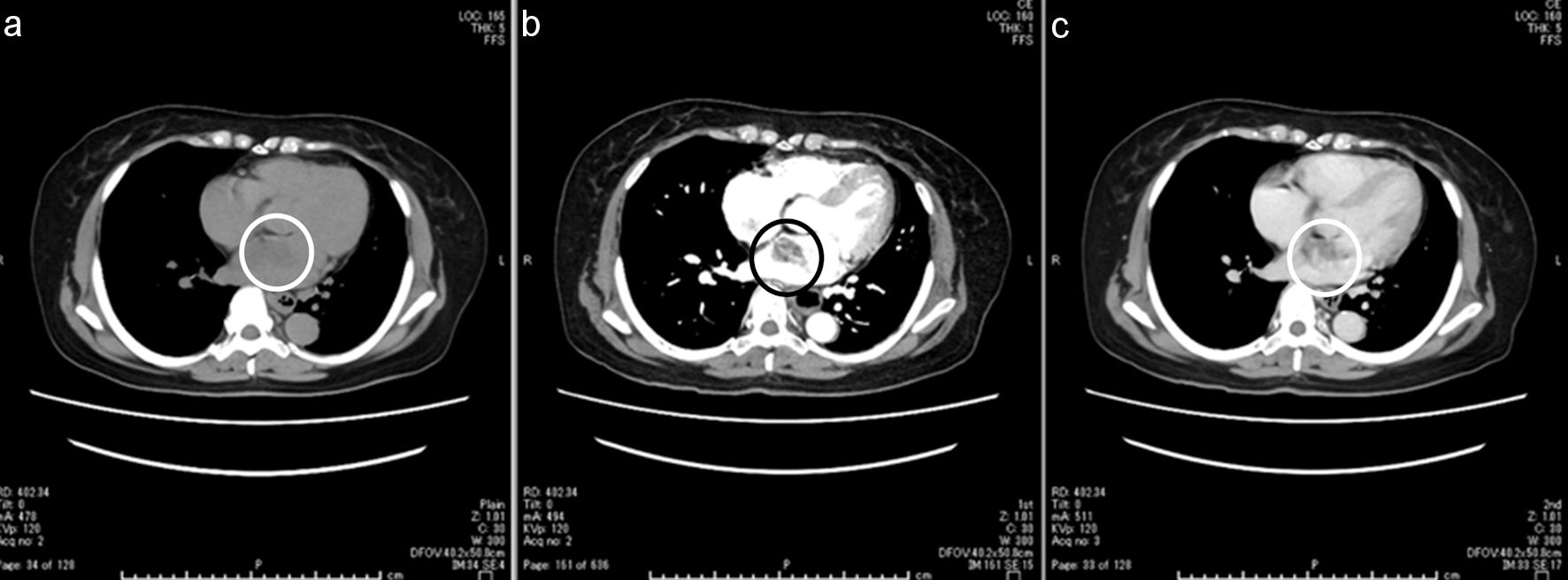 Click for large image | Figure 3. Chest computed tomography findings. (a) Non-contrast computed tomography. (b) Contrast-enhanced computed tomography in the arterial phase. (c) Contrast-enhanced computed tomography in the equilibrium phase. The left atrial mass is most clearly visualized as a low-attenuation lesion in the arterial phase (black circle) and is less clearly delineated on the non-contrast and equilibrium-phase images (white circles). |
Comprehensive transthoracic echocardiography on hospital day 2 confirmed a mobile left atrial mass measuring 37 × 21 mm with a broad attachment to the interatrial septum. Transesophageal echocardiography on hospital day 3 demonstrated a pedunculated mass measuring 20 × 30 mm arising near the fossa ovalis, without evidence of left atrial appendage thrombus. Mild mitral regurgitation was observed; mitral inflow obstruction was not quantitatively assessed.
On hospital day 14, follow-up neuroimaging demonstrated hemorrhagic transformation of the cerebellar infarction, classified as Heidelberg type 1c (PH1), involving less than 30% of the infarcted area without significant mass effect or fourth ventricular compression. No new neurological deficits were observed, and unfractionated heparin was immediately discontinued and not restarted. Therefore, surgical resection of the left atrial tumor was postponed until stabilization of the intracranial lesion. Follow-up computed tomography showed no progression of hemorrhage on hospital days 16 and 34. Repeat transthoracic echocardiography on hospital day 37 demonstrated a persistent, mobile left atrial mass measuring approximately 44 × 22 mm. After careful reassessment by the multidisciplinary team, including consideration of the anticoagulation required for cardiopulmonary bypass, the patient underwent surgical resection of the left atrial tumor on hospital day 41 with concomitant left atrial appendage closure. Intraoperatively, the tumor was found to originate from the interatrial septum near the fossa ovalis and was completely excised with its attachment site. Gross examination demonstrated a multilobulated, gelatinous mass measuring 50 × 25 × 20 mm. Histopathological examination revealed spindle- and stellate-shaped cells embedded in abundant myxoid stroma, confirming the diagnosis of cardiac myxoma. The tumor surface was friable and showed focal hemorrhagic changes, suggesting a potential for embolization but not establishing the tumor as the definitive embolic source (Fig. 4a–c).
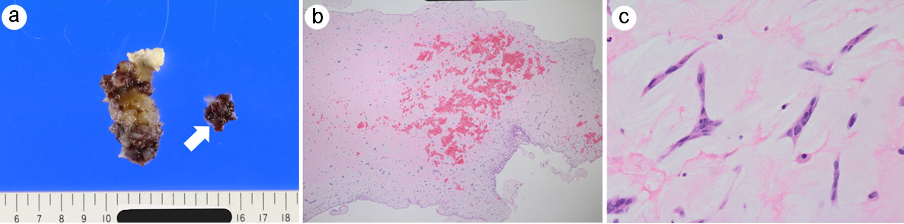 Click for large image | Figure 4. Gross and histopathological findings of the resected left atrial myxoma. (a) Gross specimen of the surgically resected left atrial mass showing a gelatinous, multilobulated appearance with focal hemorrhagic changes. The arrow indicates a detached tumor fragment. The excised tumor measured 50 × 25 × 20 mm on gross examination. Whether separation occurred in vivo or during surgical manipulation could not be determined. (b) Low-power hematoxylin and eosin (H&E) staining demonstrating abundant myxoid stroma with focal intratumoral hemorrhage. (c) High-power H&E staining showing scattered stellate and spindle-shaped myxoma cells embedded within a myxoid matrix. |
The postoperative course was uneventful, with no recurrent embolic events or neurological deterioration. Sinus rhythm was documented after surgery and during outpatient follow-up. She was discharged on hospital day 54 with a favorable functional outcome, without anticoagulation after discussion with the patient despite a CHA2DS2-VASc score of 4. At approximately 10 months after discharge, follow-up transthoracic echocardiography showed no recurrent left atrial mass, with preserved left ventricular systolic function and mild mitral regurgitation. Systematic screening for Carney complex, including genetic testing, was not performed.
| Discussion | ▴Top |
The key clinical feature of this case was the early detection of an unexpected, surgically treatable left atrial mass by emergency physician-performed POCUS before brain MRI. The patient presented with dizziness and vomiting and had an NIHSS score of 0 despite being unable to walk independently. Newly diagnosed atrial fibrillation prompted focused cardiac ultrasound, which identified a potential structural embolic source during the initial emergency department evaluation.
POCUS facilitated early recognition of the cardiac mass and prompted cardiology consultation and comprehensive echocardiography. Its contribution was diagnostic rather than a demonstrable change in immediate stroke treatment; decisions regarding thrombolysis, anticoagulation, and admission might have been unchanged without POCUS.
Cardiac myxoma is the most common primary cardiac tumor but is an uncommon cause of ischemic stroke [1, 4, 7]. Neurological presentations may delay recognition of the underlying cardiac lesion [7, 8]. Bradshaw et al reported emergency physician-performed POCUS detecting a left atrial mass in a young patient with stroke-like symptoms [10]. Quinn et al described POCUS identification of an atrial myxoma in a child presenting with stroke [9], while Chin et al reported POCUS detection of a left atrial myxoma in an adult presenting with stroke-like symptoms [11]. In contrast, our patient was an older adult with posterior circulation symptoms, and POCUS detected the mass before brain MRI. These reports illustrate the potential diagnostic role of POCUS across different clinical presentations, without establishing its routine use in stroke evaluation.
The decision to withhold intravenous thrombolysis warrants consideration. Alteplase was not administered despite presentation within the therapeutic time window, in the setting of an NIHSS score of 0 and no obvious focal neurological deficits. However, inability to walk independently was potentially disabling and may have been underestimated by the NIHSS, particularly in posterior circulation stroke.
Surgical excision is the definitive treatment for cardiac myxoma because of the risks of embolization and cardiac obstruction [4–6]. The optimal timing after ischemic stroke remains uncertain [8, 12]. Hemorrhagic transformation on hospital day 14 delayed surgery until day 41, requiring a balance between recurrent embolization and the risk of intracranial hemorrhage associated with anticoagulation during cardiopulmonary bypass.
The myxoma was a plausible embolic source given its mobility on POCUS and friable pathological appearance. Mobile, friable, villous, or papillary myxomas have been associated with greater embolic propensity [3, 4, 7, 8]. However, newly diagnosed atrial fibrillation was a competing potential source. The absence of left atrial appendage thrombus on transesophageal echocardiography did not exclude atrial fibrillation-related embolism, and the precise source could not be established.
Anticoagulation was discontinued after hemorrhagic transformation and was not resumed before discharge. Although left atrial appendage closure was performed, the CHA2DS2-VASc score of 4 indicated a continued need for thromboembolic risk assessment, rhythm surveillance, and reconsideration of anticoagulation.
This single case report cannot establish the generalizability or clinical outcome benefits of cardiac POCUS in suspected stroke. POCUS is operator-dependent and cannot replace comprehensive echocardiography. Nevertheless, it may facilitate the early recognition of structural cardiac abnormalities in selected patients with suspected cardioembolic stroke.
Conclusions
This case demonstrates that emergency physician-performed POCUS can identify an unexpected structural cardiac abnormality as a potential source of embolism at the bedside, even in a patient with an NIHSS score of 0 and disabling gait impairment. In selected patients with suspected cardioembolic stroke, focused cardiac ultrasound may facilitate early recognition of a potential cardiac source and prompt comprehensive cardiac evaluation.
Learning points
Posterior circulation infarction may present with dizziness and vomiting despite an NIHSS score of 0 and disabling gait impairment.
Newly diagnosed atrial fibrillation does not exclude a coexisting structural cardiac source of embolism.
Emergency physician-performed cardiac POCUS may facilitate early recognition of a treatable intracardiac mass, although comprehensive echocardiography remains necessary for definitive cardiac evaluation.
| Supplementary Material | ▴Top |
Video 1. Parasternal long-axis view of the left atrial mass. Emergency physician-performed cardiac POCUS demonstrating a highly mobile left atrial mass attached to the interatrial septum.
Video 2. Apical four-chamber view of the left atrial mass. Emergency physician-performed cardiac point-of-care ultrasound demonstrating a mobile left atrial mass prolapsing through the mitral valve orifice during diastole.
Acknowledgments
None to declare.
Financial Disclosure
No funding was received for this study.
Conflict of Interest
The authors declare no conflict of interest.
Informed Consent
Written informed consent for publication of this case report and accompanying images was obtained from the patient.
Author Contributions
TS performed the bedside cardiac POCUS and drafted the manuscript. RI supervised manuscript preparation and critically revised the manuscript. SS performed and interpreted the histopathological examination and contributed to the pathological description. All authors read and approved the final manuscript.
Data Availability
The data supporting the findings of this case report are available from the corresponding author upon reasonable request, subject to restrictions protecting patient privacy.
AI Use Declaration
During the preparation of this manuscript, the authors used ChatGPT (OpenAI) to assist with English-language editing and refinement of the manuscript structure and wording, particularly in the Introduction and Discussion. The authors critically reviewed and revised all AI-generated suggestions and take full responsibility for the accuracy, originality, and integrity of the final manuscript.
Abbreviations
AI: artificial intelligence; AF: atrial fibrillation; H&E: hematoxylin and eosin; MRI: magnetic resonance imaging; NIHSS: National Institutes of Health Stroke Scale; POCUS: point-of-care ultrasound
| References | ▴Top |
- Grebenc ML, Rosado de Christenson ML, Burke AP, Green CE, Galvin JR. Primary cardiac and pericardial neoplasms: radiologic-pathologic correlation. Radiographics. 2000;20(4):1073-1103.
doi pubmed - Keeling IM, Oberwalder P, Anelli-Monti M, Schuchlenz H, Demel U, Tilz GP, Rehak P, et al. Cardiac myxomas: 24 years of experience in 49 patients. Eur J Cardiothorac Surg. 2002;22(6):971-977.
doi pubmed - Wold LE, Lie JT. Cardiac myxomas: a clinicopathologic profile. Am J Pathol. 1980;101(1):219-240.
pubmed - Reynen K. Cardiac myxomas. N Engl J Med. 1995;333(24):1610-1617.
doi pubmed - Butany J, Nair V, Naseemuddin A, Nair GM, Catton C, Yau T. Cardiac tumours: diagnosis and management. Lancet Oncol. 2005;6(4):219-228.
doi pubmed - Oktaviono YH, Saputra PBT, Arnindita JN, Afgriyuspita LS, Kurniawan RB, Pasahari D, Milla C, et al. Clinical characteristics and surgical outcomes of cardiac myxoma: A meta-analysis of worldwide experience. Eur J Surg Oncol. 2024;50(2):107940.
doi pubmed - Yuan SM, Humuruola G. Stroke of a cardiac myxoma origin. Rev Bras Cir Cardiovasc. 2015;30(2):225-234.
doi pubmed - Stefanou MI, Rath D, Stadler V, Richter H, Hennersdorf F, Lausberg HF, Lescan M, et al. Cardiac myxoma and cerebrovascular events: a retrospective cohort study. Front Neurol. 2018;9:823.
doi pubmed - Quinn N, Kalichuran R, Prenderville T, McGuinness J. Point-of-Care Ultrasound echocardiography diagnosis of an atrial myxoma in a child presenting with stroke. Australas J Ultrasound Med. 2020;23(1):80-83.
doi pubmed - Bradshaw JC, Arthur J, Lewis ZB. Ultrasound-assisted diagnosis of embolic cerebrovascular accident from left atrial myxoma in the emergency department. J Emerg Med. 2021;61(4):e60-e63.
doi pubmed - Chin WV, Khaw MJ. Point-of-care ultrasound: a window into the diagnosis of atrial myxoma in stroke-like patient. J Med Ultrasound. 2025;33(4):381-384.
doi pubmed - Tu X, Tan H, Song Q, Liu L, Liu H, Cheng W. Early and intermediate outcomes of surgical treatment for atrial myxoma complicated by preoperative ischemic stroke: a retrospective study. Eur J Med Res. 2025;30(1):569.
doi pubmed
This article is distributed under the terms of the Creative Commons Attribution 4.0 International License (CC BY 4.0), which permits unrestricted use, distribution, and reproduction in any medium, including commercial use, provided the original work is properly cited.
Journal of Medical Cases is published by Elmer Press Inc.