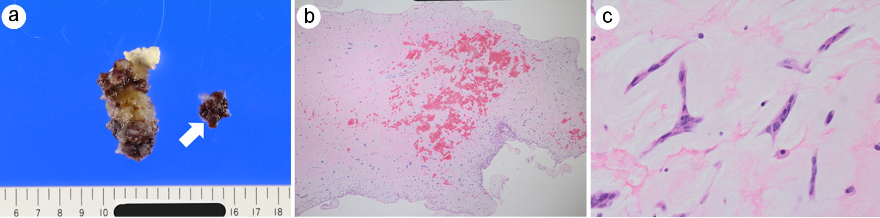
↓ Figure 1. Cardiac point-of-care ultrasound findings. (a) Parasternal long-axis view demonstrating a highly mobile left atrial mass attached to the interatrial septum. The arrow indicates the mass. Left ventricular systolic function appeared preserved. (b) Apical four-chamber view demonstrating a highly mobile left atrial mass measuring approximately 35 × 20 mm. The arrow indicates the mass prolapsing through the mitral valve orifice during diastole. LA: left atrium; LV: left ventricle; RA: right atrium; RV: right ventricle.
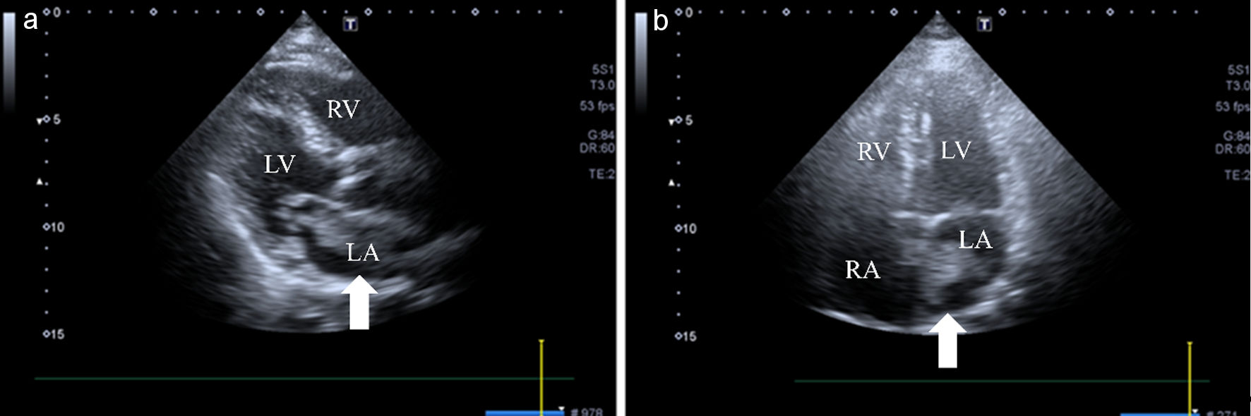
↓ Figure 2. Brain magnetic resonance imaging and magnetic resonance angiography findings. (a) Diffusion-weighted imaging shows multiple hyperintense lesions scattered in both cerebellar hemispheres, consistent with acute cerebellar infarctions. (b) Magnetic resonance angiography shows no significant stenosis, aneurysmal lesion, or large-vessel occlusion in the major intracranial arteries.
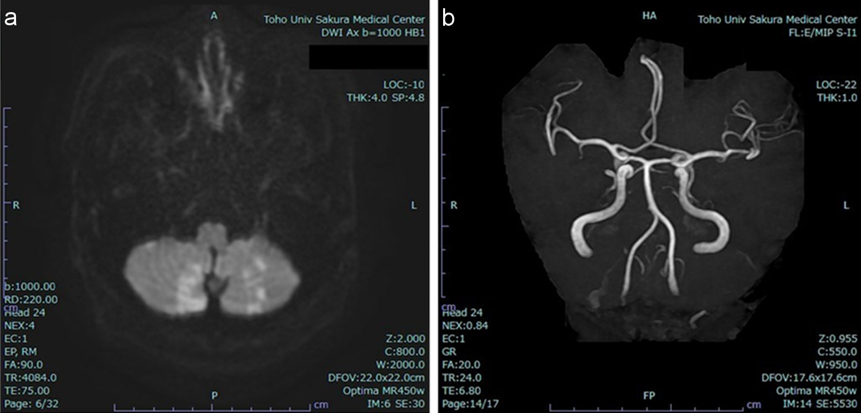
↓ Figure 3. Chest computed tomography findings. (a) Non-contrast computed tomography. (b) Contrast-enhanced computed tomography in the arterial phase. (c) Contrast-enhanced computed tomography in the equilibrium phase. The left atrial mass is most clearly visualized as a low-attenuation lesion in the arterial phase (black circle) and is less clearly delineated on the non-contrast and equilibrium-phase images (white circles).
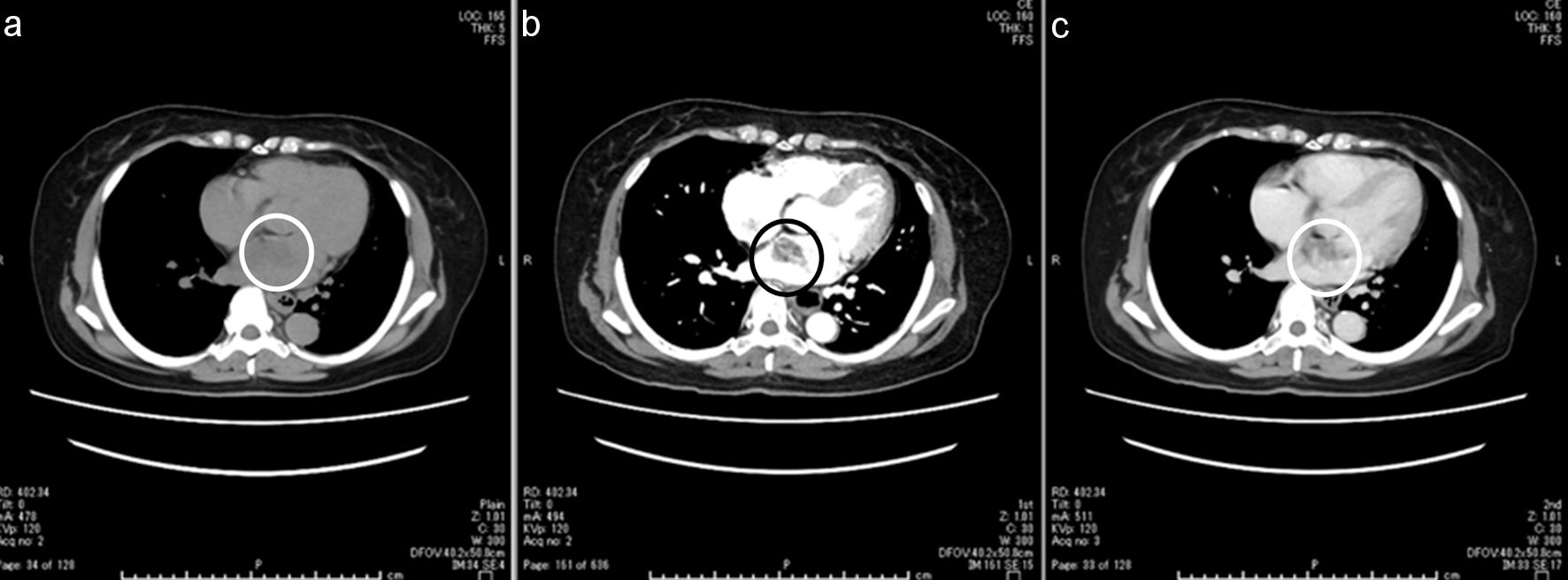
↓ Figure 4. Gross and histopathological findings of the resected left atrial myxoma. (a) Gross specimen of the surgically resected left atrial mass showing a gelatinous, multilobulated appearance with focal hemorrhagic changes. The arrow indicates a detached tumor fragment. The excised tumor measured 50 × 25 × 20 mm on gross examination. Whether separation occurred in vivo or during surgical manipulation could not be determined. (b) Low-power hematoxylin and eosin (H&E) staining demonstrating abundant myxoid stroma with focal intratumoral hemorrhage. (c) High-power H&E staining showing scattered stellate and spindle-shaped myxoma cells embedded within a myxoid matrix.