| Journal of Medical Cases, ISSN 1923-4155 print, 1923-4163 online, Open Access |
| Article copyright, the authors; Journal compilation copyright, J Med Cases and Elmer Press Inc |
| Journal website https://jmc.elmerpub.com |
Case Report
Volume 17, Number 12, December 2026, pages 750-758

Anterior Pelvic Herniation After Urethral Atresia: A Case Report and the Bladder Space-Occupying Hypothesis
Hai Nan Lia, Xiao Bao Liub, Ji Hong Shena, c
aThe First Affiliated Hospital of Kunming Medical University, Kunming 650032, China
bSchool of Mechanical and Electrical Engineering, Kunming University of Science and Technology, Kunming 650032, China
cCorresponding Author: Ji Hong Shen, The First Affiliated Hospital of Kunming Medical University, Kunming 650032, China
Manuscript submitted September 15, 2026, accepted October 22, 2026, published online November 9, 2026
Short title: Anterior Pelvic Herniation After Urethral Atresia
doi: https://doi.org/10.14740/jmc5430
| Abstract | ▴Top |
We report a case of long-standing acquired urethral atresia in an adult, accompanied by bilateral ureterovaginal fistulas, severe bladder atrophy, and an anterior pelvic hernia. We propose a hypothetical biomechanical explanation to elucidate the possible relationship among these abnormalities. We report a 71-year-old female patient who had undergone urethral tumor resection at another hospital 10 years earlier and had since experienced persistent vaginal urine leakage for more than 10 years; she presented this time with a progressively enlarging vaginal mass. The diagnosis was established based on physical examination, contrast-enhanced computed tomography (CT), and intraoperative exploration findings. Intraoperative exploration revealed complete urethral obliteration, bilateral ureterovaginal fistulae, a severely atrophic bladder remnant, and an anterior pelvic hernia containing small bowel that protruded through the vesicouterine fossa and clinically resembled a grade III cystocele. The patient underwent excision of the prolapsed vaginal mass and bilateral cutaneous ureterostomy. Based on the clinical course, imaging findings, and intraoperative observations, a three-stage biomechanical cascade hypothesis is proposed: urethral outlet obstruction may lead to elevated intravesical pressure, which in turn causes “pressure-induced perforation” at the ureterovesical junction. Direct urine diversion through the fistula results in bladder disuse atrophy. Loss of the bladder’s space-occupying function leads to reopening of the vesicouterine pouch, which then expands into an anterior pelvic hernia under repeated intra-abdominal pressure. To our knowledge, acquired urethral atresia with bilateral ureterovaginal fistulas in adults has not been previously reported. Although this hypothesis still requires further clinical and finite element modeling studies for precise validation, the proposed “bladder space-occupying effect hypothesis” may provide, in the future, a theoretical framework for subsequent research on atypical pelvic organ prolapse after long-term bladder disuse.
Keywords: Urethral atresia; Atrophy due to bladder dysfunction; Ureterovaginal fistula; Pelvic organ prolapse; Space-occupying lesion hypothesis
| Introduction | ▴Top |
Urethral atresia is a serious and debilitating postoperative complication; in addition to directly impairing the patient’s lower urinary tract function, the prolonged state of obstruction may also disrupt the biomechanical balance of the pelvic floor, gradually affecting the integrity of the remaining intact pelvic floor structures and thereby leading to a variety of secondary pelvic floor disorders [1]. However, there is currently a lack of systematic research confirming the specific progression and ultimate consequences of this long-term pathology.
Ureterovaginal fistula is one of the more serious complications among the aforementioned long-term effects; the vast majority of cases arise following gynecological pelvic surgery, whilst non-iatrogenic and non-obstetric causes are extremely rare [2]. A single case of congenital urethral atresia in a child has been reported; this was primarily due to the presence of a giant teratoma in the caudalis region, resulting in complete atresia of the mid-urethra and the formation of a urethrovaginal fistula as an alternative route for urine drainage [3]. However, there are currently very few reported cases of bilateral ureterovaginal fistula secondary to acquired urethral atresia in adults.
Furthermore, the causal relationship between bladder atrophy due to disuse and other forms of pelvic organ prolapse (POP) has not yet been systematically investigated. The classic pelvic floor support theories primarily include the three-level theory of the mid- and lower segments of the vagina proposed by DeLancey [4], the “hammock hypothesis” concerning the tissues forming the area below the mid-urethra [5], and the holistic theory of the pelvic floor proposed by Petros and Ulmsten in 1990 [6]. These approaches shift the focus from viewing pelvic floor disorders as isolated organ pathologies to considering ligaments, fascia, muscles, and nerves as an integrated whole. It primarily explains the occurrence of pelvic organ dysfunction (prolapse, incontinence, voiding disorders) in terms of damage to the pelvic floor connective tissue (fascia/ligaments). As an organ responsible for urine storage and voiding, the bladder, when full, occupies the spaces anteriorly between the symphysis pubis and posteriorly between the uterus and the vagina through its physical form, thereby sealing the vesicouterine fossa and ensuring the integrity of the anterior pelvic cavity and the physiological closure of these spaces [7]. However, none of these studies have analyzed or investigated the bladder itself as a structural-mechanical component of the anterior pelvis.
Here, unlike urethrovaginal leakage caused by congenital urethral atresia [8], we report a rare case of bilateral ureterovaginal fistulas with acquired urethral atresia in an adult, coexisting with a severely atrophic bladder and an anterior pelvic hernia. Because the evolution of this condition could only be reconstructed retrospectively from a single late assessment, we do not consider any specific sequence of events to be established. Instead, we first describe the directly observed clinical findings and then separately propose a hypothetical biomechanical cascade to explain the association among urethral atresia, fistula formation, bladder disuse atrophy, and anterior pelvic hernia. On this basis, we propose the “bladder space-occupying hypothesis” as a theoretical framework to be validated in future studies, aiming to explain atypical POP occurring after long-term bladder disuse.
| Case Report | ▴Top |
Patients and clinical data
The patient was a 71-year-old woman (gravida 3, para 2 (G3P2)) who was admitted to hospital presenting with “a subjective sensation of urine leaking from the vaginal opening for over 10 years, and a progressively enlarging vaginal mass for 1 week”. Ten years prior, the patient had undergone urethral tumor resection at another hospital due to a mass at the urethral orifice; postoperatively, she reported an inability to control her urine, with urine leaking from the vagina.
The outside hospital’s surgical records, histopathological reports, and early postoperative course records could not be retrieved; therefore, it was not possible to verify the histological nature of the urethral lesion, the extent of resection, or the exact interval between surgery and the onset of vaginal leakage. Over the subsequent 10 years, the patient experienced persistent urine leakage and required long-term use of sanitary pads. She had been diagnosed with “vesicovaginal fistula” at other hospitals on multiple occasions, and this diagnosis appeared to have been made on clinical grounds alone; this diagnosis was not confirmed at our hospital, and no targeted treatment was administered.
Over the past week, the vaginal mass had significantly enlarged to approximately the size of a “goose’s egg”, accompanied by pain, difficulty moving, and a sensation of pelvic heaviness. The patient also presented with frequent urination (10–20 times per day), urinary urgency, dysuria, and difficulty passing stools. She denied a history of pelvic radiotherapy or other pelvic surgery.
Preoperative investigations
Physical examination
A prolapsed mass measuring approximately 8 × 8 cm was visible at the vaginal opening; the mucosa was rosy in color with no obvious ulceration. The prolapse worsened with straining and could not be reduced at rest. The external urethral orifice could not be palpated. The clinical presentation was consistent with grade III cystocele [9].
Pelvic ultrasound
In the anterior view, marked protrusion of the bladder was visible, along with protrusion of the anterior vaginal wall and the vaginal fornix; in the mid-view, there was a clear echographic sign of uterine prolapse; in the posterior view, protrusion of the posterior vaginal wall was observed; there was an old anal sphincter tear at the 2 o’clock position.
Pelvic contrast-enhanced computed tomography (CT) scan
Contrast-enhanced CT showed: (1) formation of a pelvic floor hernia anterior to the uterus and vagina, with the hernial contents comprising the small intestine and its mesentery; (2) bilateral ureters descending into the hernial sac; (3) the bladder is not clearly visualized; (4) bilateral ureters are markedly thickened and tortuous throughout their course, with the left ureteral wall showing slight thickening and enhancement; (5) severe hydronephrosis of both kidneys (Fig. 1).
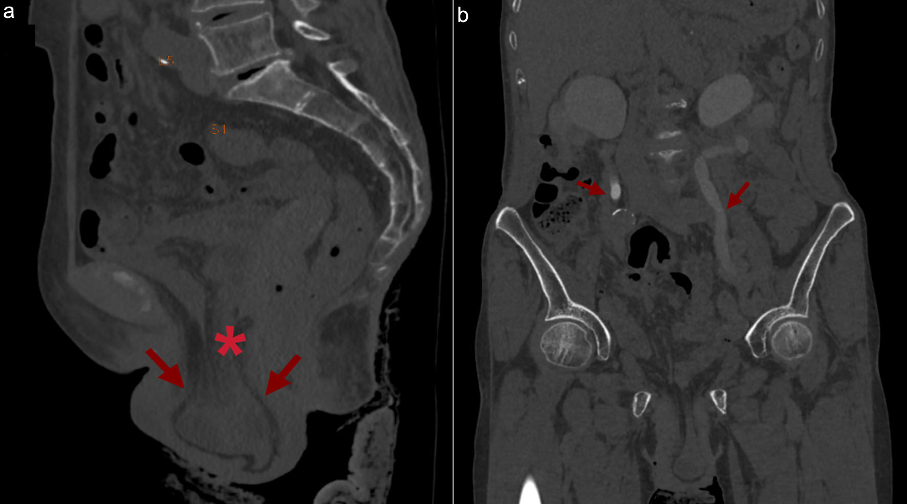 Click for large image | Figure 1. Preoperative contrast-enhanced computed tomography. (a) Sagittal reconstruction: an anterior pelvic hernia (arrow) protrudes anteriorly, anterior to the uterus and vagina; its contents are small bowel loops passing through the vesicouterine pouch. No distinct bladder is visible in its expected location (asterisk). (b) Coronal reconstruction: severe bilateral hydronephrosis, with marked dilatation and tortuosity of the ureters (arrows), which extend inferiorly into the hernial sac. Neither image directly demonstrates the fistulous tract, which was confirmed at surgery. |
Laboratory tests
Urinalysis and urine culture showed uric acid 373.3 mol/L, creatinine 129.1 µmol/L, urine occult blood 3+, and urine leukocyte esterase (3+).
Cardiac assessment
Cardiac assessment showed coronary artery calcium score of 148.62 points; a myocardial bridge in the mid-segment of the left anterior descending artery (approximately 15% stenosis); atrial septal defect with a small left-to-right shunt. A consultation with the Vascular Surgery Department confirmed mild fusiform dilation of the lower segment of the abdominal aorta (maximum diameter approximately 28 mm), with no indications for urgent intervention.
Surgical procedure
The patient underwent exploratory surgery under general anesthesia. In view of complete urethral atresia, severe bladder atrophy with no possibility of reconstruction, and bilateral ureterovaginal fistulas, vaginal mass excision, and bilateral ureterostomy were performed. Both ureters were mobilized for approximately 10 cm and transected distally. A circular skin incision was made 3 cm below the umbilical plane along the lateral margins of both rectus abdominis muscles, through which the ureters were brought out of the body. Double-J stents were placed and secured with sutures. The herniated small intestine was reduced, a pelvic drainage tube was placed at the lower right side of the incision, and the abdominal wall incision was then closed layer by layer.
| Results | ▴Top |
Intraoperative findings
The key intraoperative findings were as follows:
(1) Complete urethral atresia: The prolapsed tissue at the vaginal opening was difficult to reduce; no urethral orifice was visible, and the urethra was completely absent, replaced by a fibrous cord-like structure with no discernible lumen.
(2) Bilateral ureterovaginal fistulae: on traction, marked thickening of the anterior and lateral vaginal walls was observed; two fistula orifices, approximate visual estimate 3 cm apart and 2 mm in diameter, were visible on the anterior wall. A guidewire inserted through each fistula orifice was identified within the mobilized ureter, indicating that each tract directly communicated the distal ureter with the vaginal cavity; these fistulas were therefore classified as ureterovaginal rather than vesicovaginal. Consequently, urine from both kidneys bypassed the bladder. Based on the location of the fistula orifices, we inferred that each tract originated at or near the ureterovesical junction (UVJ); however, the exact site of perforation and its mechanism of formation could not be directly observed macroscopically.
(3) Severe bladder atrophy: Abdominal exploration revealed no distinct bladder structure; only a small, firm, thick-walled bladder remnant was observed. Intraoperatively, based on the size of the remnant, its capacity was visually estimated to be approximately 10–30 mL, which was a rough intraoperative estimate; because the urethra was atretic, formal cystometry could not be performed.
(4) Anterior pelvic hernia (bladder-uterine fossa defect): In the anterior pelvic region, a peritoneal defect measuring approximately 3–4 cm was observed; the mesentery of the small intestine and atrophic bladder tissue herniated through this defect into the anterior fornix, presenting a “fan-shaped” appearance.
(5) Bilateral ureteral hydronephrosis: The bilateral ureters were markedly dilated and tortuous, with severe hydronephrosis of both kidneys.
Postoperative course and follow-up
The patient recovered uneventfully after surgery, with unobstructed drainage from both stomas (left: 1,100–1,900 mL/day; right: 700–1,300 mL/day). She was discharged on postoperative day 7; 1 month later, a second vaginal mass excision was performed because of “residual prolapsed vaginal tissue at the excision site,” and she was discharged on postoperative day 2. Final diagnoses were: (1) bilateral ureterovaginal fistulas; (2) small bowel hernia (anterior pelvic region); (3) urinary tract infection; (4) bilateral hydronephrosis; (5) cardiac insufficiency; and (6) bilateral renal insufficiency.
One month after the first surgery, both stomas and double-J stents were functioning well; the urine was clear, and there was no infection of the skin around the stomas; and bilateral hydronephrosis and bilateral hydroureterosis were reduced compared with before.
Proposed biomechanical interpretation: a three-stage hypothesis
Based on the above findings, we propose a hypothetical sequence of events that may link these phenomena and divide it into three stages (Fig. 2). This explanation is retrospective and qualitative in nature. The temporal order of the stages was not recorded in real time or precisely measured, and the physical principles cited below are used only to examine whether this sequence is mechanically plausible. Nevertheless, the description of this unique biomechanical cascade may provide new perspectives for future research on the stability of overall pelvic mechanical balance.
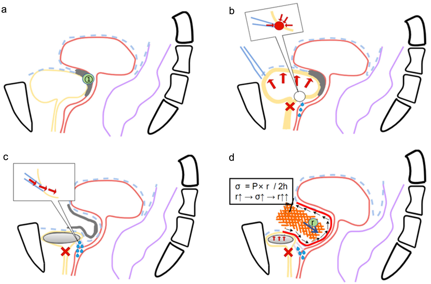 Click for large image | Figure 2. Schematic diagram of the biomechanical cascade (not drawn to scale); panels b–d present interpretations of the study findings rather than the actually observed stages. (a) Normal anatomy; ① indicates the vesicouterine pouch (closed). (b) Hypothetical stage 1: urethral atresia (red cross) and elevated intravesical pressure (red arrow); in the inset, the distal ureter (blue line) is located at the ureterovesical junction, and the red dot marks the site of perforation into the vagina (blue droplet, indicating urine leakage). (c) Hypothetical stage 2: urine flows directly from the distal ureter into the vagina (arrow in the inset), bypassing the bladder, and the bladder undergoes disuse atrophy (gray). (d) Hypothetical stage 3: the vesicouterine pouch reopens, and small bowel herniates through the defect. The expression σ = P × r/2h is shown only as a qualitative schematic: σ is wall stress; P is intra-abdominal pressure; r is the radius of curvature of the bulging wall (the width of the non-defect); h is wall thickness. |
Stage 1 (hypothesized): outlet obstruction, raised intraluminal pressure, and distal ureteral injury, with formation of bilateral ureterovaginal fistulae
Under normal physiological conditions, the bladder is a low-pressure urine-storing organ (filling pressure < 15 cm H2O) and can typically store 400–500 mL of urine prior to spontaneous voiding [10]. The UVJ acts as a one-way valve, preventing urine from the bladder from flowing back into the ureters and kidneys, while allowing urine to be pumped from the renal pelvis into the bladder under low-pressure conditions [11]. The bladder becomes a cavity capable of maintaining a balance between internal pressure and flow. When the urethra is completely occluded and the bladder outlet is obstructed, the bladder transforms from an open container that is intermittently emptied into a completely closed cavity [12].
If the outlet were completely occluded, continued urine production (approximately 700–800 mL per kidney per day) would enter a cavity with no outlet, and intravesical pressure would be expected to rise progressively [13]. The UVJ, as an area of morphological change in the bladder wall, constitutes a point of geometric discontinuity and may therefore act as a site of stress concentration, where local stresses can exceed those in the surrounding wall [14]. For the distal ureter, which can be approximated as a thin-walled tubular structure, Laplace’s law states that circumferential wall tension (T) is proportional to the transmural pressure (P) and the luminal radius (r): T = P × r [15]. This law indicates that, for the same transmural pressure, greater tension is generated in the wall of a container with a larger radius and a thinner wall. Applying this relationship qualitatively to the present case yields two inferences: first, if elevated intravesical pressure impedes ureteral emptying or is transmitted retrogradely through an incompetent UVJ, the tension borne by the distal ureteral wall would be higher; second, because both ureters were markedly dilated (Fig. 1b), a larger luminal radius under the same pressure would further increase wall tension; therefore, the dilated distal ureters are subject to greater tension. In addition, increased wall tension can compress vessels within the wall, and the reduced blood flow associated with dilation may further impair the microcirculation of the ureteral wall, leading to ischemic tissue injury [16].
Therefore, we speculate that persistently elevated intracavitary pressure may impair the microcirculation of the distal ureteral wall, predisposing it to ischemic injury [17]; prolonged ischemia combined with mechanical loading may lead to wall fibrosis and, in extreme cases, necrosis and perforation [18]. When such perforation occurs close to the vaginal wall, urine may seep along the path of least resistance and form a fistulous tract into the vagina [19]. This process comprises three conceptually distinct steps: (1) transmission of intracavitary pressure, (2) local tissue injury, and (3) formation of a specific ureterovaginal fistula. Nevertheless, although the intravesical pressure borne at the bilateral UVJs was similar in this case [20], similar pressure exposure does not necessarily produce identical tissue outcomes; wall thickness, blood supply, previous surgical scarring, and tissue tolerance may differ between the two sides, and bilateral perforation may not occur synchronously. The available data cannot determine the precise timing of the two fistulas, nor can they fully distinguish the respective contributions of mechanical perforation, direct injury related to urethral surgery, vascular interruption, or scar formation; the patient’s report of postoperative vaginal urine leakage suggests that a complete obstruction with a high-pressure state occurred, but iatrogenic factors cannot be excluded. The above mechanism is proposed in this case as one of the possibilities consistent with the intraoperative findings.
Stage 2 (hypothesized): diversion of urine through the fistulae and disuse atrophy of the bladder
Once both distal ureters drained directly into the vagina, urine from both kidneys would bypass the bladder almost entirely, so that the bladder would no longer undergo regular filling [21]. This disrupts the normal cycle of bladder filling and emptying, causing the detrusor muscle to lose its regular stretch stimulation. In experimental models in which the voiding cycle is abolished, such disuse leads to smooth-muscle atrophy, fibrosis, reduced compliance and reduced capacity [22], and a markedly reduced capacity is also well documented in long-term defunctionalized human bladders, for example in anuric patients awaiting renal transplantation [23].
Stage 3 (hypothesized): loss of the bladder’s “space-occupying” function, leading to the formation of an anterior pelvic hernia under pressure
The formation of a vaginal hernia after bladder atrophy can be qualitatively inferred using biomechanical principles. In the normal female pelvis, the full bladder occupies the space between the pubis, vagina, and uterus; when intra-abdominal pressure increases, the bladder undergoes physical deformation and displacement, causing the vesicouterine pouch—a potential peritoneal fold—to open while maintaining closer contact with surrounding organs [24]. Owing to the bladder’s good compliance, its posterior wall deforms posteroinferiorly under pressure, while surrounding supporting structures such as the pelvic floor muscles contract anterosuperiorly; these opposing displacements together constitute a “self-locking and interlocking mechanism” that closes the physiological outlet and potential spaces within the pelvic cavity [25]. After atrophy, however, the bladder no longer occupies this space, the vesicouterine pouch reopens, and the peritoneum and underlying fascia covering it are repeatedly exposed to elevated intra-abdominal pressure (P). We approximate this tissue as a thin dome-shaped membrane and adopt the Laplace relationship for a thin spherical shell: σ = P × r/2h, where σ is wall stress, r is the radius of curvature of the bulging wall, and h is wall thickness [26]. Here, r represents the radius of curvature of the bulging wall. For a bulging dome spanning an opening, a certain geometric relationship exists among the radius of curvature, the size of the opening, and the depth of bulging; when the bulging shape remains roughly similar during expansion, the opening size can reflect the radius of curvature to some extent. Therefore, enlargement of the opening is usually accompanied by an increase in the radius of curvature, and when pressure and wall thickness remain roughly unchanged, wall stress increases accordingly, which may further promote expansion [27]. Second, under daily loads such as coughing and straining during this period, pelvic floor supporting tissues are subjected to sustained loading and exhibit time-dependent creep [28], making the above deformation gradually irreversible, so that the functional opening evolves into a permanent defect and a hernial sac forms [29]. Although the above inferences are qualitative, and intra-abdominal pressure, hernial sac wall thickness, and curvature were not directly measured, irregular defect shape and tissue strain-hardening effects may also produce additional influences. Nevertheless, intraoperative findings showed that the hernial sac contained small intestine, mesentery, and atrophic bladder tissue, and the clinical presentation closely resembled bladder prolapse, indicating that this inferential process may provide a reference for subsequent validation and research through imaging reconstruction and patient-specific mechanical modeling in larger samples.
The hypothesized sequence, summarized in Table 1, comprises the following three steps:
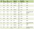 Click to view | Table 1. Summary of the Proposed Biomechanical Sequence |
(1) The initiating event in this case was complete bladder outlet obstruction caused by urethral atresia, and the resulting persistent high intravesical pressure was the main driving force of the entire cascade. According to Pascal’s principle, pressure is transmitted uniformly throughout the closed bladder, and the UVJ, as a geometric discontinuity, becomes a stress concentration area; Laplace’s law indicates that wall tension further increases in the dilated distal ureter. Persistent high wall tension may impair wall perfusion, leading to ischemic injury and perforation, and ultimately resulting in bilateral ureterovaginal fistulas. Direct measurement data are currently unavailable for these steps; however, existing clinical and imaging findings do not clearly conflict with this mechanism, and prior urethral surgery-related injury may also play a role, with the two possibly acting synergistically rather than being mutually exclusive.
(2) Once the fistulae had formed, urine from both kidneys would be diverted directly into the vagina, no longer passing through the bladder for storage and voiding. Consequently, the bladder loses its normal filling–emptying cycle, and the smooth muscle is deprived of periodic stretch stimulation. By analogy with experimental and clinical bladder disuse, smooth-muscle bundles would be expected to thin, extracellular matrix to accumulate and fibrotic remodeling to occur, converting the bladder from a compliant reservoir into the small, thick-walled remnant observed at operation; other causes of fibrosis cannot be excluded.
(3) Therefore, the bladder may lose its physical space-occupying role in the anterior pelvis, leading to reopening of the previously closed vesicouterine pouch. At this point, the tissues surrounding this opening will be directly subjected to cyclical fluctuations in intra-abdominal pressure. According to Laplace’s law for thin-shell structures (σ = P × r/2h), enlargement of the bulging area will lead to increased wall stress under the same pressure; the surrounding tissues may tear under repeated loading, and time-dependent creep under long-term cyclic loading may make the deformation permanent. Subsequently, small bowel loops and atrophic residual bladder tissue may be pushed through the defect, forming a fan-shaped, prolapsed mass that clinically closely mimics severe cystocele. This step constitutes a new hypothesis proposed on the basis of this case.
Preoperative examination and intraoperative exploration findings were consistent with each of the stages observed in the cascade described above, which supported the validity of this biomechanical model at the macroscopic morphological level (Table 2). However, such consistency does not fully equate to proof, and each abnormality may also be caused by other mechanisms. Nevertheless, this case still suggests the possibility that the mechanical effects of urethral outlet obstruction may extend beyond the bladder and lead to secondary changes in adjacent pelvic compartments.
Click to view | Table 2. Intraoperative Findings Correspond to the Mechanical Mechanism |
| Discussion | ▴Top |
This case suggests that, in addition to its traditional functions of urine storage and voiding, the bladder may also act as a space-occupying structure in the anterior pelvis, maintaining the mechanical integrity of the pelvic cavity through its own deformation and coordinated interaction with surrounding tissues. A normally filled bladder occupies the vesicouterine pouch and provides partial physical support to other pelvic tissues, helping to distribute intra-abdominal pressure [26]. Loss of this spatial support due to disease-related atrophy, surgical resection, or other causes may promote the development of an anterior pelvic hernia. Indirect clinical evidence comes from women who underwent cystectomy and subsequently developed an enterocele, in which small bowel herniates into the space previously occupied by the bladder; this is a known complication. In a recent systematic review, the mean onset time was approximately 9 months after surgery, and the typical presentation was a midline bulge of the anterior vaginal wall. Because these patients also experienced extensive surgical disruption of pelvic support structures, this does not constitute fully equivalent evidence; however, this observation is consistent with the hypothesis that the bladder itself has a supporting role.
Although this report is a single-case report, with the sequence of events mainly reconstructed retrospectively from intraoperative findings and imaging data and the biomechanical analysis being a qualitative inference, several other explanations that may participate or act jointly should also be considered. During urethral tumor resection, direct injury to the distal ureter or the bladder base may occur, disrupting tissue blood supply and leading to scar formation at the incision site; these factors may promote fistula formation, postoperative vaginal leakage, and local fibrosis. Second, the patient had clear risk factors for POP, including advanced age and a history of multiple pregnancies, and ultrasound indicated an old sphincter tear, suggesting prior obstetric-related pelvic floor injury, which may also have contributed to the pelvic floor changes. These factors may further weaken the supporting structures of the anterior pelvic wall independently of bladder atrophy. However, the above factors are not mutually exclusive but jointly contribute to the pathological process in this case together with the mechanism proposed in this article.
DeLancey’s three-level theory accurately summarizes and describes the anatomical support structures of the various regions of the pelvic floor, which are primarily composed of ligaments, fasciae, and muscles; on this basis, we propose that the bladder itself may also be regarded as a component of the pelvic support structures. The “hammock hypothesis”, on the other hand, emphasizes that the lower segment of the urethra is supported by a planar structure formed primarily by the suspensory ligament and the levator ani muscle; this case suggests that the complementary mechanisms at the level of the vaginal fornix and the vesicouterine fossa may also provide physical support to the urethra. While Petros and Ulmsten’s holistic theory emphasizes the anchoring and traction provided by ligaments and connective tissue to various organs, this case raises the possibility that the bladder contributes to the mechanical balance and stability of the surrounding pelvic region.
This finding offers a new perspective for clinical research: in patients with long-standing urethral atresia, assessment of upper urinary tract damage should be complemented by evaluation of changes in pelvic floor structure; where vaginal prolapse is present, pelvic hernia should be considered [27]. With regard to vaginal prolapse in patients with bladder dysfunction, preoperative imaging (computed tomography urography) is crucial for the definitive diagnosis of the contents of the prolapse and their relationship to the surrounding structures. In this case, the prolapsed mass was a hernia containing small intestine and atrophic bladder tissue [28]. In patients with severe bladder atrophy where bladder reconstruction is not considered viable, clinical improvements in both voiding function and bladder capacity are typically limited; attempts at bladder reconstruction in a severely atrophied and fibrotic bladder are likely to be unsuccessful [29].
The three-stage sequence organized according to intraoperative findings is reasonable at the current level of evidence and may also provide a reference for the identification and preoperative assessment of atypical POP in patients with long-term bladder disuse. This hypothesis can be validated in the future through dynamic magnetic resonance imaging (MRI), patient-specific finite element analysis, and retrospective studies including patients with long-term bladder dysfunction, thereby providing reliable evidence for long-term pelvic changes and surgical treatment decisions in patients with urethral atresia and bladder dysfunction.
Conclusion
This report describes a case of adult acquired urethral atresia accompanied by bilateral ureterovaginal fistulas, severe bladder atrophy, and an anterior pelvic hernia. Based on the clinical presentation, imaging studies, and intraoperative findings, we propose a qualitative, hypothesis-generating three-stage biomechanical framework: bladder outlet obstruction may lead to distal ureteral injury and fistula formation; long-term urinary diversion may then cause bladder disuse atrophy; and ultimately, loss of the bladder’s space-occupying effect may promote the formation of an anterior pelvic hernia under repeated intra-abdominal pressure. Iatrogenic factors such as prior surgery and known risk factors for POP, including childbirth and aging, may also be involved. If this hypothesis is further validated in future studies involving imaging, biomechanical modeling, and additional cases, the view that “the bladder is one of the important factors maintaining the stability of pelvic mechanical structures” may help extend the current pelvic floor biomechanical model, which relies mainly on ligament, fascia, and muscle traction support, and may provide an important reference for the evaluation and surgical planning of atypical POP in patients with long-term bladder disuse.
Learning points
(1) In patients with long-standing involuntary urine leakage after urethral surgery, one should not be satisfied with the diagnosis of “vesicovaginal fistula”; rather, the source of leakage should be identified and ureterovaginal fistula differentiated. In this case, both fistulas were ureterovaginal, and urine completely bypassed the bladder and flowed directly into the vagina.
(2) In patients with a nonfunctioning or atrophic bladder, if a vaginal mass resembling cystocele is present, anterior pelvic enterocele should be considered, and its contents may be small bowel; preoperative cross-sectional imaging is essential.
(3) For patients with long-standing bladder outlet obstruction or bladder disuse, upper urinary tract dilation and renal function should be assessed, and pelvic support structure function should be examined.
(4) Loss of the bladder’s space-occupying effect affects pelvic mechanical balance. This hypothesis is supported at the level of clinical observation by enterocele occurring after hysterectomy and by the present case, but it still requires quantitative validation through imaging studies and patient-specific biomechanical modeling.
(5) In most cases, a severely fibrotic bladder is generally unsuitable for reconstruction, and urinary diversion may be a more practical option.
Acknowledgments
The authors would like to express their gratitude to the Department of Urology at the First Affiliated Hospital of Kunming Medical University for their institutional support. The authors also thank the patients for their participation and for their consent to the publication of this study.
Financial Disclosure
This research did not receive any specific grant from funding agencies in the public, commercial, or not-for-profit sectors.
Conflict of Interest
The authors declare that there is no conflict of interest.
Informed Consent
Written informed consent was obtained from all individual participants included in the study. The study protocol was approved by the Institutional Review Board of the First Affiliated Hospital of Kunming Medical University. All procedures performed in studies involving human participants were in accordance with the ethical standards of the institutional and/or national research committee and with the 1964 Helsinki Declaration and its later amendments or comparable ethical standards. This article does not contain any studies with animals performed by any of the authors.
Author Contributions
Ji Hong Shen: conceptualization, methodology, supervision, writing-review and editing, surgical procedure implementation. Hai Nan Li: survey administration, data compilation, writing-initial draft preparation, visualization. Xiao Bao Li: methodology, formal analysis (qualitative biomechanical interpretation). All authors read and approved the final manuscript.
Data Availability
The data that support the findings of this study are available from the corresponding author upon reasonable request. The data are not publicly available due to privacy and ethical restrictions.
Abbreviations
CT: computed tomography; CTU: computed tomography urography; G3P2: gravida 3, para 2; MRI: magnetic resonance imaging; POP: pelvic organ prolapse; UVJ: ureterovesical junction
| References | ▴Top |
- Petros PE, Liedl B, Wenk MJ, Palma P. Etiopathogenesis of "Bladder Outlet Obstruction" in the female - inability of pelvic muscles to sufficiently open urethra. Urol Int. 2026;110(2):186-197.
doi pubmed - Bahuguna G, Panwar VK, Mittal A, Talwar HS, Mandal AK, Bhadoria AS, Chapple C. Management strategies and outcome of ureterovaginal fistulae: A systematic review and meta-analysis. Neurourol Urodyn. 2022;41(2):562-572.
doi pubmed - Yoshino K, Tanikaze S, Ueoka K, Kanazawa Y. Congenital urethral atresia with urethrovaginal fistula: report of a case. J Jpn Soc Pediatr Surg. 1990;26(7):1306-1309.
- Shkarupa D, Kubin N, Staroseltseva O, Shapovalova E, Zaytseva A, Petrov A. V13-09 The three-level uterus-sparing repair of advanced pelvic organ prolapse with the use of ultraminimesh. J Urol. 2020;203(Supplement 4):e1213.
- Yang X, Wang X, Gao Z, Li L, Lin H, Wang H, Zhou H, et al. The anatomical pathogenesis of stress urinary incontinence in women. Medicina (Kaunas). 2022;59(1):5.
doi pubmed - Quaghebeur J, Petros P, Wyndaele JJ, De Wachter S. Pelvic-floor function, dysfunction, and treatment. Eur J Obstet Gynecol Reprod Biol. 2021;265:143-149.
doi pubmed - Chikazawa K, Muro S, Imai K, Kuwata T, Akita K. Reevaluating the vesicouterine pouch: histological evidence for optimal dissection planes in oncologic surgery. J Gynecol Oncol. 2026;37(2):e16.
doi pubmed - Halleran DR, Ahmad H, Ching C, Gorra A, McCracken K, Wood RJ, Levitt MA. Acquired Urethrovaginal Fistula and Urethral Atresia in a Patient with a Sacrococcygeal Teratoma. J Pediatr Surg. 2019;54(3):612-615.
doi pubmed - Guang YL, Zhang Y, Li K. Study on the correlation between different degrees of cystocele and uterine prolapse and abnormal voiding function and clinical manifestations. Contemporary Medicine Forum. 2023;21(2):116-119.
- Schafer W, Abrams P, Liao L, Mattiasson A, Pesce F, Spangberg A, Sterling AM, et al. Good urodynamic practices: uroflowmetry, filling cystometry, and pressure-flow studies. Neurourol Urodyn. 2002;21(3):261-274.
doi pubmed - Kalayeh K, Fowlkes JB, Schultz WW, Sack BS. Ureterovesical junction deformation during urine storage in the bladder and the effect on vesicoureteral reflux. J Biomech. 2020;113:110123.
doi pubmed - Chen G, Jin X, Luo D, Ai J, Xiao K, Lai J, He Q, et al. beta-Adrenoceptor regulates contraction and inflammatory cytokine expression of human bladder smooth muscle cells via autophagy under pathological hydrostatic pressure. Neurourol Urodyn. 2020;39(8):2128-2138.
doi pubmed - Chen G, Jin X, Gao X, Ai J, Luo D, Zhou L, Xiao K, et al. Monocyte chemotactic protein-1 regulates proliferation and contractility of human bladder smooth muscle cells under hydrostatic pressure. J Interferon Cytokine Res. 2020;40(5):245-253.
doi pubmed - Razavi SE, Jouybar M. Fluid-structure interaction simulation of ureter with vesicoureteral reflux and primary obstructed megaureter. Biomed Mater Eng. 2018;29(6):821-837.
doi pubmed - Letic M. Feeling wall tension in an interactive demonstration of Laplace's law. Adv Physiol Educ. 2012;36(2):176.
doi pubmed - Dinlenc CZ, Liatsikos EN, Smith AD. Ureteral ischemia model: an explanation of ureteral dysfunction after chronic obstruction. J Endourol. 2001;15(2):213-216.
doi pubmed - Cooper JL, Francois N, Sourial MW, Miyagi H, Rose JR, Shields J, Sharma N, et al. The impact of ureteral access sheath use on the development of abnormal postoperative upper tract imaging after ureteroscopy. J Urol. 2020;204(5):976-981.
doi pubmed - Rangel EM, Morocco E, Sotelo R. Robotic surgery for urinary fistulae in females: a narrative review. Gynecol Pelvic Med. 2025;8:1.
doi pubmed - Bergstrom BS. Further insights into abdominal pressure transmission in continent and stress incontinent women. Neurourol Urodyn. 2024;43(8):2060-2064.
doi pubmed - Liu G, Lin YH, Li M, Xiao N, Daneshgari F. Temporal morphological and functional impact of complete urinary diversion on the bladder: a model of bladder disuse in rats. J Urol. 2010;184(5):2179-2185.
doi pubmed - Teimouri A, Wilcox DT, Malykhina AP, Wood D. Structural and functional bladder changes associated with disrupted voiding cycle in a murine model of cutaneous vesicostomy. J Pediatr Urol. 2026;22(1):105675.
doi pubmed - Wang XY, Xiong ZF. Application of lateral bladder dissection during total laparoscopic hysterectomy with obliterated anterior cul-de-sac. Chinese Journal of Clinical Obstetrics and Gynecology. 2021;22(2):122-124.
- Li L, Wang X, Tian D, Gao Z, Yao T, Shen J. Biomechanical analysis of female pelvic floor anatomy: A novel integrative framework. Asian J Urol. 2026.
- Ozog Y, Deprest J, Haest K, Claus F, De Ridder D, Mazza E. Calculation of membrane tension in selected sections of the pelvic floor. Int Urogynecol J. 2014;25(4):499-506.
doi pubmed - Chen S, Zhou TC. Mechanical law of abdominal wall. Chin J Gastrointest Surg. 2021;24(4):371-373.
- Yang M, Chen C, Wang Z, Long J, Huang R, Qi W, Shi R. Finite element analysis of female pelvic organ prolapse mechanism: current landscape and future opportunities. Front Med (Lausanne). 2024;11:1342645.
doi pubmed - Hakalmaz AE, Topuzlu Tekant G. Anorectal malformations and late-term problems. Turk Arch Pediatr. 2023;58(6):572-579.
doi pubmed - Fourie T, Ramphal S. Ventral pre-pubic herniation of the bladder following incontinence surgery. Afr J Urol. 2003;9(2):65-71.
- Caione P, Boldrini R, Salerno A, Nappo SG. Bladder augmentation using acellular collagen biomatrix: a pilot experience in exstrophic patients. Pediatr Surg Int. 2012;28(4):421-428.
doi pubmed
This article is distributed under the terms of the Creative Commons Attribution 4.0 International License (CC BY 4.0), which permits unrestricted use, distribution, and reproduction in any medium, including commercial use, provided the original work is properly cited.
Journal of Medical Cases is published by Elmer Press Inc.