Journal of Medical Cases, ISSN 1923-4155 print, 1923-4163 online, Open Access
Article copyright, the authors; Journal compilation copyright, J Med Cases and Elmer Press Inc
Journal website https://jmc.elmerpub.com

Case Report

Volume 17, Number 12, December 2026, pages 750-758


Anterior Pelvic Herniation After Urethral Atresia: A Case Report and the Bladder Space-Occupying Hypothesis

Figures

↓  Figure 1. Preoperative contrast-enhanced computed tomography. (a) Sagittal reconstruction: an anterior pelvic hernia (arrow) protrudes anteriorly, anterior to the uterus and vagina; its contents are small bowel loops passing through the vesicouterine pouch. No distinct bladder is visible in its expected location (asterisk). (b) Coronal reconstruction: severe bilateral hydronephrosis, with marked dilatation and tortuosity of the ureters (arrows), which extend inferiorly into the hernial sac. Neither image directly demonstrates the fistulous tract, which was confirmed at surgery.
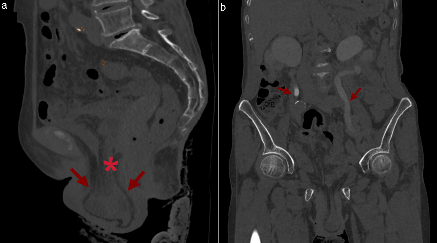
Figure 1.
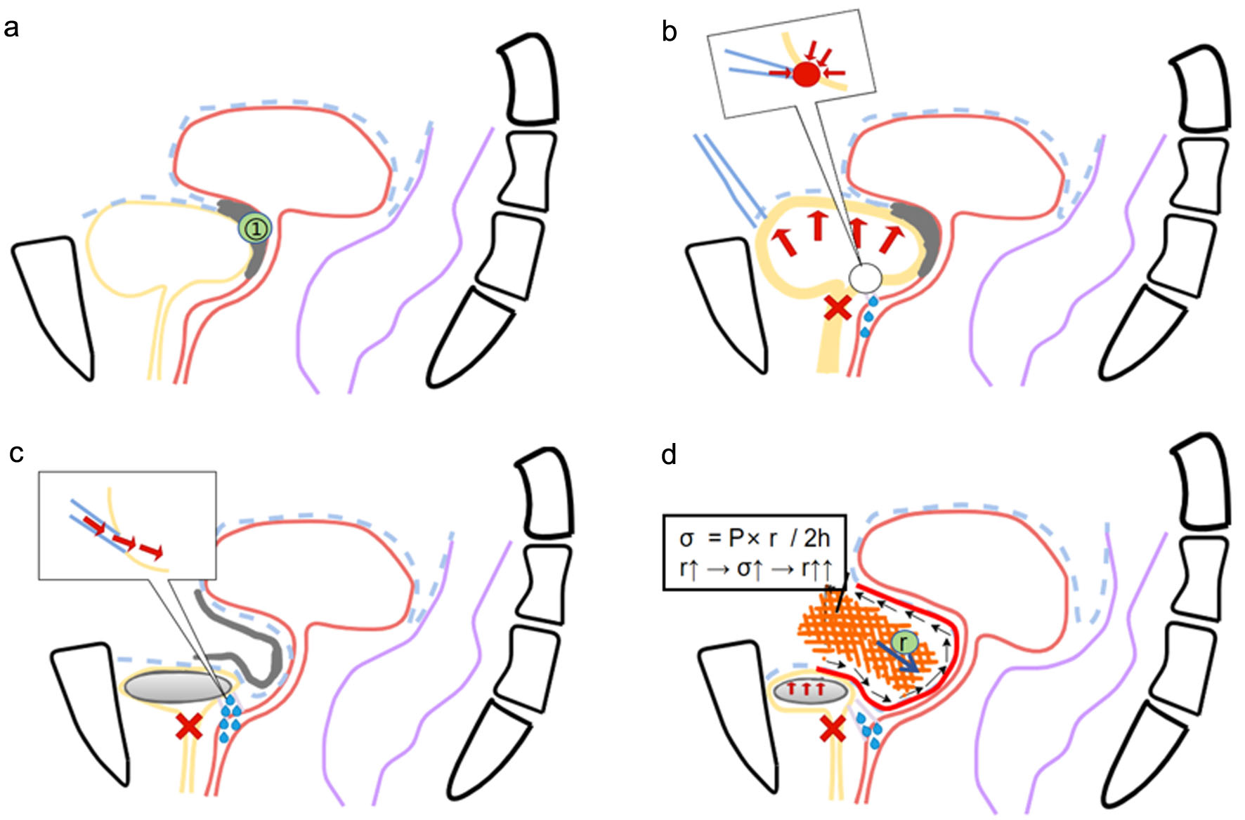
↓  Figure 2. Schematic diagram of the biomechanical cascade (not drawn to scale); panels b–d present interpretations of the study findings rather than the actually observed stages. (a) Normal anatomy; ① indicates the vesicouterine pouch (closed). (b) Hypothetical stage 1: urethral atresia (red cross) and elevated intravesical pressure (red arrow); in the inset, the distal ureter (blue line) is located at the ureterovesical junction, and the red dot marks the site of perforation into the vagina (blue droplet, indicating urine leakage). (c) Hypothetical stage 2: urine flows directly from the distal ureter into the vagina (arrow in the inset), bypassing the bladder, and the bladder undergoes disuse atrophy (gray). (d) Hypothetical stage 3: the vesicouterine pouch reopens, and small bowel herniates through the defect. The expression σ = P × r/2h is shown only as a qualitative schematic: σ is wall stress; P is intra-abdominal pressure; r is the radius of curvature of the bulging wall (the width of the non-defect); h is wall thickness.
Figure 2.

Tables

↓  Table 1. Summary of the Proposed Biomechanical Sequence
 
StageEventMechanismOutcome
Note: all stages represent a retrospective, qualitative interpretation; their timing and order could not be documented. Direct observations are distinguished from inferences in Table 2.
Stage IUrethral atresiaComplete bladder outlet obstructionChronic bladder hypertension
Pressure-induced perforation at the ureterovesical junctionIschemia and subsequent necrosis of the tissue surrounding the anastomosisBilateral ureterovaginal fistula
Stage IIUrine diversion through the fistulaThe bladder no longer stores urineBladder atrophy due to disuse
Bladder atrophyAtrophy and fibrosis of the bladder smooth muscleCapacity reduced to 10–30 mL
Stage IIILoss of “space occupancy”Uterine fossa without bladder compressionUterine pouch defect becomes open
Laplace-driven expansionPositive feedback driveProgressive expansion of the defect
Tissue creepTime-dependent irreversible deformationPermanent structural defect
Anterior pelvic herniaSmall intestine and atrophic bladder herniate through the defectClinical “fan-shaped” prolapse


↓  Table 2. Intraoperative Findings Correspond to the Mechanical Mechanism
 
Intraoperative findingSignificanceMechanical mechanism
Atresia urethralisObstruction sourceFormation of a sealed container
Bilateral ureterovaginal fistulaHigh-pressure ureteral perforation“Stress-induced perforation” mechanism
Severe bladder atrophy (approximately 10–30 mL)Structural integrity lossBladder atrophy due to disuse
Bladder-uterine pouch defect (3–4 cm)Anatomical continuity disruption“Spatial occupancy” function disabled
Small intestinal herniaClinical hernia formationLaplace-driven progressive expansion (σ = P × r/2h)