| Journal of Medical Cases, ISSN 1923-4155 print, 1923-4163 online, Open Access |
| Article copyright, the authors; Journal compilation copyright, J Med Cases and Elmer Press Inc |
| Journal website https://jmc.elmerpub.com |
Case Report
Volume 17, Number 12, December 2026, pages 745-749

A Cadaveric Case of Lusoria Artery With Calcification
Avery Savoiea, b, Dustin Rizzieria, David Jaynesa
aDepartment of Anatomy, Edward Via College of Osteopathic Medicine - Carolinas Campus, Spartanburg, SC 29303, USA
bCorresponding Author: Avery Savoie, Department of Anatomy, Edward Via College of Osteopathic Medicine - Carolinas Campus, Spartanburg, SC 29303, USA
Manuscript submitted September 5, 2026, accepted October 14, 2026, published online November 9, 2026
Short title: A Cadaveric Case of Lusoria Artery
doi: https://doi.org/10.14740/jmc5426
| Abstract | ▴Top |
The right subclavian artery is commonly derived from the first branch of the arch of the aorta, the brachiocephalic trunk. However, there have been reported cases where the right subclavian artery did not follow this typical anatomy. Several anatomical variants of the right subclavian artery point of origin have been described in literature; one uncommon variation is the lusoria artery, which is a right subclavian artery that emerges from the proximal descending aorta. This case describes a lusoria artery that originated distal to the arch of the aorta, at the level of T5, then coursed to the right across the posterior mediastinum deep to the esophagus. A bicarotid trunk was also observed originating from the aortic arch at its most superior portion. The origin of the lusoria artery exhibited areas of extensive calcification and the posterior wall was markedly attenuated, exhibiting folding with loss of structural integrity and the presence of several full-thickness perforations. Although the embryologic relationship remains unclear, this case was also notable for the presence of a persistent thyroglossal duct. Presence of the lusoria artery is rare and 2/3 of these cases are asymptomatic. The presence of symptoms likely depends on other features. Patients with a lusoria artery may present with mild respiratory distress, dysphagia, and other less frequently observed symptoms. Diminished vascular wall thickness at the lusoria artery origin can lead to a Kommerell diverticulum, which can compound the lusoria artery symptoms and present with additional symptoms due to the mass compressing adjacent tissues. This anomaly is adequately common to demand the attention of vascular surgeons confronted with a mediastinal mass accompanied by symptoms of lusoria artery.
Keywords: Developmental anatomy; Aberrant right subclavian artery; Thyroglossal duct; Embryology; Kommerell diverticulum; Dysphagia
| Introduction | ▴Top |
Normal anatomy appreciates three major branches from the aortic arch, the brachiocephalic trunk which branches into the right subclavian artery and the right common carotid artery, the left common carotid artery, and the left subclavian artery. One abnormality in this vascular development, known as an aberrant right subclavian artery (ARSA), is well documented but typically of little consequence as they rarely compromise blood flow [1]. The lusoria artery is a subtype of ARSA in which the right subclavian artery originates from the proximal descending aorta instead of from the brachiocephalic trunk. The most commonly reported ARSA variant is the lusoria artery with independently emerging common carotid arteries. The second most common variation is a lusoria artery from the proximal descending aorta with a bicarotid trunk [2]. The majority of documented ARSAs course retroesophageally, which can cause this condition to present with unique and characteristic symptoms, particularly right arm numbness or tingling following swallowing [3].
The prevalence of the lusoria artery is approximately 1% globally but is predicted to be underreported. Diagnoses have increased with advancing imaging techniques, such as ultrasonography, and a more comprehensive understanding of clinical presentation [4]. This aberrant anatomy merits concern for not only the patency of the artery, but also complication risks for thoracic procedures.
| Case Report | ▴Top |
An 84-year-old white male cadaver who died from a myocardial infarction was observed to have a lusoria artery upon dissection. The lusoria artery emerged from the proximal descending aorta at the level of T5 and coursed transversely across the posterior mediastinum deep to the esophagus and ascended to the right upper extremity (Fig. 1). Despite the abnormal origin point of the right subclavian artery in this individual, its distal branching pattern remained anatomically typical, including the vertebral artery, internal thoracic artery, thyrocervical trunk, costocervical trunk, and the dorsal scapular artery. The first branch, the vertebral artery, is visible following its normal course in Figure 2a. A bicarotid trunk was also observed originating from the aortic arch at its most cephalad aspect (Fig. 2a). Extensive calcifications and greatly reduced arterial wall thickness with focal perforations were found during excision of the origin of the lusoria artery. This section of the artery appeared folded back upon itself, which could have prevented noticeable blood loss (Fig. 3). A persistent thyroglossal duct was also observed, as shown in Figure 4.
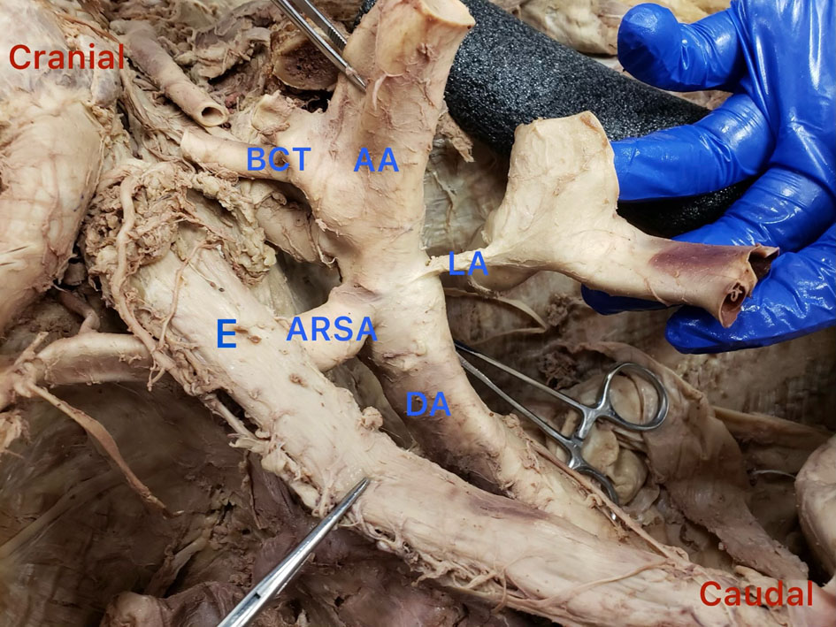 Click for large image | Figure 1. Proximal part of the arch of the aorta was cut, the heart was removed, and the aortic arch is reflected superiorly; image is of the posterior aspect of the aortic arch. Esophagus has been moved to the right to allow a clearer view. Red cranial and caudal labels were added for orientation. AA: arch of aorta; ARSA: aberrant right subclavian artery; BCT: bicarotid trunk; DA: descending aorta; E: esophagus; LA: ligamentum arteriosum. |
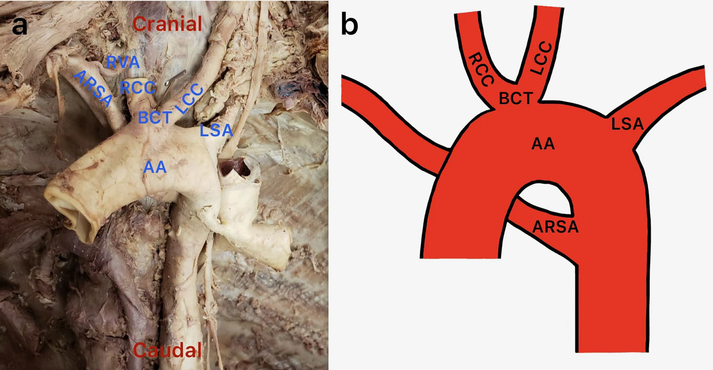 Click for large image | Figure 2. (a) Anterior view of the mediastinum showing a bicarotid trunk emerging near the apex of the aortic arch; the trunk gives rise to both common carotid arteries (the right common carotid artery has been cut near its origin). (b) Illustration by one of the authors of the Adachi and Williams classification H1. AA: arch of aorta; ARSA: aberrant right subclavian artery; BCT: bicarotid trunk; LCC: left common carotid artery; LS: left subclavian artery; RCC: right common carotid artery; RVA: right vertebral artery. |
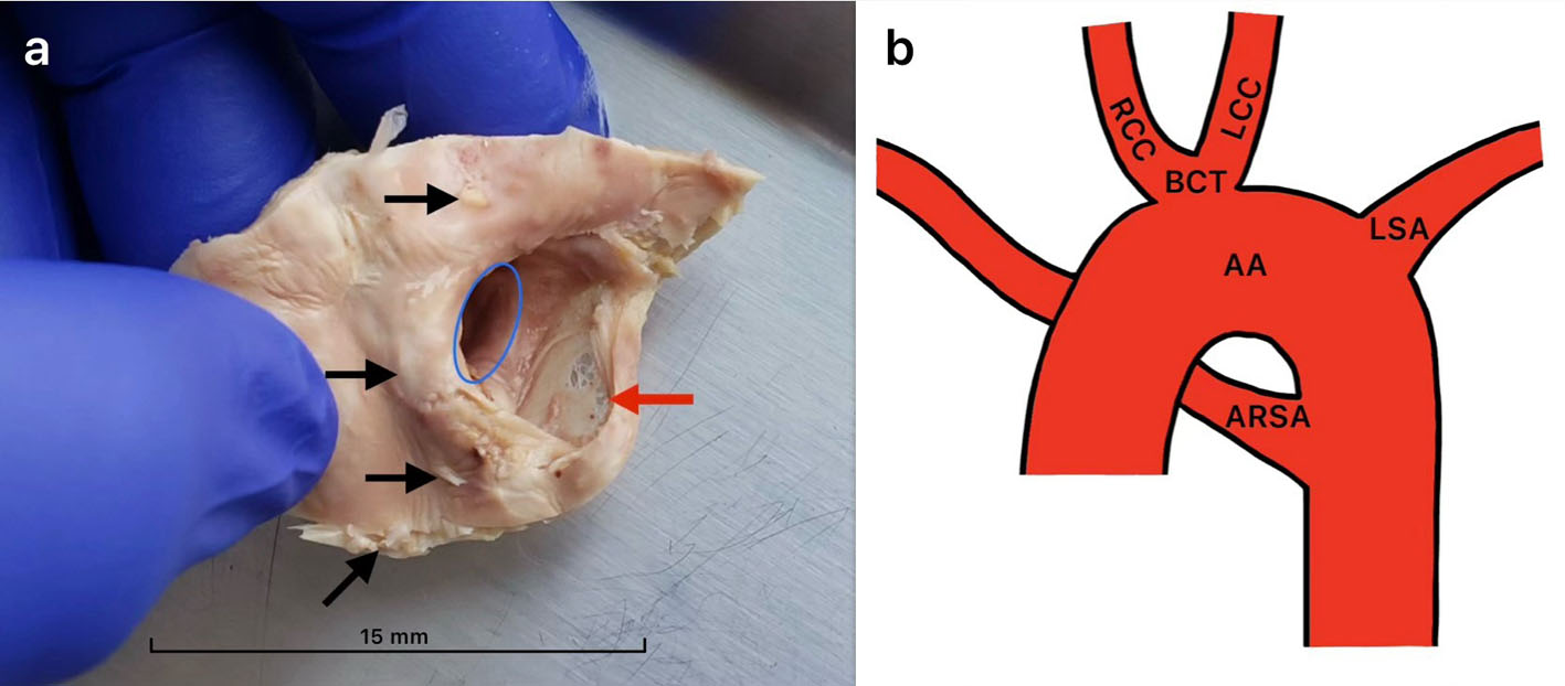 Click for large image | Figure 3. (a) Internal view of ARSA at its origin excised from aorta and then cut longitudinally and reflected to reveal the area of structural weakness, perforations, and calcifications. The blue circle shows the communication of the ARSA with the descending aorta. The black arrows indicate a few of the many calcifications at the origin of ARSA. The red arrow indicates the thinned artery wall. Very few calcifications were observed beyond the excised tissue. (b) Illustration by one of the authors of the Adachi and Williams classification H1, with yellow lines that indicate the excisions made to reveal the structures in (a). AA: arch of aorta; ARSA: aberrant right subclavian artery; BCT: bicarotid trunk; LCC: left common carotid artery; LS: left subclavian artery; RCC: right common carotid artery. |
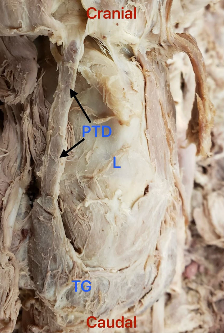 Click for large image | Figure 4. Anterior view of the lower neck. Presence of a large asymmetric right persistent thyroglossal duct. L: larynx; PTD: persistent thyroglossal duct; TG: thyroid gland. |
| Discussion | ▴Top |
ARSAs and lusoria arteries have been previously described. The name, lusoria artery, is derived from the Latin phrase lusus naturae (freak of nature) which was coined at the discovery of dysphagia secondary to a lusoria artery by Dr. David Bayford in 1794 [5]. Since 1928, the Adachi and Williams classification system has been used to describe variations in the branching patterns of the aorta. There are four different classifications for a lusoria artery (types G-1, CG-1, H-1, and N-1), which are based on the pattern of origin points of the other major branches of the aortic arch. The coexistence of a lusoria artery and bicarotid trunk is classified as type H-1 and is depicted in Figure 2b.
Close inspection of the origin of the lusoria artery revealed a vascular wall with greatly diminished thickness and calcifications visible to the naked eye. Compromised structural integrity at this aortic branch point is frequently observed in individuals with ARSAs and contributes to their increased risk of aneurysms. Kommerell diverticulum, named after the radiologist to first identify this condition, is the presence of an aneurysmal dilation at the base of an aberrant subclavian artery [6]. In this case, the lusoria artery folded back onto the area of thinned portion of the arterial wall, potentially providing mechanical reinforcement that limited blood loss and prevented Kommerell diverticulum formation.
The normal asymmetric arrangement of blood vessels associated with the arch of the aorta occurs through selective regression and remodeling of the embryonic aortic arches (vasculature of the pharyngeal arches) during the fourth to eighth weeks of development. Initially, six pairs of symmetrical aortic arches arise from the aortic sac (a primitive chamber located at the distal end of the developing heart tube); however, aortic arches 1 and 2 mostly degenerate, while aortic arch 5 completely degenerates. The common carotid arteries arise from the right and left third aortic arches. A bicarotid trunk, as seen in our case, is thought to occur when the aortic sac fails to properly bifurcate, thus causing the origins of the common carotid arteries to merge into a single median vessel. Normally the right subclavian artery is derived from the right fourth aortic arch. An ARSA, as seen in our case, forms due to abnormal regression of the right fourth aortic arch coupled with persistence of an upper section of the right dorsal aorta. These abnormalities typically occur due to one of several possible failures in the normal molecular pathways [7].
Thyroglossal duct formation occurs during week 4 of embryonic development from endoderm and guides the primitive thyroid to its final anatomic position. Persistent thyroglossal ducts are understood to be due to failure of cellular involution in embryos by week 10 of development. The exact mechanism of this failure of involution and cellular obliteration is not well understood, although a hereditary form has been reported and suggests a possible genetic component in some patients [8]. Although there is no currently known causal relationship between persistent thyroglossal ducts and ARSAs, it is reasonable to speculate that the earlier formation of the aberrant arterial structures may have interfered with normal degeneration of the thyroglossal duct. For example, aberrant vascular structures may have mechanically or developmentally disrupted normal embryologic involution of the thyroglossal duct. Another possibility is that the presence of the bicarotid trunk or ARSA may have altered local embryologic signaling environments, preventing the normal involution of the thyroglossal duct.
Although this case report describes the H-1 classification lusoria artery, the available literature revealed that no other cases have identified the findings of grossly visible calcification at the origin of the lusoria artery, or the folding habitus, which might have played an integral role in preventing Kommerell diverticulum formation in this individual. Additionally, while thyroglossal ducts might persist in up to 7% of the adult population, there are no known cases describing a persistent thyroglossal duct in the same individual as a lusoria artery [9]. Due to the anatomic proximity of these two structures and the poorly understood mechanisms behind the persistence of thyroglossal ducts, this finding might be significant in helping to garner a better understanding of the requirements for embryologic thyroglossal duct involution.
Neural crest cells play an integral role in the development of the pharyngeal arches; their activity is largely regulated by genes and are therefore largely impacted by chromosomal abnormalities. The 22q11.2 deletion syndrome, also known as CATCH 22 syndrome, is a disorder characterized by a heterozygous deletion at 22q11.2 that results in defective development of the third and fourth pharyngeal arches. Evidence from earlier studies suggests that the prevalence of the 22q11.2 deletion among patients with an aberrant right subclavian artery may be as high as 25% [10]. Increased incidence of ARSAs has also been associated with other chromosomal disorders, like trisomy 21, with an incidence of 35.7% in a study that looked at 14 individuals, and trisomy 18 [11]. ARSAs have also been reported more frequently in patients with connective tissue disorders including Loeys–Deitz syndrome, Marfan syndrome, and vascular Ehlers–Danlos syndrome [12]. It is unknown if the correlation between these connective tissue disorders and lusoria arteries are genetic, occur during development, or if the connective tissue disorders exacerbate the effects of the weakened origin of the lusoria artery, leading to Kommerell diverticulum formation, more profound symptoms, and therefore a higher rate of diagnosis. We do not believe this particular individual had any of these conditions.
While the symptomatology as it pertains to this specific anatomic anomaly in this individual cannot be known, we can attempt to understand how this variation may have impacted this person’s everyday life. It is reported that 2/3 of the population having lusoria arteries are asymptomatic [1, 13]. Females seem to be affected more than men with 69% of incidences being female. Men, however, are more likely than women to have a Kommerell’s diverticulum than women with the same anatomy [2]. Some known associations with the vascular mutation are aneurysms and other heart or vascular abnormalities, such as the bicarotid trunk, or a right-sided aortic arch. The presence of symptoms likely depends on the presence of other variants. Some signs of the abnormality may be persistent dysphagia, dyspnea, cough, retrosternal pain, or right radial pulses diminished during swallowing due to the arteries’ retroesophageal path. Dysphagia existing specifically due to the presence of a lusoria artery is known as Bayford–Autenrieth dysphagia [14]. The mean age for diagnosis of a lusoria artery is 49.9 years, likely due to an increase in symptoms as each patient ages [14]. Several hypotheses have been proposed to explain why some people are asymptomatic but later become symptomatic. One is increasing esophageal rigidity with advanced age, leading to dysphagia caused by the retroesophageal course of the artery. Another is that, in patients with the H-1 classification, the bicarotid trunk gives rise to the right and left carotid arteries in close proximity, thus preventing the trachea and esophagus from bending forward where the lusoria artery crosses. In either event, symptoms are more likely to occur when there is pressure on the artery from surrounding structures.
The presence of an aneurysm or Kommerell’s diverticulum in the proximal aorta can lead to superior vena cava syndrome and increases the risk of aortic dissections, which are the most prominent cause of death attributable to the lusoria artery [15]. Kommerell diverticulum removal should therefore be considered in patients who are appropriate surgical candidates [16]. Other medical management changes for these patients include the preferred use of a transfemoral approach for catheterization. In normal patients, the transradial catheterization approach is viewed as the gold standard. However, the risk of dissection as a complication during catheterization in these patients is significantly increased and success rates are decreased to 60% [17, 18]. The femoral approach has shown better success rates in this patient population and decreased risk of aortic dissection and is therefore the preferred method of catheterization in patients with a lusoria artery as these benefits outweigh its increased infection risk. Obtaining confirmation of an ARSA in a patient with the relevant clinical presentation or symptoms is important to ensure they are provided the correct medical care without subjecting the patient to severe complications.
The cause of death in the patient in this case was myocardial infarction (MI). To the best of our knowledge, no previous studies have established MI as an increased cause of mortality in patients with ARSAs. The most common pathological mechanisms underlying acute coronary thrombosis and MI include atherosclerotic plaque rupture, plaque erosion, and calcified nodules [19]. Calcified nodules are characterized by dense deposits of calcium that may undergo fragmentation and protrude through the overlying fibrous tissue into the arterial lumen, a process which typically occurs in areas that are subject to increased levels of mechanical stress. This can promote platelet adhesion and thrombus formation, potentially resulting in acute vascular occlusion [20]. Therefore, while a causal relationship between the observed calcifications and this patient’s MI cannot be definitively established, the presence of prominent intimal calcification at the branching point of the lusoria artery raises the possibility that localized vascular pathology may have contributed to the thrombotic event resulting in this patient's death.
Conclusion
To our best knowledge, this is the first case to present a lusoria artery with a bicarotid trunk, a weakened, thinned, and calcified vascular wall at the origin of the lusoria artery, and a persistent thyroglossal duct. Although rare, the prevalence of a lusoria artery is increasing with advancements in imaging techniques. Therefore, physicians should practice a high index of suspicion in patients with aortic variations, progressive dysphagia, aneurysms, known chromosomal abnormalities, and/or a persistent thyroglossal duct. Early diagnosis can improve long term health outcomes for patients and potentially reduce serious complications of invasive procedures.
Acknowledgments
The authors extend their sincere gratitude to the individual who generously donated his body to advance medical and anatomical education. His gift has contributed to a deeper understanding of the human body and its many variations.
Financial Disclosure
The authors have no financial disclosures to disclose and did not receive any funding for this case report.
Conflict of Interest
The authors have no conflicts of interest to disclose.
Informed Consent
Informed consent is not applicable for this case. This cadaveric case report contains no identifiable patient information and involves the anatomical examination of a donated human cadaver.
Author Contributions
Avery Savoie: conceptualization, literature review, manuscript preparation, and editing. Dustin Rizzieri: literature review, manuscript review, and editing. David Jaynes: cadaveric dissection and anatomical examination, manuscript preparation, editing, and supervision. All authors contributed to the interpretation of the findings, reviewed and approved the final manuscript, and agree to be accountable for all aspects of the work.
Data Availability
The data supporting the findings of this case report are available within the article. Additional information regarding the findings of this study is available from the corresponding author upon reasonable request.
Abbreviations
AA: arch of aorta; ARSA: aberrant right subclavian artery; BCT: bicarotid trunk; DA: descending aorta; E: esophagus; L: larynx; LA: ligamentum arteriosum; LCC: left common carotid artery; LS: left subclavian artery; MI: myocardial infarction; PTD: persistent thyroglossal duct; RCC: right common carotid artery; RVA: right vertebral artery; TG: thyroid gland
| References | ▴Top |
- Layfield DM, Nikolaidis N, Harden SP, Ohri SK. Aneurysm of aberrant right subclavian artery (arteria lusoria) presenting as cardiac dyspnoea. Thorax. 2011;66(5):456.
doi pubmed - Plotkin A, Ng B, Han SM, Weaver FA, Ham SW, Bowdish ME, Wilcox AG, et al. Association of aberrant subclavian arteries with aortic pathology and proposed classification system. J Vasc Surg. 2020;72(5):1534-1543.
doi pubmed - Choi Y, Chung SB, Kim MS. Prevalence and anatomy of aberrant right subclavian artery evaluated by computed tomographic angiography at a single institution in Korea. J Korean Neurosurg Soc. 2019;62(2):175-182.
doi pubmed - Myers PO, Fasel JH, Kalangos A, Gailloud P. Arteria lusoria: developmental anatomy, clinical, radiological and surgical aspects. Ann Cardiol Angeiol (Paris). 2010;59(3):147-154.
doi pubmed - Leite TFO, Pires LAS, Cisne R, Babinski MA, Chagas CAA. Clinical discussion of the arteria lusoria: a case report. J Vasc Bras. 2017;16(4):339-342.
doi pubmed - Hale BW, Lu JC, Romano JC, Lowery R, Yu S, Norris MD. Kommerell diverticulum: distinctions between arch side and evaluation of morphology, size, and risk. Ann Thorac Surg. 2022;114(3):848-856.
doi pubmed - Annetta R, Nisbet D, O'Mahony E, Palma-Dias R. Aberrant right subclavian artery: embryology, prenatal diagnosis and clinical significance. Ultrasound. 2022;30(4):284-291.
doi pubmed - Jenkins JR, Morgan MB. Dermal cysts: a dermatopathological perspective and histological reappraisal. J Cutan Pathol. 2007;34(11):815-829.
doi pubmed - Guzman GP, Baeza OA, Cabello VA, Montecinos LA, Leal MC, Roa SJ. [Papillary carcinoma in a thyroglossal duct cyst: report of two cases]. Rev Med Chil. 2008;136(9):1169-1174.
pubmed - Kobrynski LJ, Sullivan KE. Velocardiofacial syndrome, DiGeorge syndrome: the chromosome 22q11.2 deletion syndromes. Lancet. 2007;370(9596):1443-1452.
doi pubmed - Chaoui R, Heling KS, Sarioglu N, Schwabe M, Dankof A, Bollmann R. Aberrant right subclavian artery as a new cardiac sign in second- and third-trimester fetuses with Down syndrome. Am J Obstet Gynecol. 2005;192(1):257-263.
doi pubmed - Giuliani L, Di Toro A, Urtis M, Narula N, Grasso M, Pelenghi S, Belliato M, et al. Prevalence and complications of aberrant subclavian artery in patients with heritable and nonheritable arteriopathies. J Am Coll Cardiol. 2023;81(10):979-991.
doi pubmed - Yiu KH, Chan WS, Jim MH, Chow WH. Arteria lusoria diagnosed by transradial coronary catheterization. JACC Cardiovasc Interv. 2010;3(8):880-881.
doi pubmed - Polguj M, Chrzanowski L, Kasprzak JD, Stefanczyk L, Topol M, Majos A. The aberrant right subclavian artery (arteria lusoria): the morphological and clinical aspects of one of the most important variations—a systematic study of 141 reports. ScientificWorldJournal. 2014;2014:292734.
doi pubmed - Isselbacher EM, Preventza O, Hamilton Black Iii J, Augoustides JG, Beck AW, Bolen MA, et al. 2022 ACC/AHA guideline for the diagnosis and management of aortic disease: a report of the American Heart Association/American College of Cardiology Joint Committee on Clinical Practice Guidelines. J Am Coll Cardiol. 2022;80(24):e223-e393.
doi pubmed - Suarez AE, Slivka R. Acute superior vena cava syndrome after rupture of Kommerell's diverticulum: a case report. Ann Emerg Med. 2013;61(3):348-350.
doi pubmed - Huang IL, Hwang HR, Li SC, Chen CK, Liu CP, Wu MT. Dissection of arteria lusoria by transradial coronary catheterization: a rare complication evaluated by multidetector CT. J Chin Med Assoc. 2009;72(7):379-381.
doi pubmed - Valsecchi O, Vassileva A, Musumeci G, Rossini R, Tespili M, Guagliumi G, Mihalcsik L, et al. Failure of transradial approach during coronary interventions: anatomic considerations. Catheter Cardiovasc Interv. 2006;67(6):870-878.
doi pubmed - Choudhury T, West NE, El-Omar M. ST elevation myocardial infarction. Clin Med (Lond). 2016;16(3):277-282.
doi pubmed - Torii S, Sato Y, Otsuka F, Kolodgie FD, Jinnouchi H, Sakamoto A, Park J, et al. Eruptive calcified nodules as a potential mechanism of acute coronary thrombosis and sudden death. J Am Coll Cardiol. 2021;77(13):1599-1611.
doi pubmed
This article is distributed under the terms of the Creative Commons Attribution 4.0 International License (CC BY 4.0), which permits unrestricted use, distribution, and reproduction in any medium, including commercial use, provided the original work is properly cited.
Journal of Medical Cases is published by Elmer Press Inc.