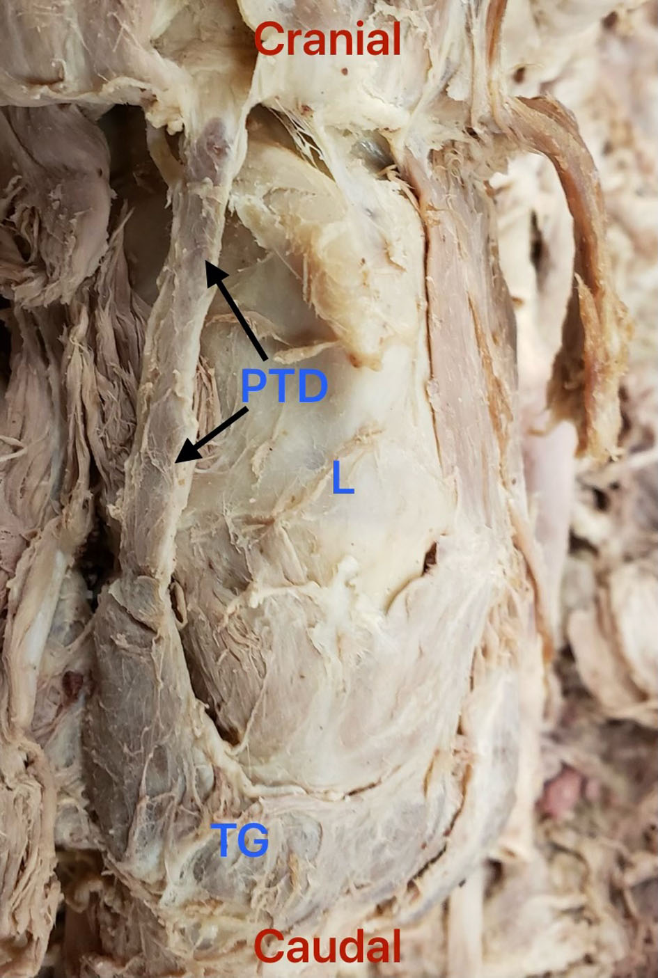
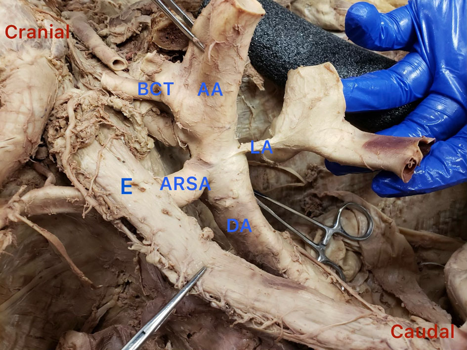
↓ Figure 1. Proximal part of the arch of the aorta was cut, the heart was removed, and the aortic arch is reflected superiorly; image is of the posterior aspect of the aortic arch. Esophagus has been moved to the right to allow a clearer view. Red cranial and caudal labels were added for orientation. AA: arch of aorta; ARSA: aberrant right subclavian artery; BCT: bicarotid trunk; DA: descending aorta; E: esophagus; LA: ligamentum arteriosum.
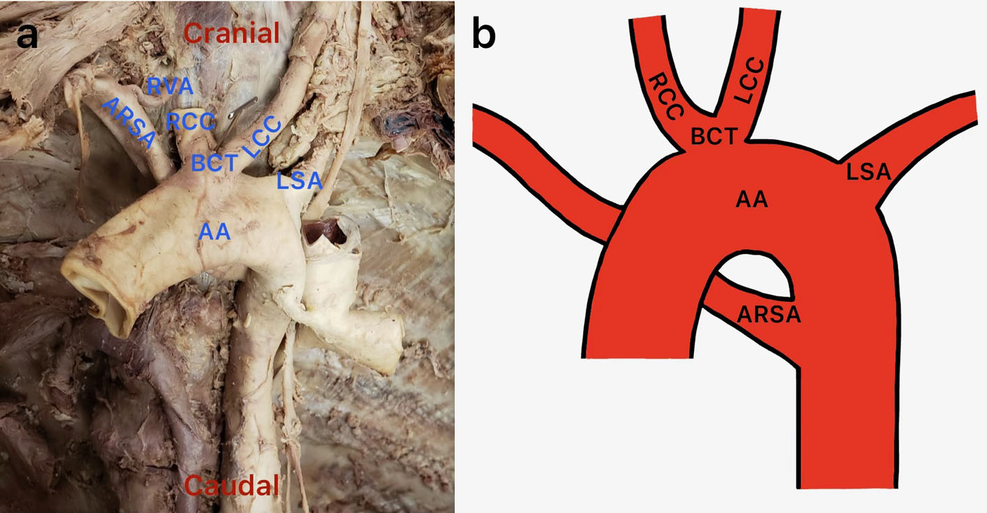
↓ Figure 2. (a) Anterior view of the mediastinum showing a bicarotid trunk emerging near the apex of the aortic arch; the trunk gives rise to both common carotid arteries (the right common carotid artery has been cut near its origin). (b) Illustration by one of the authors of the Adachi and Williams classification H1. AA: arch of aorta; ARSA: aberrant right subclavian artery; BCT: bicarotid trunk; LCC: left common carotid artery; LS: left subclavian artery; RCC: right common carotid artery; RVA: right vertebral artery.
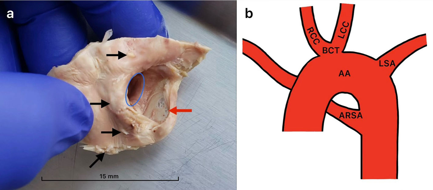
↓ Figure 3. (a) Internal view of ARSA at its origin excised from aorta and then cut longitudinally and reflected to reveal the area of structural weakness, perforations, and calcifications. The blue circle shows the communication of the ARSA with the descending aorta. The black arrows indicate a few of the many calcifications at the origin of ARSA. The red arrow indicates the thinned artery wall. Very few calcifications were observed beyond the excised tissue. (b) Illustration by one of the authors of the Adachi and Williams classification H1, with yellow lines that indicate the excisions made to reveal the structures in (a). AA: arch of aorta; ARSA: aberrant right subclavian artery; BCT: bicarotid trunk; LCC: left common carotid artery; LS: left subclavian artery; RCC: right common carotid artery.