| Journal of Medical Cases, ISSN 1923-4155 print, 1923-4163 online, Open Access |
| Article copyright, the authors; Journal compilation copyright, J Med Cases and Elmer Press Inc |
| Journal website https://jmc.elmerpub.com |
Case Report
Volume 17, Number 12, December 2026, pages 712-726

First-Attempt Video Laryngoscopic Intubation in an Adult With Down Syndrome and Severe Tracheal Compression From a Giant Retrosternal Goiter
Majlinda Nacoa, b, Esmerilda Bulkub, Rudin Domia, b, i, Alma Llukacajc, Eriselda Kurushid, Fjorba Manae, Milena Memellif, Arben Dhimaf, Krenar Lilaja, b, Eden Nacog, Alma Cania, Vedat Eljezih
aDepartment of Surgery, University of Medicine, Tirana, Albania
bService of Anesthesia and Intensive Care, University Hospital Center “Mother Theresa”, Tirana, Albania
cService of General Surgery, University Hospital Center “Mother Theresa”, Tirana, Albania
dDepartment of Anatomic Pathology, University Hospital Center “Mother Theresa”, Tirana, Albania
eService of Cardiac Surgery, University Hospital Center “Mother Theresa”, Tirana, Albania
fDepartment of Radiology, American Hospital 3, Tirana, Albania
gMedical Doctor, Tirana, Albania
hDepartment of Perioperative Medicine, CHU Gabriel-Montpied, Clermont-Ferrand, France
iCorresponding Author: Rudin Domi, Department of Surgery, University of Medicine, Tirana, Albania
Manuscript submitted August 16, 2026, accepted October 7, 2026, published online November 9, 2026
Short title: Difficult Airway in Giant Retrosternal Goiter With DS
doi: https://doi.org/10.14740/jmc5416
| Abstract | ▴Top |
Giant retrosternal goiters can cause severe tracheal compression and pose substantial challenges for airway management and surgical resection. These challenges may be further increased in adults with Down syndrome (DS), in whom craniofacial abnormalities, macroglossia, obesity, limited neck mobility, and potential cervical spine abnormalities may contribute to difficult airway management. We report the perioperative management of a 29-year-old woman with DS and a giant multinodular retrosternal goiter causing marked rightward tracheal deviation and critical tracheal narrowing to approximately 7 mm, requiring combined cervical resection and median sternotomy. Preoperative assessment identified multiple predictors of difficult mask ventilation and tracheal intubation, while severe intellectual disability made awake airway management impractical. A multidisciplinary airway and surgical strategy were therefore developed, incorporating optimized positioning and preoxygenation, video laryngoscopy as the planned primary intubation technique, flexible bronchoscopy for confirmation and rescue, and predefined escalation to surgical airway and extracorporeal support if conventional airway management failed. Following controlled intravenous induction, tracheal intubation was achieved on the first attempt with a 6.0-mm endotracheal tube without hypoxemia or hemodynamic instability. Total thyroidectomy and median sternotomy were completed uneventfully. After an adequate cuff-leak test and confirmation of full neuromuscular recovery, the patient was extubated in the operating room and received postoperative bilevel positive airway pressure and overnight intensive care monitoring. No respiratory or other major postoperative complications occurred, and she was discharged home on postoperative day 5. This case illustrates the individualized integration of established difficult-airway principles when DS coexists with severe mechanical tracheal compression from a giant retrosternal goiter. Successful management depended not on a single airway technique, but on careful preoperative anatomical and functional assessment, selection of a feasible primary approach, optimized oxygenation, and an immediately executable multidisciplinary rescue pathway.
Keywords: Down syndrome; Retrosternal goiter; Difficult airway; Median sternotomy; Video laryngoscopy
| Introduction | ▴Top |
Retrosternal (substernal) goiter is characterized by extension of thyroid tissue below the thoracic inlet and represents a clinically important subset of thyroid surgery. Although most retrosternal goiters can be removed through a cervical incision, those with extensive mediastinal involvement or deep extension below the aortic arch may require an extra-cervical approach, including median sternotomy, for safe and complete resection [1]. Giant retrosternal goiters can compress or displace the trachea, great vessels, and esophagus, creating substantial anesthetic and surgical challenges. Preoperative cross-sectional imaging is therefore essential to assess mediastinal extension, tracheal compression and deviation, and the need for a combined cervical-thoracic approach [2].
Airway management is particularly challenging when a giant retrosternal goiter occurs in a patient with Down syndrome (DS). DS is associated with craniofacial and upper-airway abnormalities, including macroglossia, midface hypoplasia, short neck, narrow upper airway, and generalized hypotonia. Atlantoaxial instability may complicate airway positioning, while obstructive sleep apnea (OSA) and congenital heart disease (CHD) may add perioperative risk [3]. These features increase the likelihood of difficult mask ventilation and tracheal intubation and, when combined with severe tracheal compression, may increase the risk of airway obstruction or collapse during induction. Video laryngoscopy can improve glottic visualization and facilitate tracheal intubation compared with conventional direct laryngoscopy [4]. However, successful glottic visualization does not eliminate the risk of airway compromise in patients with severe mediastinal compression. Adequate preoxygenation, a structured airway plan, preservation of oxygenation, immediate access to rescue techniques, and multidisciplinary preparation are therefore essential [4].
The coexistence of DS, a giant retrosternal goiter with critical tracheal compression, and the need for median sternotomy represents an uncommon and complex perioperative scenario. In this setting, airway management must address both difficult upper-airway anatomy and the potential for loss of tracheal patency after induction. Careful integration of the anatomical findings, airway characteristics, feasibility of awake techniques, and available rescue resources is therefore required.
We report the perioperative management of an adult woman with DS and a giant retrosternal goiter causing severe tracheal compression and requiring median sternotomy. This case highlights the challenges of managing an anticipated difficult airway in this setting and the importance of individualized airway assessment, multidisciplinary preparation, and anticipation of potential airway deterioration. Rather than proposing a novel airway technique, it adds practical experience by illustrating how established airway-management principles can be integrated and adapted when severe mechanical tracheal compression coexists with DS and conventional awake airway management is not feasible.
| Case Report | ▴Top |
Investigations
A 29-year-old woman with DS and intellectual disability, height 154 cm and weight 89 kg (BMI 37.5 kg/m2), presented with an 18-month history of progressively enlarging anterior neck swelling, associated with intermittent positional dyspnea and orthopnea. Computed tomography (CT) of the neck and chest demonstrated a giant multinodular retrosternal goiter extending deeply into the superior mediastinum, below the aortic arch (Figs. 1 and 2). The mass caused marked rightward tracheal deviation and severe extrinsic tracheal compression, with a minimum luminal diameter of approximately 7 mm (Figs. 1 and 2). Comprehensive preoperative laboratory, cardiovascular, airway, and thyroid assessment findings are summarized in Table 1.
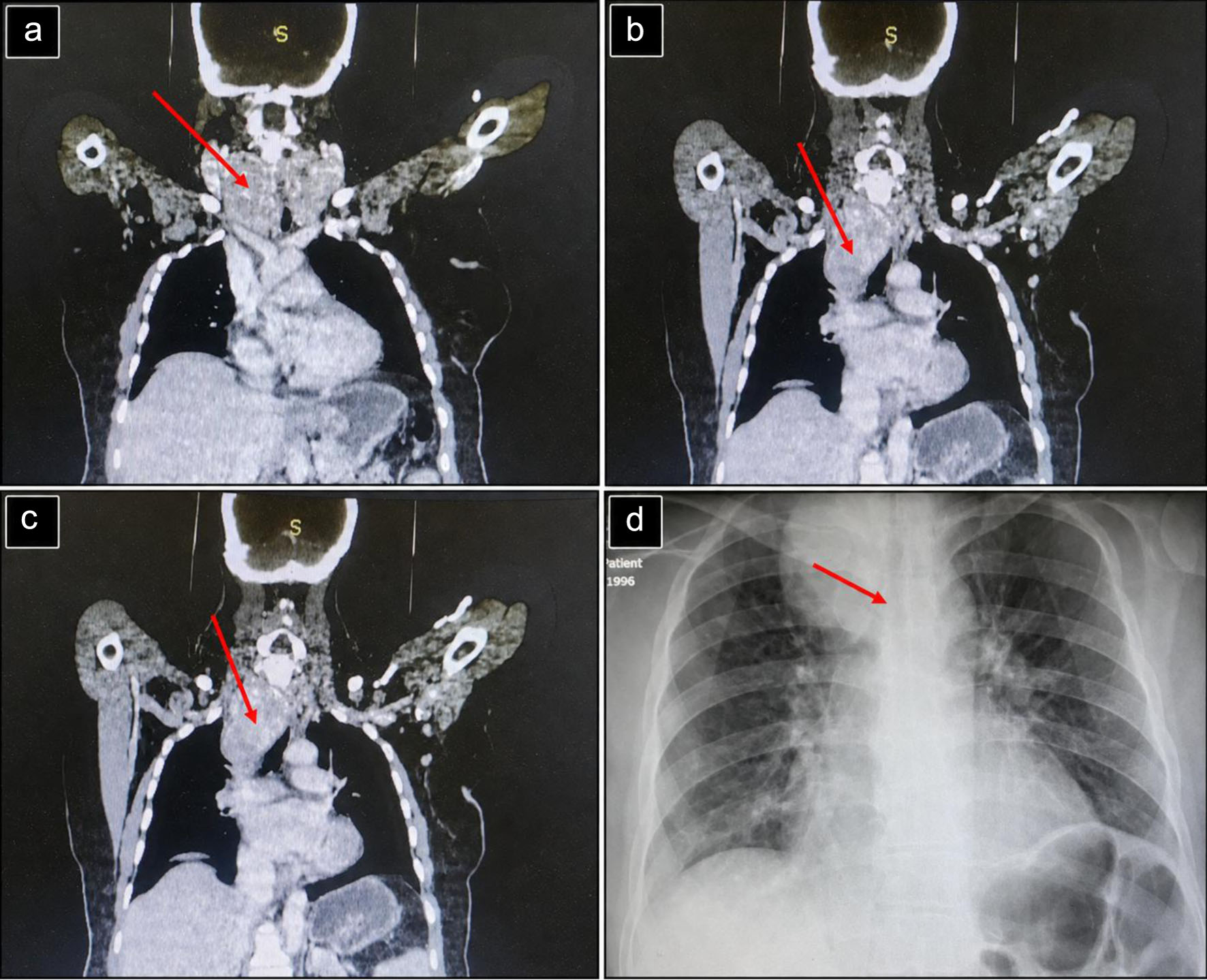 Click for large image | Figure 1. Preoperative imaging demonstrating extensive retrosternal extension of a multinodular goiter. (a–c) Coronal contrast-enhanced CT images demonstrating a markedly enlarged multinodular thyroid gland with substantial retrosternal extension into the superior mediastinum (red arrows), causing narrowing and displacement of the trachea. (d) Preoperative chest radiograph demonstrating widening of the superior mediastinum with rightward tracheal deviation (red arrow), consistent with the retrosternal extension of the goiter. CT: computed tomography. |
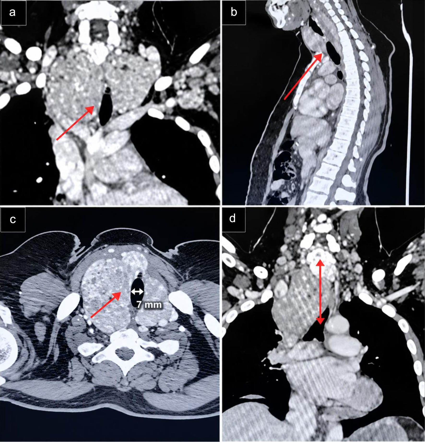 Click for large image | Figure 2. Contrast-enhanced CT demonstrating giant retrosternal goiter with tracheal compression and deviation. (a) Coronal CT images demonstrating the longitudinal extent of the tracheal stenosis, measuring approximately 10 mm in length. (b) Sagittal CT images demonstrating the longitudinal extent of the tracheal stenosis, measuring approximately 10 mm in length. (c) Axial chest CT image demonstrating severe narrowing of the tracheal lumen, most pronounced at the proximal tracheal segment. (d) Coronal CT image demonstrating the distance between the proximal margin of the tracheal stenosis and the tracheal bifurcation (carina), measuring approximately 60 mm. CT: computed tomography. |
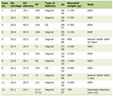 Click to view | Table 1. Comprehensive Preoperative Assessment of Our Patient With Down Syndrome and Giant Retrosternal Goiter |
Preoperatively, the patient’s airway was assessed and found to have relative macroglossia, a high-arched palate, a short neck with a neck circumference of 43 cm, a sternomental distance of < 12 cm, mouth opening of 4 cm, a thyromental distance of < 6 cm, a modified Mallampati class IV view, reduced neck mobility, and a positive upper lip bite test. Based on these findings, the patient was considered to have both a potentially difficult airway for intubation and difficult mask ventilation. Given the patient’s severe intellectual disability and anticipated inability to cooperate, awake fiberoptic intubation even under sedation was considered unsuitable. Preoperative laryngoscopy was not performed; therefore, baseline vocal-cord mobility could not be objectively documented. No clinical signs suggestive of pre-existing vocal-cord dysfunction were identified before surgery.
The clinical course and major diagnostic and therapeutic events are summarized chronologically in Table 2.
Click to view | Table 2. Timeline of the Patient’s Clinical Presentation, Diagnostic Assessment, Airway Management, Surgery, and Postoperative Course |
Diagnosis
The patient was diagnosed with a giant retrosternal multinodular goiter causing critical tracheal narrowing and mediastinal extension below the aortic arch. The combination of severe airway compression, DS-associated difficult airway features, and the potential for airway collapse following induction constituted a high-risk anesthetic and surgical scenario. Because complete cervical delivery was considered unsafe, a combined cervical approach with median sternotomy was planned.
Treatment
A multidisciplinary airway and surgical strategy was developed involving anesthesiologists, endocrine surgeons, cardiothoracic surgeons, and perfusionists. All relevant team members were informed in advance of the patient’s severe extrinsic tracheal compression, the anticipated airway risks, and the planned airway and rescue strategy. Given the reported orthopnea, positional tolerance was assessed before induction. Given the reported orthopnea, the patient’s tolerance of the ramped position was assessed before induction. The position was well tolerated, without clinically significant respiratory deterioration or desaturation. On arrival in the operating room, saturation of oxygen (SpO2) was 92% on room air and increased to 94% after positioning in the ramped position, also without supplemental oxygen. Standard American Society of Anesthesiologists (ASA) monitoring and invasive arterial pressure monitoring were then established, and induction was performed with the patient maintained in the ramped position. High-flow nasal cannula (HFNC) oxygenation with a fraction of inspired oxygen (FiO2) 1.0 at 40 L/min was initiated 20 min before induction assuring SpO2 100%.
The airway strategy was based on balancing the risks associated with loss of spontaneous ventilation against the feasibility of awake airway management. Video laryngoscopy was selected as the primary technique for tracheal intubation, with flexible bronchoscopy available for immediate airway rescue. Completely awake fiberoptic or videolaryngoscopic intubation was considered impractical because the patient was unable to cooperate adequately. Conversely, deep or heavily sedated awake intubation was avoided because of concern for respiratory depression and further compromise of the already compressed trachea. Inhalational induction with preservation of spontaneous ventilation was also considered but was not selected because of the patient’s inability to cooperate and the need for a predictable and prompt definitive airway.
A stepwise rescue plan was established before induction. A supraglottic airway device (laryngeal mask airway (LMA)) was immediately available as a rescue option, and the surgical team was prepared to establish a surgical airway if adequate ventilation could not be maintained. If conventional airway rescue failed but oxygenation and hemodynamic stability were maintained sufficiently to permit controlled transfer, the patient would be awakened if clinically feasible and transferred to a separate operating room equipped for emergency femoro-femoral venoarterial extracorporeal membrane oxygenation (VA-ECMO) and cardiopulmonary bypass (CPB). Immediate VA-ECMO/CPB was not available or primed in the initial operating room; therefore, extracorporeal support was planned as an escalation pathway rather than as an immediately deployable rescue during catastrophic airway collapse. If controlled transfer was not clinically feasible, emergency airway and cardiopulmonary rescue would be undertaken according to the available institutional capabilities. The cardiothoracic surgeons and perfusion team were informed of this escalation pathway and prepared accordingly before induction.
After multidisciplinary discussion, controlled intravenous induction was selected despite the severe extrinsic tracheal compression. Anesthesia was induced with propofol 150 mg and fentanyl 150 µg, followed by succinylcholine 100 mg. Tracheal intubation was performed immediately as the first-line approach using a video laryngoscope. Intubation was successfully achieved on the first attempt, with no hypoxemia or hemodynamic instability. Video laryngoscopy was performed using a STORZ C-MAC videolaryngoscope (Karl Storz, Tuttlingen, Germany) with a Macintosh #3 blade, providing a Cormack–Lehane Grade I view, and no bougie was required (Fig. 3). The endotracheal tube was advanced beyond the stenotic segment and positioned appropriately in relation to the tracheal bifurcation. Given the approximately 7-mm luminal diameter observed on preoperative imaging, endotracheal tubes of sizes 5.0, 5.5, and 6.0 were prepared to allow stepwise selection according to the actual airway dimensions encountered during intubation. A 6.0-mm tube (external diameter 8.2 mm) was successfully advanced through the stenotic segment without resistance or deterioration in oxygenation or hemodynamic status and was therefore retained as the definitive airway. Correct tube position and patency beyond the area of tracheal compression were subsequently confirmed by flexible fiberoptic bronchoscopy performed through the endotracheal tube. Vecuronium 10 mg was administered only after definitive airway control had been established.
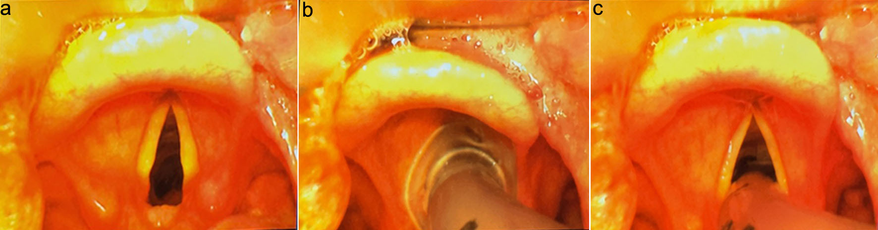 Click for large image | Figure 3. Sequential video laryngoscopy views during airway management in our patient with a large retrosternal goiter. (a) Initial video laryngoscopy view demonstrating Cormack–Lehane grade 1. (b) Video laryngoscopy view during advancement of the endotracheal tube toward the glottic opening, demonstrating the tube approaching the vocal cords under direct video guidance. (c) Improved visualization of the glottis with clear identification of the vocal cords, facilitating controlled endotracheal intubation. |
Total intravenous anesthesia (TIVA) was used for maintenance, with propofol (100–150 µg/kg/min, based on total body weight) and remifentanil (0.3–0.5 µg/kg/min, based on ideal body weight) administered by continuous infusion. Anesthetic depth was monitored using processed electroencephalography (pEEG) and the patient state index (PSi; Masimo) (Fig. 4).
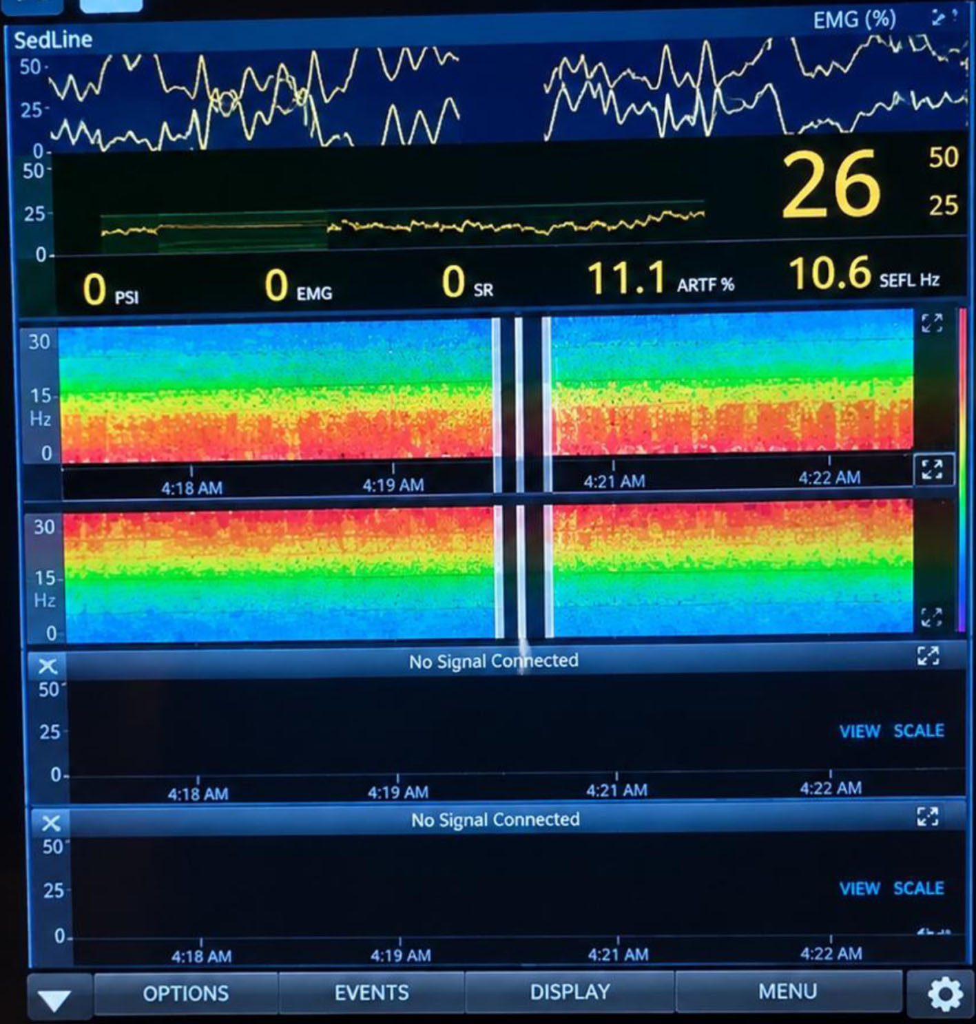 Click for large image | Figure 4. Intraoperative processed EEG monitoring during general anesthesia: SedLine display showing bilateral density spectral array (DSA) trends and processed EEG monitoring, with a PSI of 26, demonstrating continued adequate anesthesia depth and bilateral cerebral electrical activity. EEG: electroencephalography; PSI: patient state index. |
Total thyroidectomy was performed through a combined low transverse cervical incision and full median sternotomy (Fig. 5). Intraoperative nerve monitoring was used to identify and preserve both recurrent laryngeal nerves. Estimated blood loss was 250 mL, and operative time was 185 min. Following resection, a cuff-leak test was performed to assess for clinically significant upper-airway obstruction before extubation and demonstrated an adequate leak. No formal dynamic assessment for tracheomalacia, such as fiberoptic bronchoscopy evaluation during spontaneous and positive-pressure breathing, flow–volume loop analysis, or direct dynamic assessment of the trachea after decompression, was performed. The patient was extubated in the operating room after demonstrating full recovery of consciousness, adequate spontaneous ventilation, stable oxygenation, hemodynamic stability, and a quantitative train-of-four (TOF) ratio of 0.9 following reversal with neostigmine. Neuromuscular recovery was objectively confirmed by a TOF ratio of 0.9; therefore, additional reversal with sugammadex was not considered necessary.
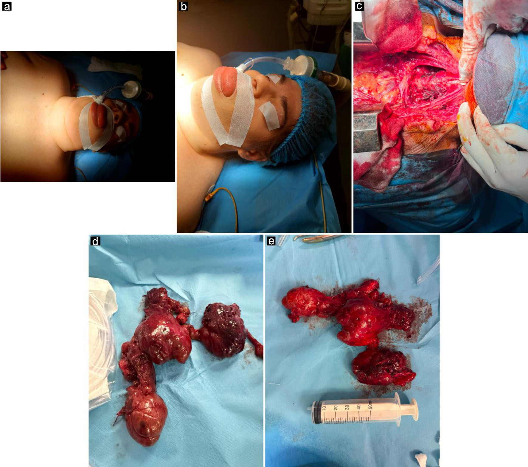 Click for large image | Figure 5. Perioperative airway features and surgical findings. (a) Patient intubated and positioned for surgery, demonstrating marked anterior neck enlargement caused by the giant goiter. (b) Macroglossia identified during airway assessment, representing an additional anatomical challenge for airway management. (c) Surgical field following thyroid gland removal and sternotomy, demonstrating the operative site after resection of the retrosternal goiter. (d, e) Macroscopic views of the resected thyroid specimen, demonstrating its large size and multinodular appearance. |
Follow-up and outcome
The postoperative course was uneventful. The patient was monitored in the intensive care unit (ICU) overnight and subsequently transferred to the ward. Following extubation, BiPAP was applied for 6 h with an FiO2 of 50%, pressure support (PS) of 16 cm H2O, and positive end-expiratory pressure (PEEP) of 8 cm H2O. Thoracic drainage tubes were removed on the first postoperative day. Respiratory exercises and chest physiotherapy were initiated early and continued throughout the postoperative period.
There was no evidence of hypocalcemia or postoperative voice changes. Levothyroxine replacement therapy was initiated at 100 µg daily, with ongoing endocrinology follow-up. Her chronic risperidone therapy (2 mg daily) was also resumed.
Postoperative analgesia consisted of paracetamol and a single dose of ibuprofen. The patient was considered clinically suitable for a single dose of a nonsteroidal anti-inflammatory drug (NSAID), with no contraindication related to bleeding risk, renal function, or the concomitant median sternotomy. No further NSAID administration was required. No clinically significant cervical hematoma, postoperative bleeding, renal dysfunction, or sternal complication occurred. Regional analgesic techniques, including superficial cervical plexus, parasternal, or serratus plane blocks, were not used. Analgesia was adequate without the need for postoperative opioids, facilitating effective respiratory exercises and chest physiotherapy while minimizing the risk of pulmonary complications.
The patient remained clinically stable and was discharged home on postoperative day 5.
Final histopathological examination demonstrated a 1.2-cm noninvasive follicular thyroid neoplasm with papillary-like nuclear features (NIFTP) arising within a background of multinodular goiter. The lesion showed a predominantly follicular growth pattern and characteristic papillary-type nuclear features, with no evidence of capsular or vascular invasion. The available pathology report did not specifically document the extent of capsular sampling. Given the absence of identified invasion and the low-risk nature of NIFTP, postoperative radioactive iodine therapy was not administered following total thyroidectomy. Levothyroxine replacement was continued without intentional thyroid-stimulating hormone (TSH) suppression, with ongoing endocrinology follow-up and thyroid-function monitoring.
| Discussion | ▴Top |
DS and anesthetic implications
DS is usually recognized in childhood, and consequently, much of the perioperative literature has focused on pediatric patients. This emphasis reflects the substantial congenital and multisystem comorbidity associated with DS, the frequent need for anesthesia for surgical and diagnostic procedures during childhood, and the progressive improvement in survival resulting from advances in medical and surgical care [5–8].
Importantly, many DS-associated abnormalities persist into adulthood and remain relevant to perioperative airway management. Craniofacial and upper-airway features, including macroglossia, midface hypoplasia, a short neck, a high-arched palate, and a relatively narrow upper airway, may contribute to difficult airway management. OSA, respiratory disease, cervical spine abnormalities, and laryngotracheal or subglottic abnormalities may further increase perioperative risk [9, 10]. Cardiovascular disease, including congenital or residual cardiac disease and pulmonary hypertension (PH), remains an important consideration in adults with DS [11]. Accordingly, preoperative assessment should be individualized, considering airway anatomy, respiratory and cardiovascular status, cervical spine considerations, previous anesthetic or airway difficulties, and the patient’s ability to cooperate with the planned airway technique [12].
Improved survival has resulted in an increasingly important adult perioperative population with DS. Adult-focused literature indicates that many individuals with DS now live well into adulthood, with life expectancy approaching 60 years, although outcomes remain influenced by associated comorbidities. Survival among individuals with DS and CHD has also improved across successive birth cohorts [13, 14]. Thus, airway characteristics recognized during childhood remain clinically relevant when patients with DS present for major surgery in adulthood. In the present patient, relative macroglossia, a high-arched palate, a short neck, limited neck mobility, obesity, and a Mallampati class IV airway represented persistent DS-associated challenges that were further compounded by severe mechanical tracheal compression from the giant retrosternal goiter.
Overall, perioperative care should be multidisciplinary and individualized, with emphasis on difficult-airway preparedness, cardiovascular optimization, cervical-spine protection, prevention of pulmonary hypertensive events, and vigilant postoperative monitoring. Preoperative assessment should include CHD, PH, OSA, recent respiratory infection, aspiration risk, cervical spine instability, and previous airway difficulties. Echocardiography is important when cardiac disease or PH is suspected. Intraoperatively, careful positioning, appropriate monitoring, judicious fluid management, and avoidance of factors increasing pulmonary vascular resistance are essential. Postoperatively, respiratory and cardiovascular monitoring, opioid-sparing analgesia, and higher-acuity observation should be considered in patients with severe OSA, PH, or significant cardiac disease [13].
Perioperative airway management in large retrosternal goiter
Retrosternal goiters can cause clinically significant airway compromise, ranging from delayed postoperative respiratory failure to acute, life-threatening obstruction. Reported cases have described severe tracheal compression with relatively limited preoperative respiratory symptoms, including postoperative respiratory deterioration related to tracheal compression and thyroid edema [15]. Other reports have demonstrated that substernal multinodular goiters may remain clinically stable before progressing to acute airway obstruction [16]. More recently, severe tracheal compression from a benign multinodular goiter was associated with acute hypercapnic respiratory failure, which was successfully managed with awake fiberoptic intubation followed by urgent thyroidectomy [17]. Collectively, these reports emphasize that the clinical severity of a retrosternal goiter cannot be determined solely from symptoms and that airway planning should consider the degree and location of tracheal compression, mediastinal extension, and the patient’s respiratory reserve.
Preoperative airway assessment should therefore be comprehensive rather than dependent on a single bedside predictor. Studies evaluating combinations of the Wilson Sum Score, Mallampati classification, thyromental and sternomental distances, and the upper lip bite test have demonstrated variable predictive performance, supporting their use as complementary components of a broader airway assessment [18–21]. A Cochrane diagnostic accuracy review similarly found that commonly used bedside tests generally have limited sensitivity despite relatively high specificity, reinforcing the importance of integrating anatomical findings, previous airway history, functional characteristics, and the overall clinical context [22]. In the present patient, these clinical assessments were interpreted together with the markedly abnormal cross-sectional imaging and the known anatomical features of DS and obesity, rather than being used as independent determinants of the airway strategy.
Ultrasound may provide an additional objective component of airway assessment, particularly when external anatomical measurements are difficult to interpret. Studies have examined anterior neck soft-tissue thickness and hyoid-to-skin distance as potential predictors of difficult laryngoscopy, although the available evidence supports ultrasound as an adjunct rather than a standalone screening modality [23, 24]. Airway ultrasound was not performed in our patient; therefore, the clinical examination and, importantly, CT provided the principal anatomical information for airway planning.
When difficult tracheal intubation is anticipated, video laryngoscopy represents an important option for securing the upper airway. In a randomized study of 140 adults with simulated difficult airways, the McGrath MAC and King Vision demonstrated comparable laryngeal views and first-attempt success, with differences in the time required for visualization and intubation [25]. Although this evidence does not specifically address severe intrathoracic tracheal compression, it supports the role of video laryngoscopy as part of a planned difficult-airway strategy. In our patient, its selection was based not on the expectation that video laryngoscopy would relieve the distal obstruction, but on the need to obtain rapid and reliable upper-airway access in the presence of both DS-associated difficult-airway features and severe mechanical tracheal narrowing.
Airway management does not end with successful intubation, particularly in patients with obesity, prolonged tracheal compression, and anticipated difficult reintubation. Post-extubation complications may include airway obstruction, desaturation, apnea, inadequate neuromuscular recovery, and the need for reintubation [26]. Accordingly, extubation should be regarded as a planned phase of difficult-airway management, with appropriate positioning, oxygenation, respiratory monitoring, and access to non-invasive respiratory support or reintubation when clinically indicated. In the present patient, the risk of postoperative airway compromise was considered in the decision to perform tracheal extubation only after full recovery and objective neuromuscular recovery, followed by close postoperative respiratory monitoring and non-invasive ventilatory support.
Our case in the context of the current literature
DS and the anticipated difficult airway
Our patient presented with several of the characteristic airway features described in patients with DS, including relative macroglossia, a high-arched palate, a short neck, limited neck mobility, and a Mallampati IV airway. These findings, together with obesity and the large anterior neck mass, raised concerns about both mask ventilation and tracheal intubation. Cervical spine radiographs did not show atlantoaxial instability, which allowed airway management to proceed without evidence of an additional cervical spine-related risk, although excessive neck extension was still avoided. This is in keeping with the literature, which emphasizes careful airway assessment and cervical positioning in patients with trisomy 21 because of their characteristic craniofacial anatomy and the potential for cervical spine abnormalities.
Additional airway complexity from the giant retrosternal goiter
The main challenge in our patient, however, was the combination of these DS-related airway features with severe mechanical airway obstruction caused by the giant retrosternal goiter. CT scan showed marked rightward tracheal deviation, a minimum tracheal diameter of approximately 7 mm, and extension of the goiter below the aortic arch. Therefore, the concern was not limited to a potentially difficult laryngoscopy view. The critical issue was whether the airway would remain patent during induction and after the onset of positive-pressure ventilation. Experience reported in patients with giant retrosternal goiters similarly highlights the need for individualized airway planning when substantial tracheal compression is present, with bronchoscopy and extracorporeal support considered when the risk of airway loss is particularly high [27, 28]. In our case, the combination of severe anatomical compression and a potentially difficult upper airway justified preparation for advanced rescue strategies. Based on these anatomical and clinical considerations, a stepwise airway-management strategy was developed, progressing from the planned primary approach to conventional rescue measures and, when necessary, extracorporeal rescue (Fig. 6).
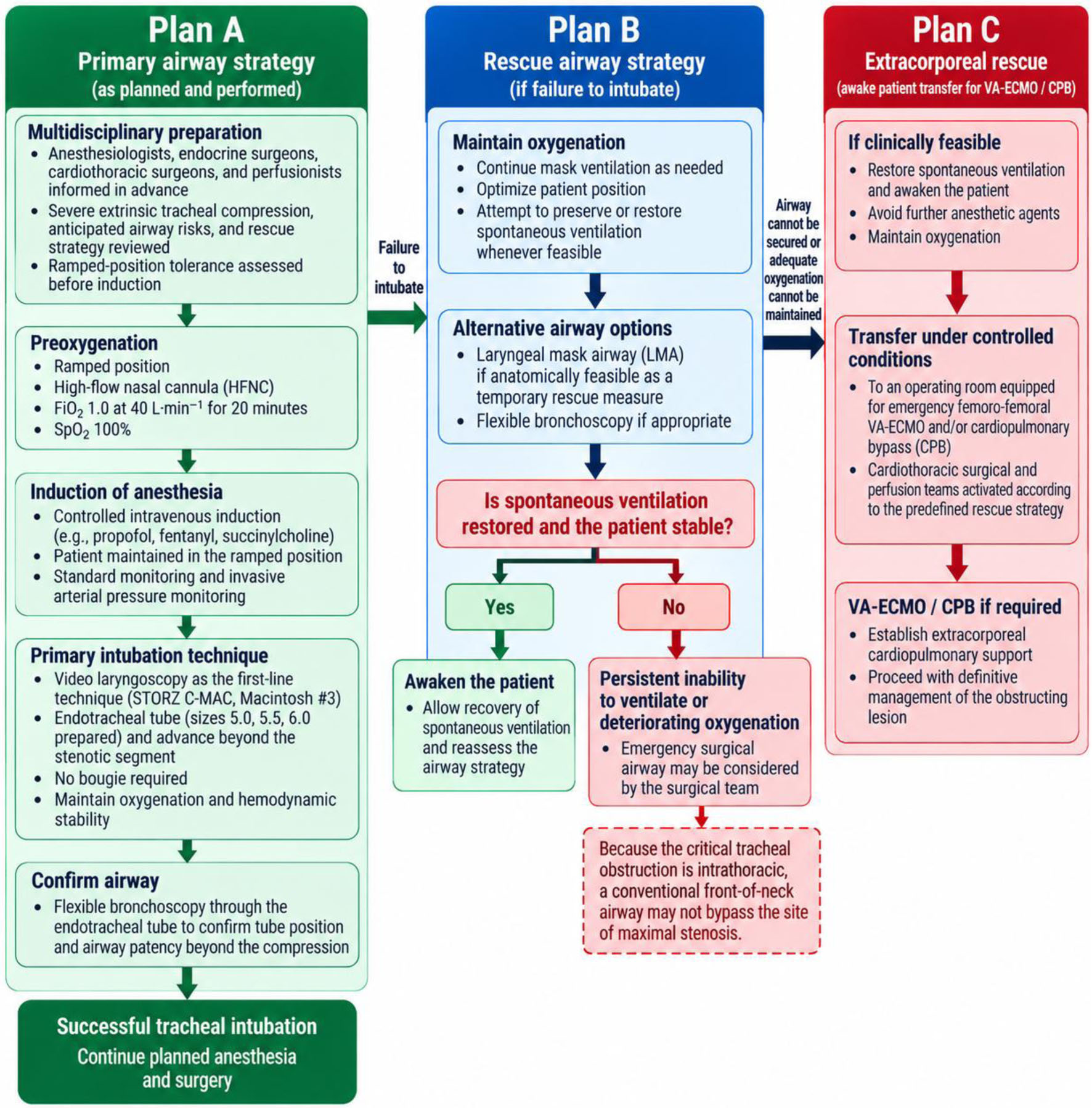 Click for large image | Figure 6. Stepwise airway management algorithm for severe intrathoracic tracheal compression. |
Selection of the intubation strategy
Video laryngoscopy was selected as the primary intubation technique, consistent with reported experience in patients undergoing surgery for substernal goiter. A large retrospective series of 179 patients reported successful airway management with direct or video laryngoscopy in 162 patients (90.5%), whereas 17 patients (9.5%) required fiberoptic intubation [29]. In our patient, however, the choice of technique required consideration of both the severe 7-mm tracheal narrowing and established predictors of difficult upper-airway management, including obesity, limited neck mobility, macroglossia, and a Mallampati IV airway.
Awake fiberoptic intubation would ordinarily be considered in the presence of severe tracheal compression. In this case, severe intellectual disability made adequate cooperation with an awake technique unlikely. Completely awake video laryngoscopy was similarly considered impractical, while heavily sedated awake intubation was avoided because of the potential for respiratory depression and further compromise of the compressed airway. Inhalational induction with maintenance of spontaneous ventilation was also considered but was not selected because it could not provide the predictable and prompt airway control required in a patient unable to cooperate. After multidisciplinary discussion, controlled intravenous induction was therefore chosen, accepting the potential risk associated with loss of spontaneous ventilation but with immediate tracheal intubation and an established rescue pathway.
These considerations are consistent with contemporary Difficult Airway Society (DAS) guidelines, which emphasizes pre-induction airway assessment and planning, optimization of patient positioning and oxygenation, multidisciplinary communication, and preparation for failure. In patients with anticipated difficult airway management, awake tracheal intubation should be considered [30, 31]. The 2025 DAS guidelines also recommend video laryngoscopy for tracheal intubation whenever possible, together with continuous oxygenation and objective confirmation of successful tracheal intubation [30]. In our case, these principles were applied through advance multidisciplinary planning, HFNC preoxygenation, video laryngoscopy as the primary technique, flexible bronchoscopy confirmation, and a predefined rescue strategy with surgical and extracorporeal support available if airway control failed. Although awake intubation was considered, it was not feasible because of the patient’s inability to cooperate, supporting the need for an individualized approach.
Preoxygenation and positioning were optimized before induction. The patient tolerated the ramped position without clinically significant respiratory deterioration, and HFNC oxygenation with FiO2 1.0 was initiated before induction, achieving SpO2 of 100%. Following induction with propofol, fentanyl, and succinylcholine, video laryngoscopy provided a Cormack–Lehane grade I view, and the trachea was successfully intubated on the first attempt without hypoxemia or hemodynamic instability. Although CT demonstrated a minimum luminal diameter of approximately 7 mm, the 6.0-mm internal-diameter tube, with an external diameter of 8.2 mm, passed through the stenotic segment without resistance. This apparent discrepancy may reflect differences between static imaging and the dynamic airway dimensions encountered during instrumentation; however, the exact explanation cannot be established retrospectively. Correct tube position and patency beyond the compressed segment were subsequently confirmed by flexible bronchoscopy. Correct tube position and patency beyond the compressed segment were subsequently confirmed by flexible bronchoscopy.
The availability of immediate rescue options was an integral part of this strategy. Flexible bronchoscopy and a supraglottic airway were prepared for immediate rescue, while surgical airway access and escalation to femoro-femoral VA-ECMO/CPB were planned if conventional measures failed. Thus, the airway strategy was individualized according to the severity of tracheal compression, the patient’s ability to cooperate, and the available rescue resources. The successful first-attempt intubation without respiratory or hemodynamic deterioration supports the feasibility of video laryngoscopy as a primary approach in selected patients with severe retrosternal goiter when comprehensive rescue planning is established in advance.
Extubation and assessment for postoperative airway instability
Tracheal extubation was an important consideration because prolonged compression by a large retrosternal goiter may weaken the tracheal cartilaginous framework and predispose to airway collapse after thyroidectomy [32, 33]. Although clinically significant post-thyroidectomy tracheomalacia appears to be uncommon, it remains a potentially serious complication in patients with large retrosternal goiters and marked tracheal compression [34]. In our patient, an adequate cuff-leak test following thyroid resection, together with full recovery of consciousness, adequate spontaneous ventilation, stable oxygenation and hemodynamics, and a quantitative TOF ratio of 0.9 after reversal with neostigmine, supported extubation in the operating room. In the event of clinically significant post-extubation respiratory deterioration, immediate reoxygenation and reintubation using the same difficult-airway strategy employed during induction would have been undertaken, with video laryngoscopy as the primary technique and flexible bronchoscopy available for airway assessment and confirmation. Extubation was performed only after confirming satisfactory neuromuscular recovery and the absence of clinically significant residual blockade. Sugammadex was not administered because adequate recovery had already been achieved with neostigmine. No formal dynamic assessment for tracheomalacia, including bronchoscopic evaluation during spontaneous and positive-pressure breathing, was performed, as noted in the “Limitations” section. The subsequent absence of clinically evident airway collapse or respiratory compromise was reassuring, although tracheomalacia could not be definitively excluded. Given the combination of DS, obesity, severe preoperative airway compression, and the extent of surgery, postoperative respiratory support and overnight ICU observation were considered appropriate.
Postoperative respiratory management and outcome
The uneventful postoperative respiratory course was particularly reassuring given the multiple factors that could have contributed to respiratory compromise. DS may be associated with reduced upper-airway and respiratory reserve, while obesity, median sternotomy, thoracic drainage, and postoperative pain can further impair ventilation and effective airway clearance. In our patient, BiPAP was used after extubating, together with early respiratory exercises, chest physiotherapy, adequate analgesia, and overnight ICU monitoring. These measures were especially important after the combined cervical and thoracic procedure, as effective pain control allowed the patient to participate in respiratory exercises despite the extensive surgical incision and chest drains. The absence of significant respiratory complications, hypocalcemia, or postoperative voice changes, followed by discharge home on postoperative day 5, reflects a favorable short-term outcome despite the considerable airway and perioperative challenges presented by this case. Table 3 summarizes the key principles of treatment and airway management applied in our case, highlighting the main perioperative strategies used to anticipate and address the complex anatomical and respiratory challenges.
Click to view | Table 3. Summary of Perioperative Management and Clinical Outcome in Our Patient With Down Syndrome and Giant Retrosternal Goiter |
An incidental NIFTP was identified on final pathology in this patient. NIFTP is a low-risk thyroid neoplasm characterized by a predominantly follicular growth pattern and papillary-type nuclear features, with absence of capsular or vascular invasion and other exclusionary features [35]. Because the lesion was completely removed by total thyroidectomy and no invasion was identified, no postoperative radioactive iodine therapy was administered. Levothyroxine replacement was continued without intentional TSH suppression, with ongoing endocrinology follow-up and thyroid-function monitoring. These findings are clinically relevant because NIFTP does not require management as conventional differentiated thyroid carcinoma when the defining noninvasive criteria are fulfilled.
Limitations
A limitation of this case is that tracheomalacia was not formally assessed using a dynamic diagnostic technique. Although an adequate cuff leak was demonstrated and the patient was successfully extubated without immediate respiratory compromise, the cuff-leak test does not assess dynamic tracheal collapse and therefore cannot exclude tracheomalacia. Dynamic bronchoscopic assessment during spontaneous and positive-pressure breathing, or another dedicated functional assessment, would have provided a more definitive evaluation. Another limitation was the absence of preoperative laryngoscopy, which precluded objective documentation of baseline vocal-cord mobility.
Conclusion
This case demonstrates that airway management in a patient with DS and a giant retrosternal goiter causing critical tracheal compression requires a patient-specific strategy that addresses both difficult upper-airway anatomy and the risk of loss of distal tracheal patency after induction. Preoperative CT assessment of tracheal narrowing, deviation, and mediastinal extension, combined with assessment of the patient’s ability to cooperate, was essential for selecting an appropriate airway strategy. Although awake fiberoptic intubation would ordinarily be considered in severe tracheal compression, it was not feasible in this patient because of severe intellectual disability. Controlled intravenous induction with immediate video-laryngoscopy intubation was therefore undertaken only after optimization of positioning and oxygenation and establishment of a clearly defined rescue pathway.
The successful first-attempt intubation, passage of a suitably selected endotracheal tube through the stenotic segment, and subsequent fiberoptic bronchoscopy confirmation illustrate that video laryngoscopy can be a feasible primary technique in selected patients when awake airway management is impractical, provided that comprehensive rescue capability is established before induction. Equally important, airway management did not end with intubation: assessment for postoperative airway instability, confirmation of neuromuscular recovery, cuff-leak testing, planned respiratory support, and close postoperative observation were integral to safe extubating.
The principal lesson from this case is therefore not that one airway technique should be universally preferred, but that successful management depends on anticipating where airway failure may occur, selecting the least hazardous feasible primary approach, and preparing an immediately executable escalation pathway before induction. In patients with critical retrosternal tracheal compression, particularly when additional difficult-airway features or inability to cooperate are present, multidisciplinary preparation and rescue readiness should be regarded as integral components of the airway technique itself.
Learning points
Integrate difficult-airway anatomy with CT-defined tracheal compression and deviation when planning airway management.
Individualize the intubation technique according to airway anatomy, severity of compression, patient cooperation, and available expertise.
Establish a multidisciplinary rescue plan before induction, including bronchoscopy, supraglottic airway, surgical airway, and extracorporeal rescue when appropriate.
Optimize positioning and oxygenation before induction and prepare appropriately sized endotracheal tubes for critical stenosis.
Treat extubation as a second high-risk airway event, with assessment of airway patency, neuromuscular recovery, and postoperative respiratory support.
A cuff-leak test does not exclude tracheomalacia; dynamic assessment may be required when clinically indicated.
Acknowledgments
None to declare.
Financial Disclosure
None to declare.
Conflict of Interest
None to declare.
Informed Consent
Written informed consent for publication of the case report and identifiable facial and oral photographs was obtained from the patient’s legal guardian. The case report was reviewed by the institutional ethics committee, which determined that formal ethical approval was not required for publication of this single case report (Protocol Number 2319/1).
Author Contributions
RD and MN contributed to the conceptualization and design of the case report, interpretation of the clinical course, drafting of the manuscript, and critical revision of the manuscript for important intellectual content; RD, EN, and EB contributed to the literature review, synthesis, and interpretation of relevant evidence; MM, AD, KL, AC, and AL contributed to the acquisition, analysis, and interpretation of clinical and perioperative data; RD, VE, EK, and FM contributed to manuscript drafting, language editing, and critical revision. All authors reviewed and approved the final version of the manuscript and agreed to be accountable for all aspects of the work, ensuring that questions related to the accuracy or integrity of any part of the manuscript were appropriately investigated and resolved.
Data Availability
The authors declare that data supporting the findings of this study are available within the article.
| References | ▴Top |
- Hedayati N, McHenry CR. The clinical presentation and operative management of nodular and diffuse substernal thyroid disease. Am Surg. 2002;68(3):245-251; discussion 251-242.
pubmed - Al Dulaimi QA, Abu-Sa'da MO, Fatayerji RN, Alaboud A, Al Masalmeh F. Management of giant recurrent retrosternal thyroid goiter in a low-volume surgical center: a case report. Cureus. 2025;17(12):e100194.
doi pubmed - Pelli M, Loffredo C, Menna C, Fiorelli S, Massullo D. Anesthetic management of an adult patient with Down syndrome for thoracic surgery. Cureus. 2021;13(8):e17130.
doi pubmed - Apfelbaum JL, Hagberg CA, Connis RT, Abdelmalak BB, Agarkar M, Dutton RP, Fiadjoe JE, et al. 2022 American Society of Anesthesiologists Practice Guidelines for management of the difficult airway. Anesthesiology. 2022;136(1):31-81.
doi pubmed - Kucik JE, Shin M, Siffel C, Marengo L, Correa A, Congenital Anomaly Multistate P, Survival C. Trends in survival among children with Down syndrome in 10 regions of the United States. Pediatrics. 2013;131(1):e27-36.
doi pubmed - Ciccozzi A, Pizzi B, Vittori A, Piroli A, Marrocco G, Della Vecchia F, Cascella M, et al. The perioperative anesthetic management of the pediatric patient with special needs: an overview of literature. Children (Basel). 2022;9(10):1438.
doi pubmed - Glinianaia SV, Rankin J, Pierini A, Coi A, Santoro M, Tan J, Reid A, et al. Ten-year survival of children with congenital anomalies: a EUROPEAN cohort study. Pediatrics. 2022;149(3):e2021053793.
doi pubmed - Hore K, Ali U. Anaesthesia for the child with trisomy 21. BJA Educ. 2024;24(12):440-446.
doi pubmed - Shott SR. Down syndrome: common otolaryngologic manifestations. Am J Med Genet C Semin Med Genet. 2006;142C(3):131-140.
doi pubmed - Hines CB, Simmons SA. Down syndrome: a review of key perioperative implications. AORN J. 2022;116(1):4-20.
doi pubmed - Aboud MA, Arya VK, Dutta V, Ducas R, Al-Moustadi W, Niyogi SG. Anesthetic considerations for adult patients with both Down syndrome and congenital heart disease undergoing noncardiac surgery: a review article. J Cardiothorac Vasc Anesth. 2023;37(4):613-626.
doi pubmed - Lewanda AF, Matisoff A, Revenis M, Harahsheh A, Futterman C, Nino G, Greenberg J, et al. Preoperative evaluation and comprehensive risk assessment for children with Down syndrome. Paediatr Anaesth. 2016;26(4):356-362.
doi pubmed - Malinzak EB. Perioperative care of adults with Down syndrome: a narrative review. Can J Anaesth. 2021;68(10):1549-1561.
doi pubmed - Wright LK, Stallings EB, Cragan JD, Pabst LJ, Alverson CJ, Oster ME. Narrowing the survival gap: trends in survival of individuals with down syndrome with and without congenital heart defects born 1979 to 2018. J Pediatr. 2023;260:113523.
doi pubmed - Cagli K, Ulas MM, Hizarci M, Sener E. Substernal goiter: an unusual cause of respiratory failure after coronary artery bypass grafting. Tex Heart Inst J. 2005;32(2):224-227.
pubmed - Sato H, Masuda R, Iwazaki M, Inomoto C, Shoji S, Osamura YR. Airway obstruction caused by substernal thyrotoxic multinodular goiter. Tokai J Exp Clin Med. 2016;41(4):181-184.
pubmed - Khalil A, Liyanage S, Ishaq H, Babu A. Benign multinodular goitre presenting as acute decompensated type two respiratory failure. BMJ Case Rep. 2024;17(4):e259462.
doi pubmed - Domi R. A comparison of Wilson sum score and combination Mallampati, tiromental and sternomental distances for predicting difficult intubation. Maced J Med Sci. 2009;2(2):141-144.
doi - Varghese A, Mohamed T. A comparison of Mallampati scoring, upper lip bite test and sternomental distance in predicting difficult intubation. Int J Res Med Sci. 2016;4(7):2645-2648.
doi - Domi R. The best prediction test of difficult intubation. J Anaesthesiol Clin Pharmacol. 2010;26(2):193-196.
doi - Raju NV, N K, Meenakshisundaram S, Narayanan S. Comparative evaluation of airway assessment parameters for predicting difficult intubation: a prospective observational study. Cureus. 2026;18(1):e101087.
doi pubmed - Roth D, Pace NL, Lee A, Hovhannisyan K, Warenits AM, Arrich J, Herkner H. Bedside tests for predicting difficult airways: an abridged Cochrane diagnostic test accuracy systematic review. Anaesthesia. 2019;74(7):915-928.
doi pubmed - Vidhya S, Swain BP, Dash A, Swain A, Sahu S. Ultrasound-guided airway evaluation: Is it a game changer for predicting difficult endotracheal intubation? Cureus. 2024;16(1):e52557.
doi pubmed - Eksert S, Kahraman E, Inan S, Simsek F, Ince ME, Celik C, Aydin GS, et al. Comparison of focused upper airway ultrasonographic and anthropometric measurements for predicting difficult intubation: a prospective observational study. Medicina (Kaunas). 2026;62(7):1408.
doi pubmed - Kumar A, Taluja A, Saxena B, Dwivedi P. A comparative evaluation of 2 videolaryngoscopes as an intubation aid in a simulated difficult airway: a prospective randomised study. Turk J Anaesthesiol Reanim. 2022;50(5):340-345.
doi pubmed - Baftiu N, Krasniqi I, Haxhirexha K, Domi R. Survey about the Extubation Practice among Anaesthesiologists in Kosovo. Open Access Maced J Med Sci. 2018;6(2):350-354.
doi pubmed - Mahajan S, L S, Kumar S. Airway management in a patient with retrosternal goiter: a context-sensitive airway management strategy. Indian J Otolaryngol Head Neck Surg. 2022;74(Suppl 2):2214-2216.
doi pubmed - Sun X, Chen C, Zhou R, Chen G, Jiang C, Zhu T. Anesthesia and airway management in a patient with acromegaly and tracheal compression caused by a giant retrosternal goiter: a case report. J Int Med Res. 2021;49(4):300060521999541.
doi pubmed - Tasche KK, Dorneden AM, Swift WM, Boyd NH, Shonka DC, Pagedar NA. Airway management in substernal goiter surgery. Ann Otol Rhinol Laryngol. 2022;131(2):225-228.
doi pubmed - Ahmad I, El-Boghdadly K, Iliff H, Dua G, Higgs A, Huntington M, Mir F, et al. Difficult Airway Society 2025 guidelines for management of unanticipated difficult tracheal intubation in adults. Br J Anaesth. 2026;136(1):283-307.
doi pubmed - Ahmad I, El-Boghdadly K, Bhagrath R, Hodzovic I, McNarry AF, Mir F, O'Sullivan EP, et al. Difficult Airway Society guidelines for awake tracheal intubation (ATI) in adults. Anaesthesia. 2020;75(4):509-528.
doi pubmed - Ren W, Shang X, Fu H, Peng Z. Prolonged endotracheal intubation: a feasible option for tracheomalacia after retrosternal goitre surgery. Ann Palliat Med. 2020;9(4):1764-1769.
doi pubmed - Sulaiman A, Lutfi A, Ikram M, Fatimi S, Bin Pervez M, Shamim F, Abbas SA, et al. Tracheomalacia after thyroidectomy for retrosternal goitres requiring sternotomy- a myth or reality? Ann R Coll Surg Engl. 2021;103(7):504-507.
doi pubmed - Ghaloo SK, Afzal SS, Abbas SA, Ansari S, De M, Iftikhar H. Tracheomalacia in patients undergoing thyroid surgery-What is the true estimate: a systematic review and meta-analysis. World J Otorhinolaryngol Head Neck Surg. 2025;11(1):125-137.
doi pubmed - Paniza ACJ, Mendes TB, Viana MDB, Thomaz DMD, Chiappini PBO, Colozza-Gama GA, Lindsey SC, et al. Revised criteria for diagnosis of NIFTP reveals a better correlation with tumor biological behavior. Endocr Connect. 2019;8(11):1529-1538.
doi pubmed
This article is distributed under the terms of the Creative Commons Attribution 4.0 International License (CC BY 4.0), which permits unrestricted use, distribution, and reproduction in any medium, including commercial use, provided the original work is properly cited.
Journal of Medical Cases is published by Elmer Press Inc.