Journal of Medical Cases, ISSN 1923-4155 print, 1923-4163 online, Open Access
Article copyright, the authors; Journal compilation copyright, J Med Cases and Elmer Press Inc
Journal website https://jmc.elmerpub.com

Case Report

Volume 17, Number 12, December 2026, pages 712-726


First-Attempt Video Laryngoscopic Intubation in an Adult With Down Syndrome and Severe Tracheal Compression From a Giant Retrosternal Goiter

Figures

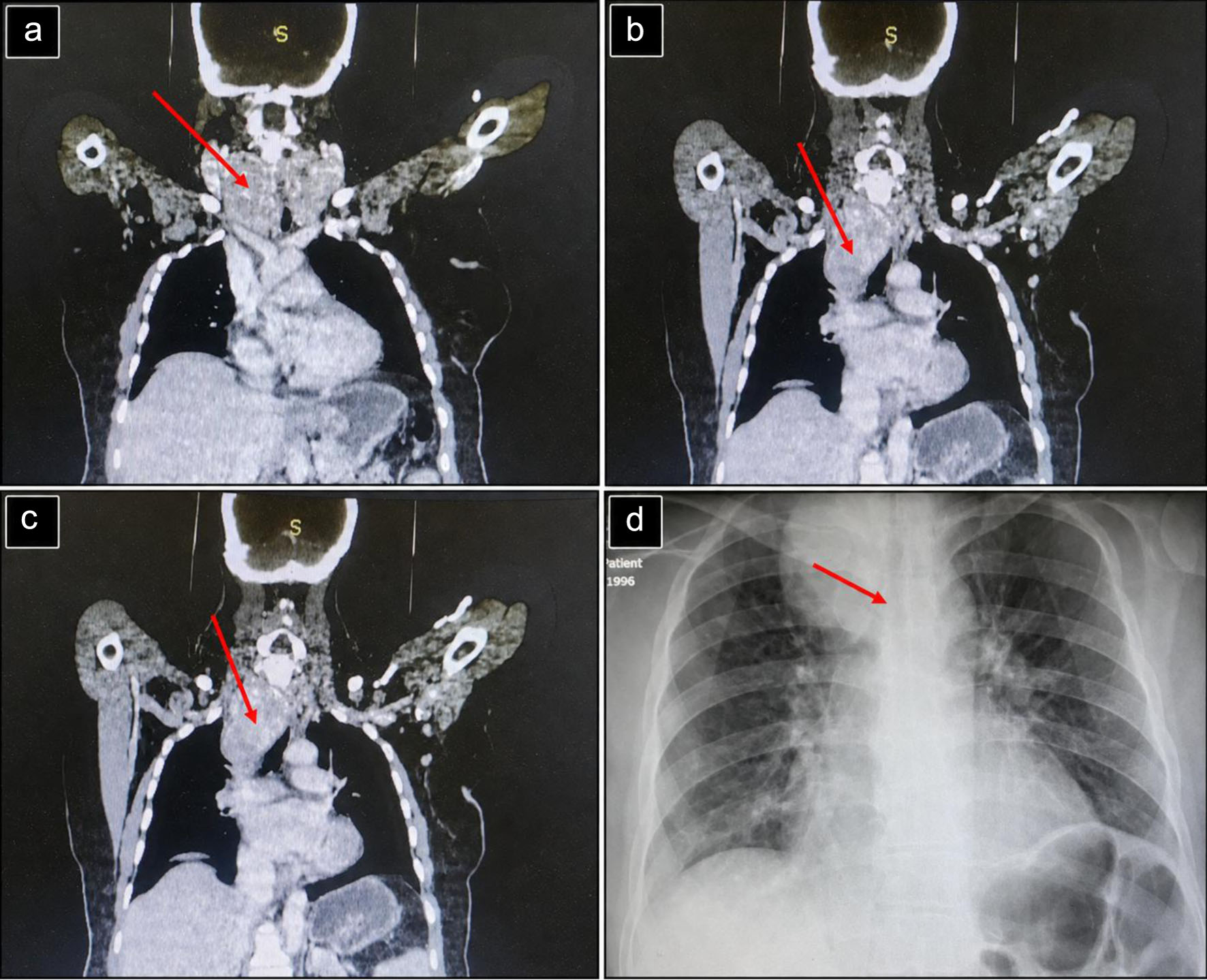
↓  Figure 1. Preoperative imaging demonstrating extensive retrosternal extension of a multinodular goiter. (a–c) Coronal contrast-enhanced CT images demonstrating a markedly enlarged multinodular thyroid gland with substantial retrosternal extension into the superior mediastinum (red arrows), causing narrowing and displacement of the trachea. (d) Preoperative chest radiograph demonstrating widening of the superior mediastinum with rightward tracheal deviation (red arrow), consistent with the retrosternal extension of the goiter. CT: computed tomography.
Figure 1.
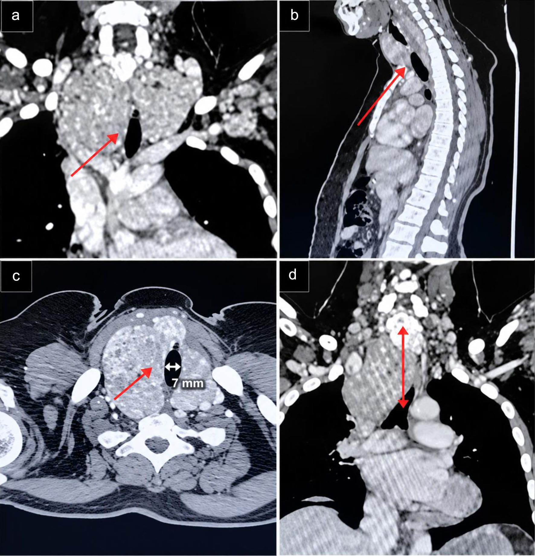
↓  Figure 2. Contrast-enhanced CT demonstrating giant retrosternal goiter with tracheal compression and deviation. (a) Coronal CT images demonstrating the longitudinal extent of the tracheal stenosis, measuring approximately 10 mm in length. (b) Sagittal CT images demonstrating the longitudinal extent of the tracheal stenosis, measuring approximately 10 mm in length. (c) Axial chest CT image demonstrating severe narrowing of the tracheal lumen, most pronounced at the proximal tracheal segment. (d) Coronal CT image demonstrating the distance between the proximal margin of the tracheal stenosis and the tracheal bifurcation (carina), measuring approximately 60 mm. CT: computed tomography.
Figure 2.
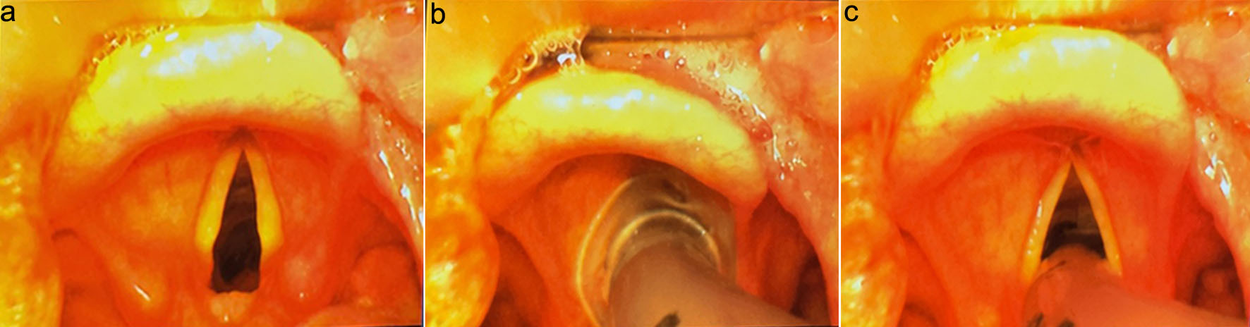
↓  Figure 3. Sequential video laryngoscopy views during airway management in our patient with a large retrosternal goiter. (a) Initial video laryngoscopy view demonstrating Cormack–Lehane grade 1. (b) Video laryngoscopy view during advancement of the endotracheal tube toward the glottic opening, demonstrating the tube approaching the vocal cords under direct video guidance. (c) Improved visualization of the glottis with clear identification of the vocal cords, facilitating controlled endotracheal intubation.
Figure 3.
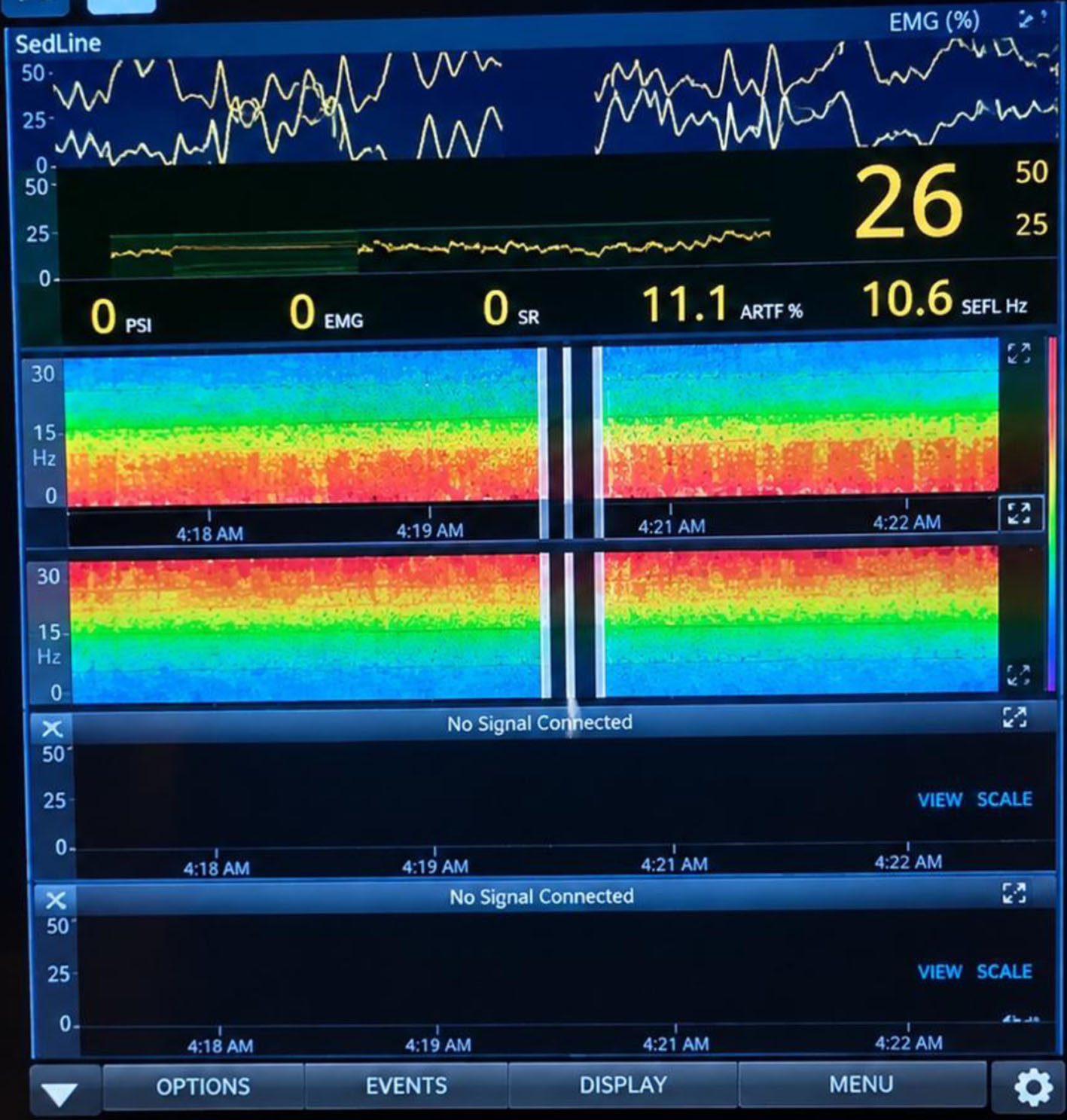
↓  Figure 4. Intraoperative processed EEG monitoring during general anesthesia: SedLine display showing bilateral density spectral array (DSA) trends and processed EEG monitoring, with a PSI of 26, demonstrating continued adequate anesthesia depth and bilateral cerebral electrical activity. EEG: electroencephalography; PSI: patient state index.
Figure 4.
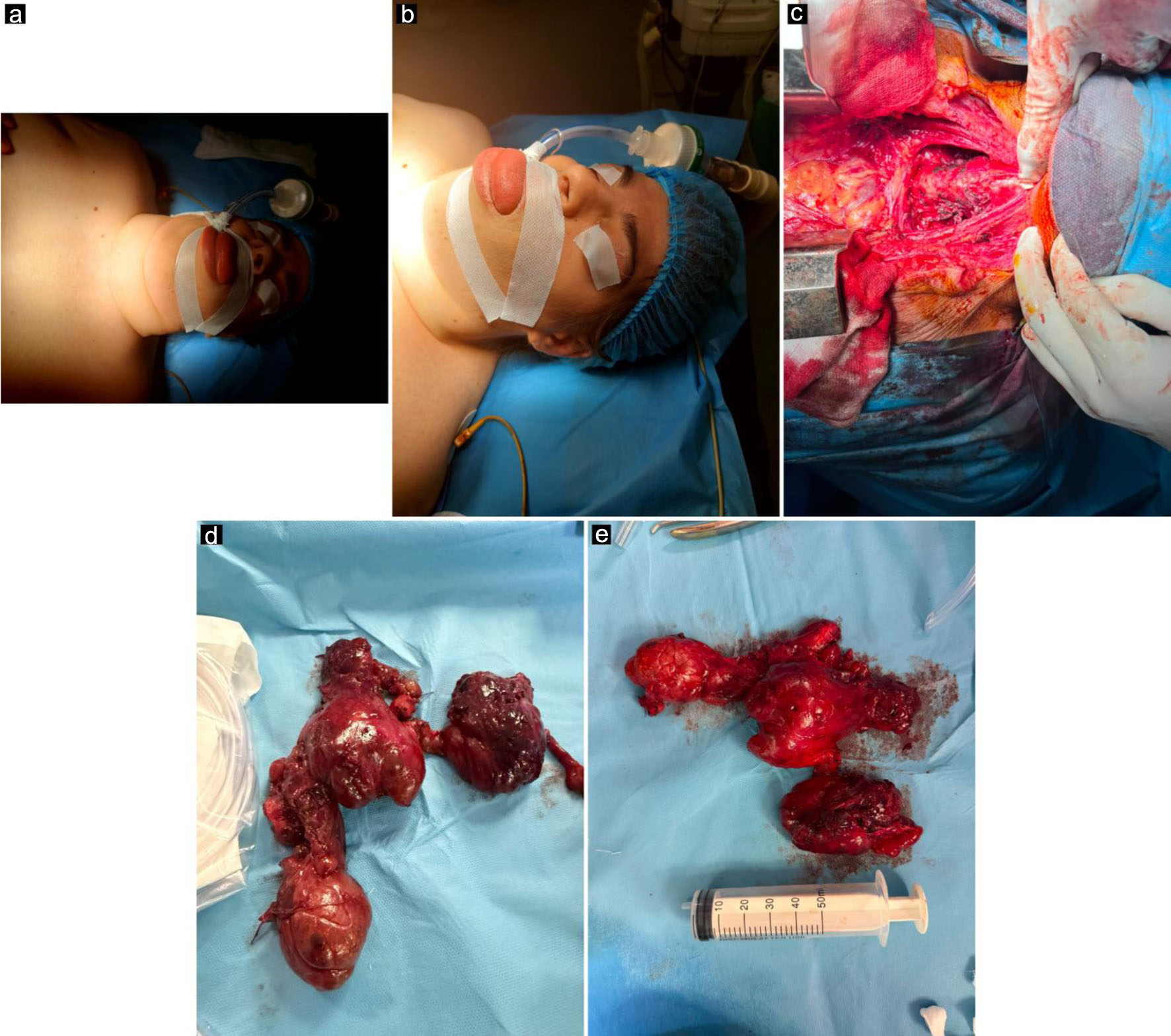
↓  Figure 5. Perioperative airway features and surgical findings. (a) Patient intubated and positioned for surgery, demonstrating marked anterior neck enlargement caused by the giant goiter. (b) Macroglossia identified during airway assessment, representing an additional anatomical challenge for airway management. (c) Surgical field following thyroid gland removal and sternotomy, demonstrating the operative site after resection of the retrosternal goiter. (d, e) Macroscopic views of the resected thyroid specimen, demonstrating its large size and multinodular appearance.
Figure 5.
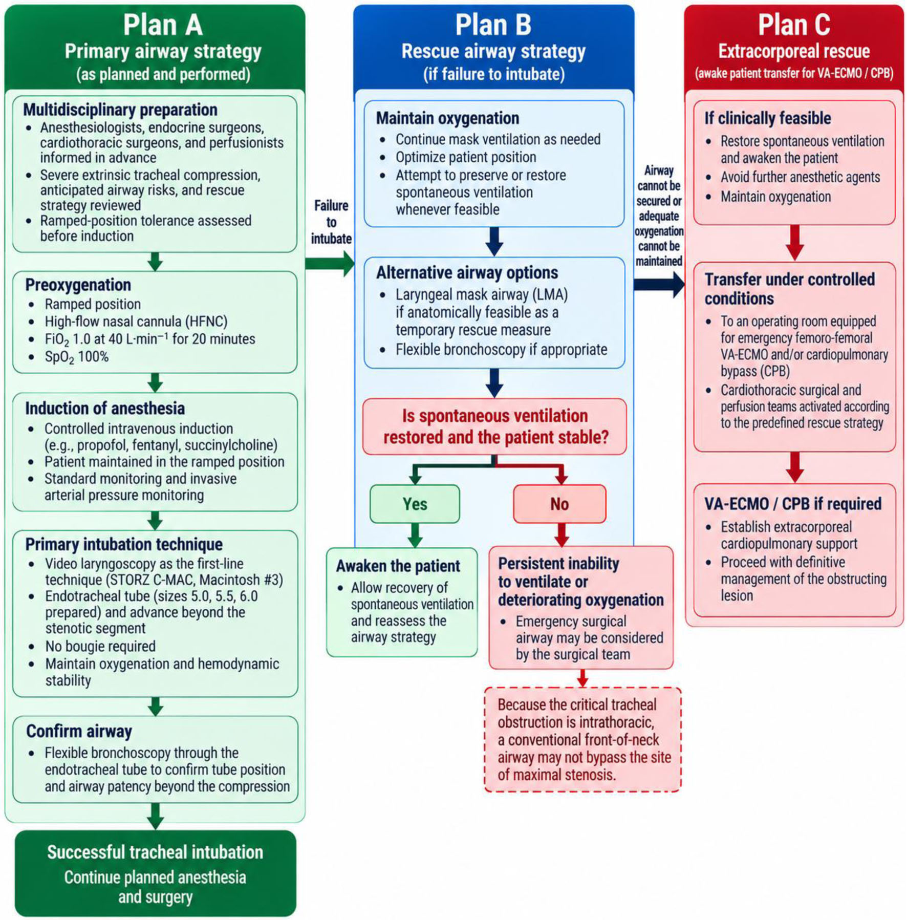
↓  Figure 6. Stepwise airway management algorithm for severe intrathoracic tracheal compression.
Figure 6.

Tables

↓  Table 1. Comprehensive Preoperative Assessment of Our Patient With Down Syndrome and Giant Retrosternal Goiter
 
ParameterValueNormal limits/interpretation
Anti-TPO: anti-thyroid peroxidase antibody; Anti-Tg: anti-thyroglobulin antibody; TSH: thyroid-stimulating hormone; FT4: free thyroxine; 25-OH vitamin D3: 25-hydroxyvitamin D3; PTH: parathyroid hormone; LVEF: left ventricular ejection fraction; PASP: pulmonary artery systolic pressure; OSA: obstructive sleep apnea; STOP-BANG: Snoring, Tiredness, Observed apnea, high blood Pressure, BMI, Age, Neck circumference, and Gender; BMI: body mass index; SVC: superior vena cava; CT: computed tomography; FNA: fine-needle aspiration; ECG: electrocardiogram, bpm: beats per minute.
Anti-TPO15.2 IU/mL< 34 IU/mL; negative
Anti-TG25 IU/mL< 115 IU/mL; negative
Thyroglobulin473 ng/mL< 77 ng/mL; elevated
23-OH-vitamin D353.44 ng/mL30–100 ng/mL; normal
TSH1.37 µIU/mL0.27–4.2 µIU/mL; normal
FT41.4 ng/dL0.93–1.7 ng/dL; normal
Calcium (total)8.4 mg/dL8.4–10.2 mg/dL; low-normal
Calcium (ionized)1.30 mmol/L1.13–1.32 mmol/L; normal
PTH37.5 pg/mL15–65 pg/mL; normal
Albumin3.8 g/dL3.5–5.2 g/dL; normal
EchocardiographyLVEF 62%; no right-sided dilatation; estimated PASP 31 mm Hg, ECG 71 bpm sinus rhythmPreserved LV systolic function; no evidence of pulmonary hypertension
OSASTOP-BANG 3 points (BMI, increased neck circumference, snoring)Intermediate risk for OSA
Great vessels compressionNo clinical or CT evidenceNo evidence of great-vessel or SVC compression
Thyroid ultrasoundMultinodular goiterStructural thyroid abnormality
Preoperative FNANot performed; relatives declined the procedureNo preoperative cytological/Bethesda classification available


↓  Table 2. Timeline of the Patient’s Clinical Presentation, Diagnostic Assessment, Airway Management, Surgery, and Postoperative Course
 
TimeClinical event
BiPAP: bilevel positive airway pressure; PEEP: positive end-expiratory pressure.
18 months before surgeryProgressive anterior neck swelling developed, associated with intermittent positional dyspnea and orthopnea.
Preoperative assessmentCT of the neck and chest demonstrated a giant multinodular retrosternal goiter extending deeply into the superior mediastinum, below the aortic arch, with marked rightward tracheal deviation and severe extrinsic tracheal compression. The minimum tracheal luminal diameter was approximately 7 mm. Comprehensive preoperative laboratory, cardiovascular, airway, and thyroid assessments were performed.
Preoperative airway planningAirway assessment identified relative macroglossia, a high-arched palate, short neck, neck circumference of 43 cm, sternomental distance < 12 cm, mouth opening of 4 cm, thyromental distance < 6 cm, Modified Mallampati class IV, reduced neck mobility, and a positive upper lip bite test. Cervical spine radiographs excluded atlantoaxial instability. Awake fiberoptic intubation, even under sedation, was considered unsuitable because of severe intellectual disability and anticipated inability to cooperate.
Day of surgery before inductionA multidisciplinary airway and surgical strategy was established involving anesthesiologists, endocrine surgeons, cardiothoracic surgeons, and perfusionists. Tolerance of the ramped position was assessed and was well tolerated without clinically significant respiratory deterioration or desaturation. On arrival in the operating room, SpO2 was 92% on room air and increased to 94% after ramped positioning without supplemental oxygen. High-flow nasal cannula oxygen at FiO2 1.0 and 40 L/min was initiated 20 min before induction, achieving SpO2 100%.
Induction and intubationControlled intravenous induction was performed with propofol 150 mg, fentanyl 150 µg, and succinylcholine 100 mg. Video laryngoscopy using a STORZ C-MAC Macintosh #3 blade provided a Cormack–Lehane grade I view. A 6.0-mm internal-diameter endotracheal tube was passed successfully on the first attempt without hypoxemia or hemodynamic instability. Flexible bronchoscopy confirmed tube position and airway patency beyond the compressed segment. Vecuronium was administered only after definitive airway control.
SurgeryTotal thyroidectomy was performed through a low transverse cervical incision with full median sternotomy and intraoperative nerve monitoring. Operative time was 185 min and estimated blood loss was 250 mL.
End of surgery/extubationAn adequate cuff leak was demonstrated. After full consciousness, adequate spontaneous ventilation, stable oxygenation and hemodynamics, and quantitative neuromuscular recovery with a train-of-four ratio of 0.9 after neostigmine, the patient was safely extubated in the operating room. No formal dynamic assessment for tracheomalacia was performed.
Postoperative days 0–1The patient was monitored overnight in the intensive care unit and received BiPAP for 6 h with FiO2 50%, pressure support 16 cm H2O, and PEEP 8 cm H2O. Thoracic drains were removed on postoperative day 1. Respiratory exercises and chest physiotherapy were provided. No respiratory or other major postoperative complications occurred.
Postoperative day 5The patient was discharged home in stable clinical condition. No clinically significant cervical hematoma, bleeding, renal dysfunction, hypocalcemia, voice change, or sternal complication was reported.
1-month follow-upThe patient remained clinically well without recurrent respiratory symptoms or other reported postoperative complications.


↓  Table 3. Summary of Perioperative Management and Clinical Outcome in Our Patient With Down Syndrome and Giant Retrosternal Goiter
 
PhaseChallengeManagement in our caseOutcome
BiPAP: bilevel positive airway pressure; CPB: cardiopulmonary bypass; CT: computed tomography; ETT: endotracheal tube; ICU: intensive care unit; IV: intravenous; LMA: laryngeal mask airway; TIVA: total intravenous anesthesia; TOF: train-of-four; VA-ECMO: veno-arterial extracorporeal membrane oxygenation.
Preoperative assessmentGiant retrosternal goiter with severe tracheal compression (∼7 mm), rightward deviation, and extension below the aortic archMultidisciplinary airway and surgical assessment; CT-based airway and mediastinal evaluationCritical airway narrowing identified; combined cervical–sternotomy approach planned
Airway assessmentDown syndrome, macroglossia, short neck, Mallampati IV, reduced neck mobility, obesity, and anticipated difficult mask ventilation/intubationComprehensive multifactorial airway assessment; cervical spine radiographsHigh-risk airway recognized; atlantoaxial instability excluded
Airway strategySevere tracheal compression with potential airway compromise after induction; awake fiberoptic intubation considered unsuitable because of inability to cooperateVideo laryngoscopy selected as primary technique; flexible bronchoscopy and rescue strategies preparedIndividualized primary and rescue airway plan established before induction
Rescue preparednessPotential inability to ventilate or intubate after inductionLMA and bronchoscopy immediately available; surgical airway and transfer to a facility equipped for femoro-femoral VA-ECMO/CPB planned if requiredAdvanced rescue options available throughout airway management
Induction and intubationCritical tracheal narrowing and risk of respiratory/hemodynamic deteriorationIV induction with propofol, fentanyl, and succinylcholine; STORZ C-MAC video laryngoscopy; smaller ETT sizes preparedFirst-attempt intubation with grade I view; 6.0-mm ETT passed beyond stenosis without hypoxemia or hemodynamic instability; position confirmed by bronchoscopy
Surgery and maintenanceExtensive cervical and mediastinal disease requiring major surgeryTIVA with propofol/remifentanil; invasive arterial monitoring; total thyroidectomy through cervical incision and median sternotomyOperative time 185 min; blood loss 250 mL; no major intraoperative complications
ExtubationRisk of postoperative airway obstruction and possible tracheomalacia after long-standing compressionCuff-leak test; full consciousness, adequate spontaneous ventilation, stable oxygenation/hemodynamics, and TOF ratio 0.9 confirmed before extubationSuccessful operating-room extubation without immediate airway compromise; tracheomalacia not formally excluded
Postoperative care and outcomeDown syndrome, obesity, sternotomy, and risk of postoperative respiratory compromiseBiPAP for 6 h, overnight ICU monitoring, early respiratory exercises/chest physiotherapy, multimodal analgesiaUneventful respiratory recovery; no significant postoperative respiratory complication; discharge on postoperative day 5