| Journal of Medical Cases, ISSN 1923-4155 print, 1923-4163 online, Open Access |
| Article copyright, the authors; Journal compilation copyright, J Med Cases and Elmer Press Inc |
| Journal website https://jmc.elmerpub.com |
Case Report
Volume 17, Number 12, December 2026, pages 702-706

Ductal Carcinoma In Situ in a Complex Fibroadenoma
Apostolos Mitrousiasa, Sandra Maria Tsotib, c, Ioannis Papapanagiotoub, Aristotelis Nikitarasa, Vasileios Kallesb
aBreast Surgery Unit, Euroclinic, Athens, Greece
bBreast Surgery Unit, Mediterraneo Hospital, Athens, Greece
cCorresponding Author: Sandra Maria Tsoti, Breast Surgery Unit, Mediterraneo Hospital, Athens, Greece
Manuscript submitted June 18, 2026, accepted September 24, 2026, published online November 9, 2026
Short title: DCIS in a Complex Fibroadenoma
doi: https://doi.org/10.14740/jmc5412
| Abstract | ▴Top |
Fibroadenomas are common benign breast lesions in young women, whereas carcinoma arising within a fibroadenoma is exceptionally rare and is usually identified incidentally following excision. Preoperative recognition can be challenging because clinical and imaging appearances may remain largely benign and small malignant foci may not be captured by core needle biopsy. We report an uncommon presentation of low-grade ductal carcinoma in situ (DCIS) arising within a complex fibroadenoma in a 29-year-old woman. The patient presented with an enlarging palpable breast mass demonstrating interval growth and mild sonographic heterogeneity. Core needle biopsy showed a fibroadenomatous lesion with adenosis but no atypia or malignancy. Excision was undertaken because of the lesion’s interval growth and atypical features, and histopathological examination demonstrated low-grade cribriform DCIS with atypical ductal hyperplasia confined within a complex fibroadenoma, with clear margins and no invasive disease. Following multidisciplinary assessment, observation without adjuvant therapy was recommended. This case highlights the diagnostic limitations of core biopsy in an enlarging or atypical fibroadenoma and the importance of clinical, radiological and pathological correlation when determining the need for excision. Although rare, in situ carcinoma should remain a diagnostic consideration when a presumed fibroadenoma demonstrates discordant or changing features.
Keywords: Complex fibroadenoma; Ductal carcinoma in situ; Breast neoplasms; Young patient
| Introduction | ▴Top |
Fibroadenomas are the most common benign breast tumors, typically occurring in women in their 20s and 30s [1]. Histologically, fibroadenomas are biphasic tumors composed of stromal and epithelial elements. They have very low malignant potential, and simple fibroadenomas do not significantly increase long-term breast cancer risk [2]. Complex fibroadenomas, defined by cysts (> 3 mm), sclerosing adenosis, epithelial calcifications, or papillary apocrine change, carry a slightly higher subsequent breast cancer risk than simple fibroadenomas [3, 4]. Most fibroadenomas are managed conservatively with surveillance because many remain stable or regress [5]. However, interval growth or atypical features warrant closer evaluation.
Carcinoma arising within a fibroadenoma is rare. Reports date back to 1931, with relatively few cases documented [6]. The estimated incidence of carcinoma (in situ or invasive) developing inside a fibroadenoma is about 0.02–0.3%. Most are discovered incidentally on histologic examination after excision rather than by clinical or radiologic suspicion [7]. Among malignant transformations, ductal carcinoma in situ (DCIS) is most frequent, followed by invasive carcinoma and lobular carcinoma in situ (LCIS) [5]. About 75% of reported lesions are in situ (DCIS or LCIS) without invasion [8]. These foci are often low grade and hormone receptor–positive; in one series, > 90% expressed estrogen receptor (ER) and/or progesterone receptor and were largely human epidermal growth factor receptor 2 (HER2)-negative (luminal subtype) [9].
Patients with carcinoma in fibroadenoma tend to be older than typical fibroadenoma patients (mean early-to-mid 40s) [1, 8], though cases in women in their 20s and even teenagers exist [9]. Imaging is usually nonspecific; microcalcifications or irregular margins may raise suspicion [10]. Definitive diagnosis relies on histopathological examination, highlighting the value of excision for discordant or changing lesions.
In this report, we present low-grade DCIS arising within a pathologically confirmed complex fibroadenoma in a 29-year-old woman and discuss clinical implications. This case report is presented in accordance with the CARE guidelines [11].
| Case Report | ▴Top |
A 29-year-old woman presented with a 6-month history of a right-breast lump. The mass, in the upper-outer quadrant (10 o’clock), was superficial, mobile, well-circumscribed, and non-tender, measuring approximately 3cm on clinical examination. Clinical examination of the contralateral breast and both axillae was unremarkable. She had no personal breast history or family history of breast/ovarian cancer. Menstrual/reproductive history was unremarkable; she had not used hormonal contraception. Ultrasound showed a lobulated, hypoechoic nodule with mild heterogeneity and vascularity, measuring 2.8 cm (previously 1.9 cm 4 months earlier), compatible with a suspicious fibroadenoma (Fig. 1). Ultrasound-guided core biopsy demonstrated a fibroadenomatous lesion with proliferative changes, including foci of adenosis, without atypia or malignancy. The combination of interval growth, mild heterogeneity, vascularity, and adenosis on core raised suspicion for a complex fibroadenoma or a fibroepithelial tumor, and surgical excision was advised. Wide local excision yielded a well-circumscribed, ovoid mass (3.0 × 2.5 × 1.9 cm) with a rubbery, whitish cut surface and lobulated architecture.
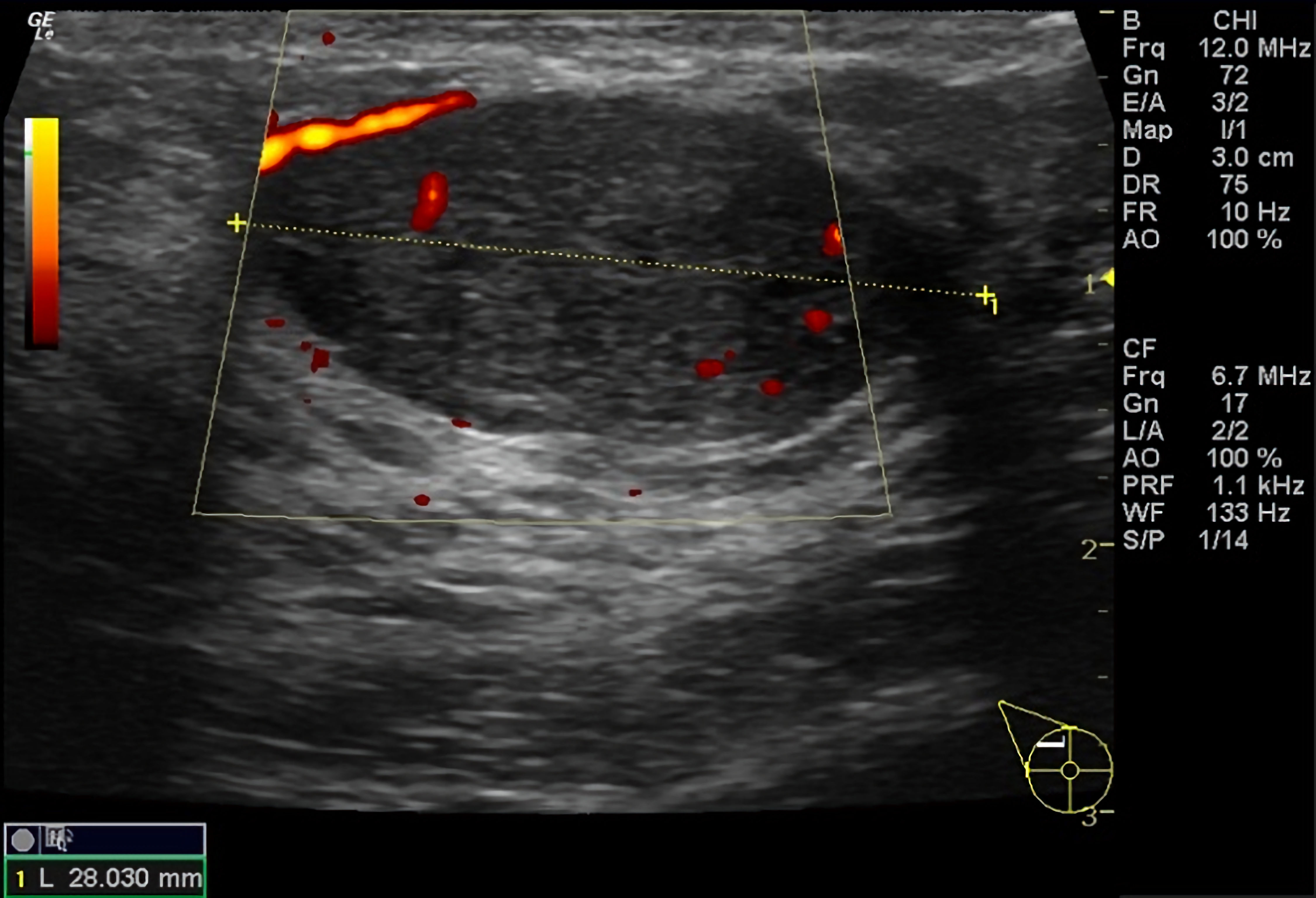 Click for large image | Figure 1. Color Doppler ultrasound of the right breast demonstrating a 2.8 cm, well-circumscribed, oval, hypoechoic mass with mild lobulation, mild internal heterogeneity, parallel orientation, and mild internal and external vascularity. |
Histopathologic examination confirmed a complex fibroadenoma containing foci of DCIS (Fig. 2). Microscopically, typical fibroadenoma architecture (stromal proliferation with benign glands) coexisted with atypical epithelial proliferations ranging from atypical ductal hyperplasia (ADH) to low-grade cribriform DCIS. The DCIS comprised monomorphic cells forming sieve-like cribriform spaces without necrosis or marked pleomorphism (nuclear grade 1). The foci measured only a few millimeters and were completely confined within the fibroadenoma, without extension into surrounding breast tissue. Margins were negative; DCIS was > 2 mm from the closest inked margin. No invasive carcinoma was identified on extensive sampling. Adjacent non-lesional tissue showed fibrocystic change only.
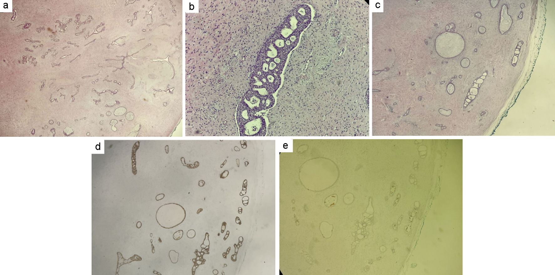 Click for large image | Figure 2. Histopathological and immunohistochemical findings of ductal carcinoma in situ arising within a complex fibroadenoma. (a) H&E section demonstrating fibroadenomatous architecture. (b, c) H&E sections demonstrating atypical ductal hyperplasia and low-grade ductal carcinoma in situ. (d) Estrogen receptor (ER) immunostaining. (e) CK5/6 immunostaining. H&E: hematoxylin and eosin stain; CK: cytokeratin. |
Immunohistochemistry showed strong estrogen-receptor positivity (100% nuclei) and complete loss of cytokeratin (CK)5/6 expression in the neoplastic cells, supporting a clonal process and aiding distinction from usual ductal hyperplasia. These findings confirmed low-grade DCIS arising within a complex fibroadenoma.
Following multidisciplinary review and counselling, axillary surgery was not indicated for pure in-situ disease. No adjuvant radiotherapy or endocrine therapy was recommended given the very small volume of low-grade DCIS, clear margins, and the patient’s fertility plans. Comprehensive germline multigene testing was negative. Following multidisciplinary review, an individualized surveillance plan was adopted, consisting of clinical examination every 6 months for 2 years, annual ultrasound, magnetic resonance imaging (MRI) every 1–2 years, and initiation of mammography at age 30. This strategy reflected an institution-specific multidisciplinary decision in the absence of established surveillance guidelines for carcinoma arising within a fibroadenoma. At the most recent follow-up, 3 months after surgery, the patient remained disease-free.
| Discussion | ▴Top |
DCIS arising within a fibroadenoma is an uncommon pathological finding and presents a diagnostic challenge because malignant foci may coexist with otherwise benign clinical, radiological, and core-biopsy findings [1, 9]. The present case is notable for the occurrence of low-grade DCIS confined within a complex fibroadenoma in a 29-year-old woman, following a core biopsy that showed no atypia or malignancy. Although carcinoma arising within fibroadenomas has been previously reported, it more commonly occurs at an older age and is frequently diagnosed incidentally after excision. This case therefore adds to the limited literature on carcinoma arising within fibroadenomas in younger women and highlights the importance of reassessing enlarging or atypical lesions despite initially benign core-biopsy findings.
Carcinoma (in situ or invasive) arising in a fibroadenoma is well documented yet rare. Incidence estimates range from 0.02% to 0.125% in pathologic series [1, 9]. Contemporary series reinforce the rarity; in a 16-patient cohort, such cases represented a tiny fraction of breast tumors over multiple years. Most lesions are in situ; in that series, 12/16 (75%) were DCIS or LCIS within the fibroadenoma, and 4/16 (25%) had an invasive component [9]. Our case aligns with the predominant in-situ pattern.
Reported carcinomas within fibroadenomas are frequently low grade and hormone receptor–positive, often HER2-negative (luminal subtype) [9]. Our patient’s ER-positive, CK5/6-negative DCIS with coexisting ADH within the fibroadenoma is compatible with a possible stepwise progression from ADH to DCIS within a fibroepithelial milieu [12]. These observations align with cohort data demonstrating a higher breast cancer risk in women with complex fibroadenomas and in those with associated proliferative disease, particularly atypical hyperplasia [4]. While most fibroadenomas occur in younger women, malignant transformation is typically described in the early 40s [9, 13]; nevertheless, younger patients, including teenagers, have also been reported [14]. Complex fibroadenomas warrant particular attention and a low threshold for excision if enlarging or atypical [3].
Preoperative detection of DCIS within a fibroadenoma is difficult: clinical and imaging features are often benign-appearing, and core biopsy may undersample atypical foci. In the present case, the benign core-biopsy findings were interpreted in the context of significant interval growth and atypical sonographic features rather than in isolation. The resulting concern for a complex fibroadenoma or other fibroepithelial lesion supported surgical excision rather than continued surveillance. Microcalcifications may be the only clue; small foci can be missed on core biopsy and identified only on excision [15]. Thorough sectioning and adequate sampling are therefore essential.
Management follows principles for DCIS not otherwise specified. Complete excision with negative margins is the cornerstone. When margins are clear and DCIS is small/low-grade, omission of radiotherapy can be considered in selected patients [16–18]. Endocrine therapy may be considered after breast-conserving treatment for ER-positive DCIS to reduce the risk of local recurrence and subsequent breast cancer; however, its potential benefits should be weighed against treatment-related adverse effects and individual considerations, including fertility and family planning [19]. Some reported cases received tamoxifen [1], whereas ER-negative cases are treated with surgery ± radiation in accordance with contemporary DCIS management guidelines [18, 20]. If invasion is present (reported in approximately 20–25%), management follows invasive breast cancer protocols, including consideration of sentinel node biopsy and subtype-directed systemic therapy [8]. Nodal positivity appears uncommon, and outcomes appear favorable in small-volume disease [9].
Overall prognosis for DCIS confined within a fibroadenoma is excellent with appropriate local therapy. Continued surveillance is prudent given a slightly elevated risk of future primaries in some patients [21].
Conclusions
Carcinoma, most often DCIS, can rarely arise within fibroadenoma. This case in a 29-year-old woman emphasizes that complex or changing fibroadenomas merit careful assessment. Complete excision and meticulous histopathology enable diagnosis of small in-situ foci. In appropriately selected, low-risk cases with negative margins, omission of adjuvant therapy may be considered following multidisciplinary assessment, with subsequent structured surveillance.
Acknowledgments
None to declare.
Financial Disclosure
No specific funding was obtained for this case report.
Conflict of Interest
The authors declare that they have no conflict of interest.
Informed Consent
Written informed consent was obtained from the patient for publication of this case report and the accompanying image.
Author Contributions
Apostolos Mitrousias: investigation, resources, and writing – original draft. Sandra Maria Tsoti: writing – original draft, writing – review and editing, and project administration. Ioannis Papapanagiotou: investigation. Aristotelis Nikitaras: writing – review and editing. Vasileios Kalles: investigation and supervision. All authors read and approved the final version of the manuscript.
Data Availability
All data supporting the findings of this case report are included within this article.
Abbreviations
ADH: atypical ductal hyperplasia; CK: cytokeratin; DCIS: ductal carcinoma in situ; ER: estrogen receptor; HER2: human epidermal growth factor receptor 2; MRI: magnetic resonance imaging
| References | ▴Top |
- Wu YT, Chen ST, Chen CJ, Kuo YL, Tseng LM, Chen DR, Kuo SJ, et al. Breast cancer arising within fibroadenoma: collective analysis of case reports in the literature and hints on treatment policy. World J Surg Oncol. 2014;12:335.
doi pubmed - Hartmann LC, Sellers TA, Frost MH, Lingle WL, Degnim AC, Ghosh K, Vierkant RA, et al. Benign breast disease and the risk of breast cancer. N Engl J Med. 2005;353(3):229-237.
doi pubmed - Nassar A, Visscher DW, Degnim AC, Frank RD, Vierkant RA, Frost M, Radisky DC, et al. Complex fibroadenoma and breast cancer risk: a Mayo Clinic Benign Breast Disease Cohort Study. Breast Cancer Res Treat. 2015;153(2):397-405.
doi pubmed - Dupont WD, Page DL, Parl FF, Vnencak-Jones CL, Plummer WD, Jr., Rados MS, Schuyler PA. Long-term risk of breast cancer in women with fibroadenoma. N Engl J Med. 1994;331(1):10-15.
doi pubmed - Gordon PB, Gagnon FA, Lanzkowsky L. Solid breast masses diagnosed as fibroadenoma at fine-needle aspiration biopsy: acceptable rates of growth at long-term follow-up. Radiology. 2003;229(1):233-238.
doi pubmed - Cheatle GL, Cutler M. Tumours of the breast: their pathology, symptoms, diagnosis and treatment. London: Edward Arnold & Co.; 1931.
- Ni XH, An R, Shi QW, Wang CL. Low-grade ductal carcinoma in situ within a fibroadenoma of the breast: a rare case report and review of the literature. Onco Targets Ther. 2023;16:399-406.
doi pubmed - Krishnamurthy K, Alghamdi S, Gyapong S, Kaplan S, Poppiti RJ. A clinicopathological study of fibroadenomas with epithelial proliferation including lobular carcinoma in-situ, atypical ductal hyperplasia, DCIS and invasive carcinoma. Breast Dis. 2019;38(3-4):97-101.
doi pubmed - Xu L, Luo S, Mao Q, Gao Y, Luo L, Qu W, Cao Y. Breast carcinoma arising in a fibroadenoma: A case series of 16 patients and review of the literature. Oncol Lett. 2024;27(1):39.
doi pubmed - Tagliati C, Lanni G, Cerimele F, Di Martino A, Calamita V, Lucidi Pressanti G, Mingliang Y, et al. Low diffusion level within a fibroadenoma as the sole sign of ductal carcinoma in situ: A case report. Breast Dis. 2021;40(4):283-286.
doi pubmed - Riley DS, Barber MS, Kienle GS, Aronson JK, von Schoen-Angerer T, Tugwell P, Kiene H, et al. CARE guidelines for case reports: explanation and elaboration document. J Clin Epidemiol. 2017;89:218-235.
doi pubmed - Kader T, Hill P, Rakha EA, Campbell IG, Gorringe KL. Atypical ductal hyperplasia: update on diagnosis, management, and molecular landscape. Breast Cancer Res. 2018;20(1):39.
doi pubmed - Diaz NM, Palmer JO, McDivitt RW. Carcinoma arising within fibroadenomas of the breast. A clinicopathologic study of 105 patients. Am J Clin Pathol. 1991;95(5):614-622.
doi pubmed - Hung WY, Lee CL, Lin CY. A juvenile female with ductal carcinoma in situ arising from a fibroadenoma. J Cancer Res Pract. 2023;10(2):82-84.
doi - Zeeshan S, Siddique S, Riaz N. Ductal carcinoma-in-situ within fibroadenoma of the breast: a case report with literature review. Int J Case Rep Images. 2018;9:100916Z01SZ2018.
doi - Early Breast Cancer Trialists' Collaborative G, Correa C, McGale P, Taylor C, Wang Y, Clarke M, Davies C, et al. Overview of the randomized trials of radiotherapy in ductal carcinoma in situ of the breast. J Natl Cancer Inst Monogr. 2010;2010(41):162-177.
doi pubmed - Rakovitch E, Nofech-Mozes S, Hanna W, Sutradhar R, Baehner FL, Miller DP, Fong C, et al. Multigene expression assay and benefit of radiotherapy after breast conservation in ductal carcinoma in Situ. J Natl Cancer Inst. 2017;109(4):djw256.
doi pubmed - Gilleard O, Goodman A, Cooper M, Davies M, Dunn J. The significance of the Van Nuys prognostic index in the management of ductal carcinoma in situ. World J Surg Oncol. 2008;6:61.
doi pubmed - Loibl S, Andre F, Bachelot T, Barrios CH, Bergh J, Burstein HJ, Cardoso MJ, et al. Early breast cancer: ESMO Clinical Practice Guideline for diagnosis, treatment and follow-up. Ann Oncol. 2024;35(2):159-182.
doi pubmed - Wu YT, Wu HK, Chen ST, Chen CJ, Chen DR, Lai HW. Fibroadenoma progress to ductal carcinoma in situ, infiltrating ductal carcinoma and lymph node metastasis? Report an unusual case. J Surg Case Rep. 2017;2017(5):rjx064.
doi pubmed - Limite G, Esposito E, Sollazzo V, Ciancia G, Formisano C, Di Micco R, De Rosa D, et al. Lobular intraepithelial neoplasia arising within breast fibroadenoma. BMC Res Notes. 2013;6:267.
doi pubmed
This article is distributed under the terms of the Creative Commons Attribution 4.0 International License (CC BY 4.0), which permits unrestricted use, distribution, and reproduction in any medium, including commercial use, provided the original work is properly cited.
Journal of Medical Cases is published by Elmer Press Inc.