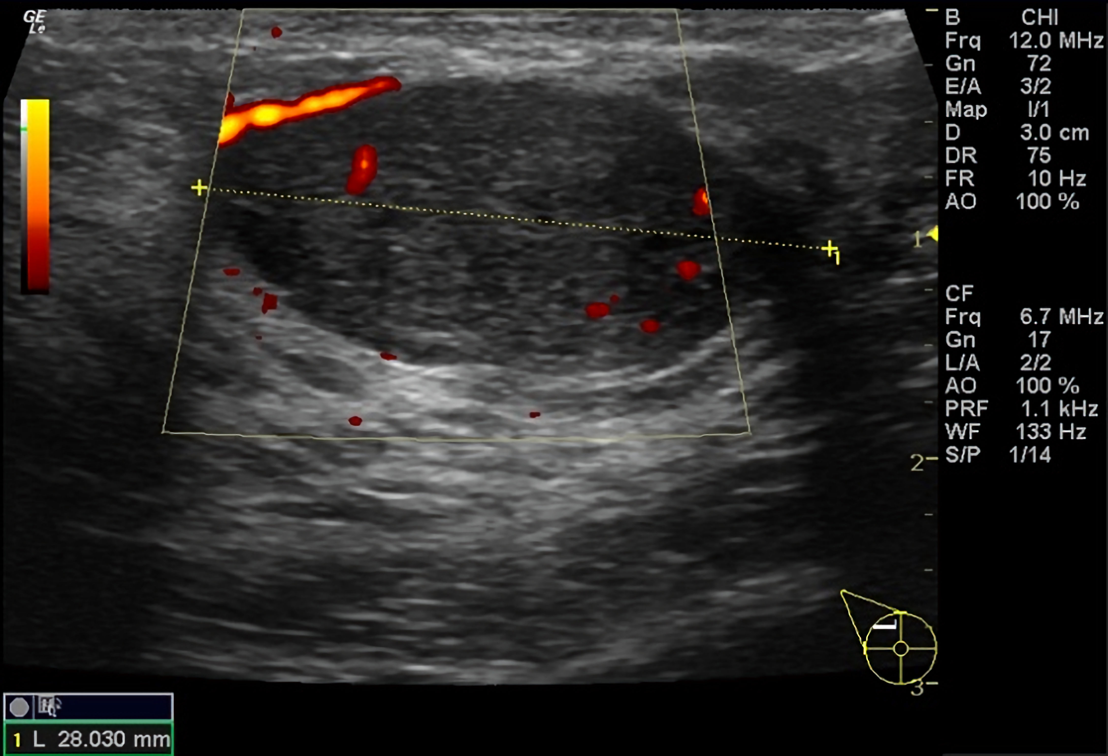
↓ Figure 1. Color Doppler ultrasound of the right breast demonstrating a 2.8 cm, well-circumscribed, oval, hypoechoic mass with mild lobulation, mild internal heterogeneity, parallel orientation, and mild internal and external vascularity.
| Journal of Medical Cases, ISSN 1923-4155 print, 1923-4163 online, Open Access |
| Article copyright, the authors; Journal compilation copyright, J Med Cases and Elmer Press Inc |
| Journal website https://jmc.elmerpub.com |
Case Report
Volume 17, Number 12, December 2026, pages 702-706
Ductal Carcinoma In Situ in a Complex Fibroadenoma
Figures