| Journal of Medical Cases, ISSN 1923-4155 print, 1923-4163 online, Open Access |
| Article copyright, the authors; Journal compilation copyright, J Med Cases and Elmer Press Inc |
| Journal website https://jmc.elmerpub.com |
Case Report
Volume 17, Number 11, November 2026, pages 655-659

Asymptomatic Intracardiac Migration of a Cane-Shaped Polymethylmethacrylate Cement Embolus Following Percutaneous Vertebroplasty
Jinchul Noha, Sanghoun Leea, b, Youngmin Oha, Jongpil Euna, b
aDepartment of Neurosurgery, Research Institute of Clinical Medicine, Biomedical Research Institute, Jeonbuk National University Medical School and Hospital, Jeonju 54907, Korea
bCorresponding Authors: Sanghoun Lee and Jongpil Eun, Department of Neurosurgery, Research Institute of Clinical Medicine, Biomedical Research Institute, Jeonbuk National University Medical School and Hospital, Jeonju 54907, Korea
Manuscript submitted July 22, 2026, accepted September 14, 2026, published online October 2, 2026
Short title: Intracardiac Cement Embolus After PVP
doi: https://doi.org/10.14740/jmc5403
| Abstract | ▴Top |
Percutaneous vertebroplasty (PVP) is a minimally invasive and effective treatment for osteoporotic vertebral compression fractures. Although generally safe, cement leakage is a relatively common complication and may rarely result in intracardiac embolism. We report the case of a 75-year-old woman who underwent PVP for an L2 compression fracture after failed conservative treatment. Postoperative imaging revealed venous cement leakage, but she remained asymptomatic. Six months later, during a routine health examination, echocardiography incidentally detected an abnormal structure in the right atrium. Cardiac computed tomography demonstrated a linear, cane-shaped polymethylmethacrylate embolus extending from the right atrium to the right ventricle. Despite the absence of cardiopulmonary symptoms, surgical removal was performed because of the elongated and sharp morphology of the embolus and the associated risk of cardiac perforation. Open-heart surgery was successfully completed without major complications. Although many intracardiac cement embolisms are asymptomatic and can be managed conservatively, embolus morphology and location should guide treatment decisions. Early recognition and individualized management are essential to prevent life-threatening complications.
Keywords: Percutaneous vertebroplasty; Polymethylmethacrylate; Bone cement embolism; Intracardiac embolism
| Introduction | ▴Top |
Percutaneous vertebroplasty (PVP) was developed in 1987 by Galibert and Deramond in France and has been used for various spinal disorders (osteoporotic vertebral fractures, traumatic fractures, benign tumors, and malignant tumors) over the past four decades [1]. Various types of bone cements have been developed, but polymethylmethacrylate (PMMA) remains the most commonly utilized in PVP [2].
PVP provides mechanical stabilization of the vertebral body and reduces pain. The rate of pain reduction has been reported to be between about 70% and 100% [3, 4].
PVP is considered a relatively safe and simple procedure owing to its minimally invasive nature; however, complications occur in approximately 1–10% of cases [5]. These complications are mostly asymptomatic but can be classified into three main categories based on their severity: mild, moderate, and severe [6, 7].
The main cause of these complications is cement leakage, reported to occur in about 30–65% of cases [8]. Among these, venous leakage is the most concerning, as it can lead to pulmonary and cardiac problems owing to venous cement embolism. We report a case of an incidentally discovered intracardiac PMMA embolism approximately 6 months after PVP, in which the patient remained asymptomatic.
| Case Report | ▴Top |
A 75-year-old female presented to our outpatient clinic with lower back pain. She had a history of osteoporosis at another hospital and had previously undergone vertebroplasty for a compression fracture at L1. Lumbar spine magnetic resonance imaging revealed a new compression fracture at L2 (Fig. 1).
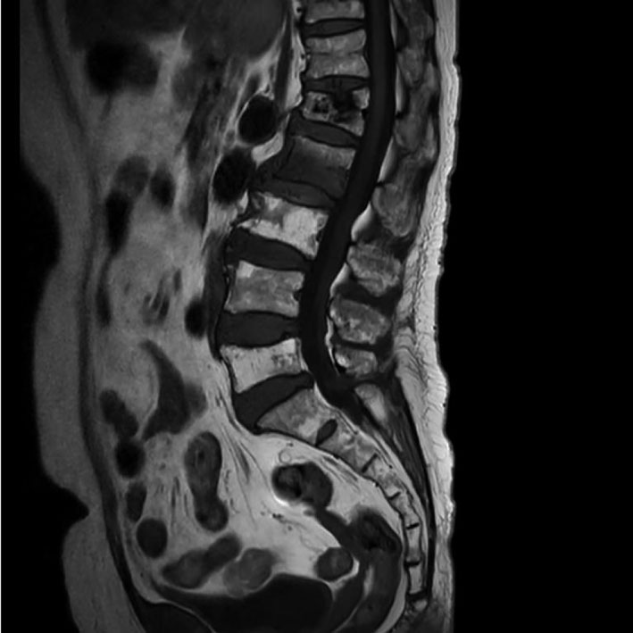 Click for large image | Figure 1. Sagittal lumbar spine MRI. The T1-weighted image demonstrates a new acute compression fracture at the L2 vertebral body. MRI: magnetic resonance imaging. |
After 2 weeks of conservative treatment, the patient continued to complain of persistent lower back pain; therefore, PVP was performed. Under local anesthesia in the prone position, a Jamshidi needle was used, and PVP was performed on the right side of L2 using a transpedicular approach. PMMA (3 mL) was then injected. Postoperative radiography revealed venous leakage of the bone cement (Fig. 2a, b). The patient remained asymptomatic without cardiopulmonary symptoms, and no additional imaging was performed to evaluate possible cement migration at that time. Her back pain improved significantly and she was discharged 2 days later.
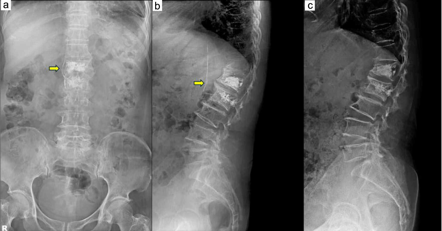 Click for large image | Figure 2. Postoperative and follow-up lumbar radiographs. (a, b) Postoperative anteroposterior and lateral radiographs showing venous leakage of the bone cement following percutaneous vertebroplasty at L2. (c) Follow-up lumbar radiograph obtained 2 weeks later, showing that the previously observed venous cement leakage was no longer visible. |
Two weeks after discharge, she returned to the outpatient clinic for follow-up. A lumbar radiograph showed that the previously observed venous cement leakage was no longer visible (Fig. 2c). Because the patient remained asymptomatic, no additional imaging studies were performed at that time to evaluate possible migration of the cement into the inferior vena cava, right heart, or pulmonary circulation.
Six months later, during a routine health checkup at another hospital, echocardiography revealed abnormal findings in the right atrium. Cardiac computed tomography showed a linear foreign body (bone cement) extending across the right atrium (RA) and right ventricle (RV) (Fig. 3a, b).
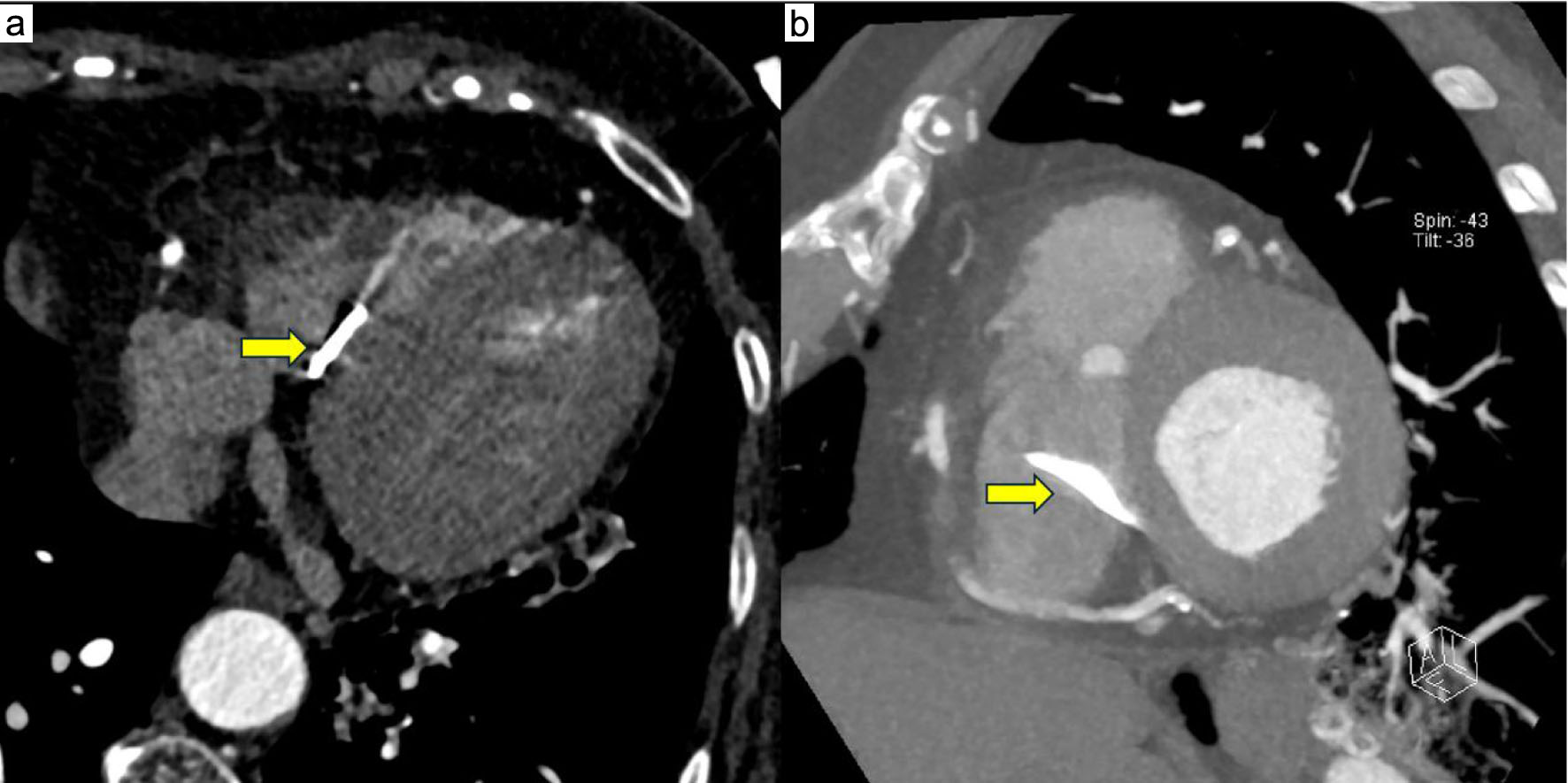 Click for large image | Figure 3. Cardiac CT scans. (a, b) Axial and reconstructed views showing a linear, cane-shaped hyperdense foreign body extending from the RA into the RV. CT: computed tomography; RA: right atrium; RV: right ventricle. |
The embolus was cane-shaped and extended from the RA into the RV. Although the patient remained asymptomatic, the elongated and sharp morphology of the embolus and its extension into the right ventricle raised concern for cardiac perforation and valvular injury. Following multidisciplinary discussion involving an interventional cardiologist, a cardiac surgeon, and a neurosurgeon, surgical removal was favored because of the embolus morphology, location, and the potential risk of serious cardiac complications.
Open-heart surgery was performed for cement removal. After median sternotomy, heparinization, and pericardiotomy, bicaval cannulation and cardiopulmonary bypass were established, and systemic cooling was initiated. The cement extended into the septal leaflet and penetrated the RV side. Cane-shaped cement was removed with partial division of the septal leaflet (Fig. 4). Tricuspid valve ring annuloplasty and right atriotomy were performed. The procedure was completed without major complications, and the patient was discharged 1 week later.
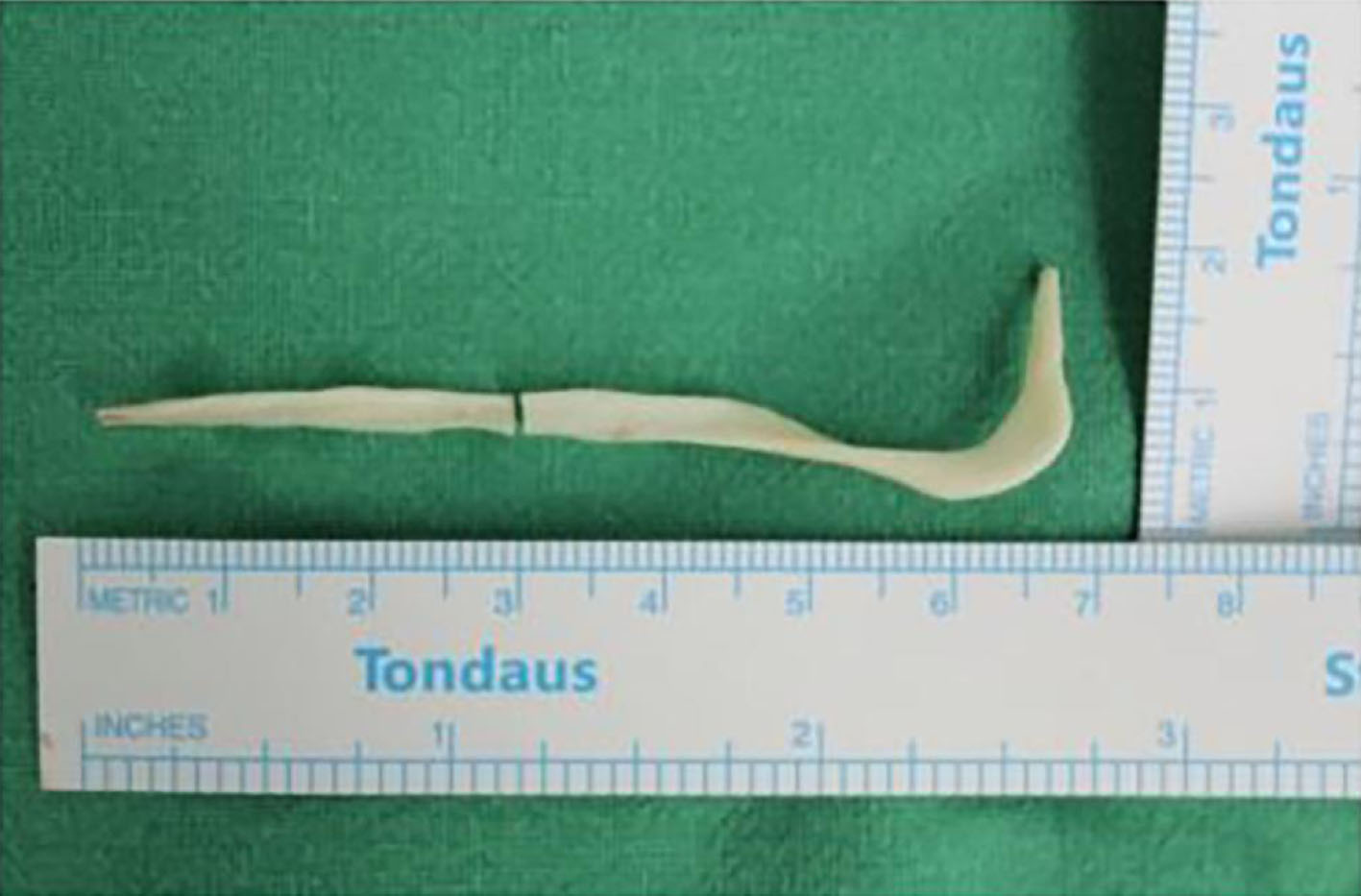 Click for large image | Figure 4. Intraoperative photograph of the removed bone cement. The extracted cane-shaped bone cement after successful surgical removal via open-heart surgery. |
| Discussion | ▴Top |
PVP is a safe and effective procedure for pain relief; however, complications owing to cement leakage may occur. The frequency of complications is reportedly 1–10% [5], with cement leakage occurring in 30–65% of cases [8]. Venous cement leakage occurs in about 24% [9]. Complications can be categorized as mild, moderate, or severe. Mild complications include transient hypotension and cement leakage into the intervertebral disc space or paravertebral soft tissue. Moderate complications include infection and cement leakage into the epidural or foraminal spaces. Severe complications include cement extravasation into the paravertebral vein, potentially leading to pulmonary and cardiac embolisms [6, 7].
Risk factors for cement leakage include intravertebral clefts, cortical disruption, low cement viscosity, and high injection volume [10]. In our case, no definite patient-specific risk factor for venous cement leakage could be identified retrospectively. To reduce the risk of venous cement leakage and subsequent embolization, careful fluoroscopic monitoring during cement injection, use of appropriately viscous cement, avoidance of excessive injection pressure or volume, and immediate cessation of injection when venous leakage is observed are important procedural considerations [6].
Intracardiac and pulmonary cement embolisms are of particular concern. Cement may migrate through the paravertebral veins into the azygos vein or inferior vena cava, then through the right atrium and ventricle, eventually reaching the pulmonary arteries [11]. In our case, migration from the L2 venous leakage to the right atrium and right ventricle was presumed based on the anatomical venous pathway but was not directly demonstrated on serial imaging. The incidence of intracardiac cement embolism is approximately 3.9% and it is usually asymptomatic [12]. The most common symptoms include chest pain and dyspnea; in severe cases, tamponade or perforation may occur [13, 14].
In a systematic review by Hatzantonis et al, conservative treatment was recommended for asymptomatic intracardiac cement embolisms, while surgical intervention was advised for symptomatic cases. Treatment may involve either open-heart surgery or percutaneous approaches, with generally favorable outcomes. Open surgical removal is particularly considered when the cement is located in the right ventricle because of the risk of perforation and other severe complications [15]. Conservative management in asymptomatic cases may include anticoagulation therapy, such as warfarin, until endothelialization reduces the thrombogenic risk [16].
When venous cement leakage is identified after vertebroplasty, further evaluation should be guided by the patient’s symptoms and the extent and morphology of the leakage. Additional imaging, such as chest radiography, computed tomography, or echocardiography, may be considered when migration into the cardiopulmonary circulation is suspected [12, 15]. In asymptomatic patients without evidence of high-risk intracardiac embolization, close clinical and imaging follow-up may be appropriate [15, 16]. In contrast, intervention should be considered when the embolus is intracardiac, elongated or sharp, associated with right ventricular involvement, or considered to carry a substantial risk of cardiac perforation or valvular injury. Accordingly, management should be individualized through multidisciplinary assessment based on symptoms, embolus morphology, location, and the potential risk of future complications [15]. Our case demonstrates that the absence of cardiopulmonary symptoms does not exclude delayed cement migration, emphasizing the need for continued clinical vigilance when venous cement leakage is identified.
However, several reports describe cardiac perforation caused by sharp or elongated cement embolisms [14, 17]. Intracardiac cement embolism may also present after a prolonged latent period and remain clinically silent until significant cardiac injury develops. Guo et al reported an ultra-delayed intracardiac cement embolism 2.5 years after vertebroplasty, with multiple rod-shaped cement fragments located in the right ventricle, including one embedded in the ventricular myocardium [18]. Setoguchi et al described a patient who remained asymptomatic for 6 months after vertebroplasty before presenting with cardiac tamponade due to right ventricular perforation by migrated bone cement [19]. A similar concern for valvular injury was illustrated by Hamade et al, who reported an intracardiac cement embolus causing extensive tricuspid valve damage that required surgical removal and valve replacement [20]. In our case, the patient remained asymptomatic for 6 months and the embolus was detected incidentally. Nevertheless, its long, sharp, cane-shaped morphology extending from the right atrium to the right ventricle raised concern for cardiac perforation or valvular injury. Intraoperative findings demonstrating involvement of the tricuspid septal leaflet and penetration toward the right ventricular side further supported the decision for surgical removal despite the absence of symptoms.
Conclusion
PVP is generally a safe and effective procedure for pain relief. However, cement leakage can lead to serious complications, including intracardiac embolism. While many cases remain asymptomatic and may be managed conservatively, elongated and sharp emboli pose a risk of cardiac perforation. Treatment should be individualized based on embolus morphology, location, and patient symptoms. In our case, despite the absence of symptoms, surgical removal was performed owing to the high risk associated with the embolus shape and location.
Acknowledgments
None to declare.
Financial Disclosure
The authors received no financial support for the research, authorship, and/or publication of this article.
Conflict of Interest
The authors declare no conflict of interest.
Informed Consent
Written informed consent was obtained from the patient for publication of this case report and the accompanying images.
Author Contributions
Jinchul Noh, Sanghoun Lee, Youngmin Oh, and Jongpil Eun contributed to the conception and design of the study. Jinchul Noh collected the clinical data and drafted the manuscript. All authors reviewed and revised the manuscript, approved the final version for publication, and agreed to be accountable for all aspects of the work.
Data Availability
The data generated during this study are not publicly available because of patient privacy but are available from the corresponding author upon reasonable request.
| References | ▴Top |
- Galibert P, Deramond H, Rosat P, Le Gars D. [Preliminary note on the treatment of vertebral angioma by percutaneous acrylic vertebroplasty]. Neurochirurgie. 1987;33(2):166-168.
pubmed - Beall DP. Vertebral augmentation: the comprehensive guide to vertebroplasty, kyphoplasty, and implant augmentation. Georg Thieme Verlag. 2020.
- Barr JD, Barr MS, Lemley TJ, McCann RM. Percutaneous vertebroplasty for pain relief and spinal stabilization. Spine (Phila Pa 1976). 2000;25(8):923-928.
doi pubmed - Watts NB, Harris ST, Genant HK. Treatment of painful osteoporotic vertebral fractures with percutaneous vertebroplasty or kyphoplasty. Osteoporos Int. 2001;12(6):429-437.
doi pubmed - Manaster B. Invited commentary authors’ response. Radiographics. 2003;23(6):1384-1387.
- Cavka M, Delimar D, Rezan R, Zigman T, Duric KS, Cimic M, Dumic-Cule I, et al. Complications of percutaneous vertebroplasty: a pictorial review. Medicina (Kaunas). 2023;59(9):1536.
doi pubmed - Al-Nakshabandi NA. Percutaneous vertebroplasty complications. Ann Saudi Med. 2011;31(3):294-297.
doi pubmed - Laredo J-D, Hamze B. Complications of percutaneous vertebroplasty and their prevention. New Techniques in Interventional Musculoskeletal Radiology. 2007:273-288.
- Kim YJ, Lee JW, Park KW, Yeom JS, Jeong HS, Park JM, Kang HS. Pulmonary cement embolism after percutaneous vertebroplasty in osteoporotic vertebral compression fractures: incidence, characteristics, and risk factors. Radiology. 2009;251(1):250-259.
doi pubmed - Zhan Y, Jiang J, Liao H, Tan H, Yang K. Risk factors for cement leakage after vertebroplasty or kyphoplasty: a meta-analysis of published evidence. World Neurosurg. 2017;101:633-642.
doi pubmed - Groen RJ, du Toit DF, Phillips FM, Hoogland PV, Kuizenga K, Coppes MH, Muller CJ, et al. Anatomical and pathological considerations in percutaneous vertebroplasty and kyphoplasty: a reappraisal of the vertebral venous system. Spine (Phila Pa 1976). 2004;29(13):1465-1471.
doi pubmed - Fadili Hassani S, Cormier E, Shotar E, Drir M, Spano JP, Morardet L, Collet JP, et al. Intracardiac cement embolism during percutaneous vertebroplasty: incidence, risk factors and clinical management. Eur Radiol. 2019;29(2):663-673.
doi pubmed - Lee V, Patel R, Meier P, Lawrence D, Roberts N. Conservative management of inferior vena cava cement spike after percutaneous vertebroplasty causes fatal cardiac tamponade. J Rheumatol. 2014;41(1):141-142.
doi pubmed - Kim SY, Seo JB, Do KH, Lee JS, Song KS, Lim TH. Cardiac perforation caused by acrylic cement: a rare complication of percutaneous vertebroplasty. AJR Am J Roentgenol. 2005;185(5):1245-1247.
doi pubmed - Hatzantonis C, Czyz M, Pyzik R, Boszczyk BM. Intracardiac bone cement embolism as a complication of vertebroplasty: management strategy. Eur Spine J. 2017;26(12):3199-3205.
doi pubmed - Krueger A, Bliemel C, Zettl R, Ruchholtz S. Management of pulmonary cement embolism after percutaneous vertebroplasty and kyphoplasty: a systematic review of the literature. Eur Spine J. 2009;18(9):1257-1265.
doi pubmed - Zhang Y, Liu X, Liu H. Cardiac Perforation Caused by Cement Embolism after Percutaneous Vertebroplasty: A Report of Two Cases. Orthop Surg. 2022;14(2):456-460.
doi pubmed - Guo J, Lu X, Lu L, Xue C. Ultra-delayed intracardiac cement embolism after percutaneous vertebroplasty: a case report and review of management strategies. J Vis Exp. 2026;233.
doi pubmed - Setoguchi T, Fukamachi D, Matsumoto N, Okumura Y. Right ventricular perforation after vertebroplasty. JACC Case Rep. 2026;31(33):109332.
doi pubmed - Hamade A, Darwish M, Shoumar B, Sayyed A, Najjar CG, Moussa M. Intracardiac cement embolism following vertebroplasty. Cureus. 2025;17(7):e87535.
doi pubmed
This article is distributed under the terms of the Creative Commons Attribution 4.0 International License (CC BY 4.0), which permits unrestricted use, distribution, and reproduction in any medium, including commercial use, provided the original work is properly cited.
Journal of Medical Cases is published by Elmer Press Inc.