Journal of Medical Cases, ISSN 1923-4155 print, 1923-4163 online, Open Access
Article copyright, the authors; Journal compilation copyright, J Med Cases and Elmer Press Inc
Journal website https://jmc.elmerpub.com

Case Report

Volume 17, Number 11, November 2026, pages 655-659


Asymptomatic Intracardiac Migration of a Cane-Shaped Polymethylmethacrylate Cement Embolus Following Percutaneous Vertebroplasty

Figures

↓  Figure 1. Sagittal lumbar spine MRI. The T1-weighted image demonstrates a new acute compression fracture at the L2 vertebral body. MRI: magnetic resonance imaging.
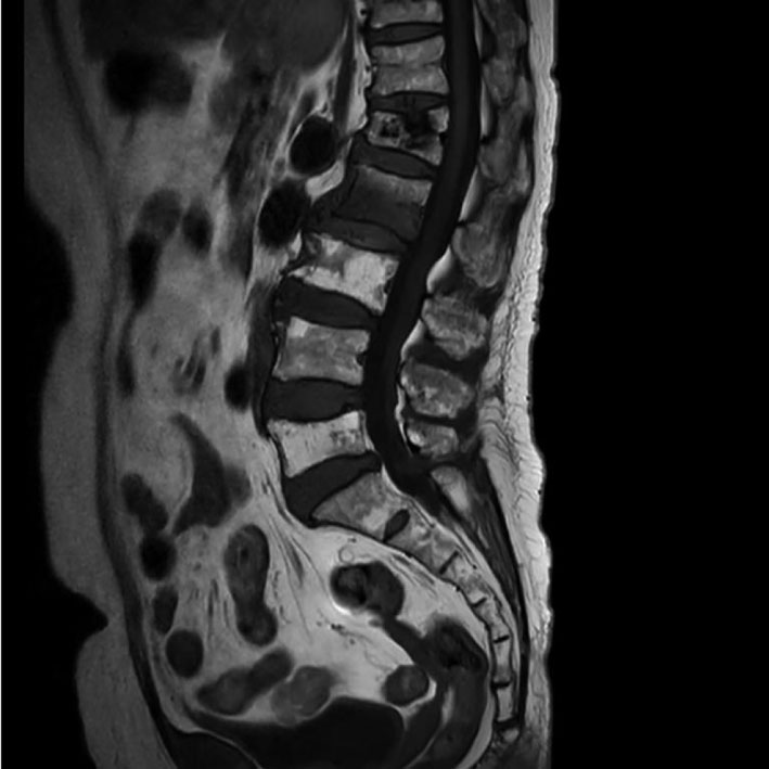
Figure 1.
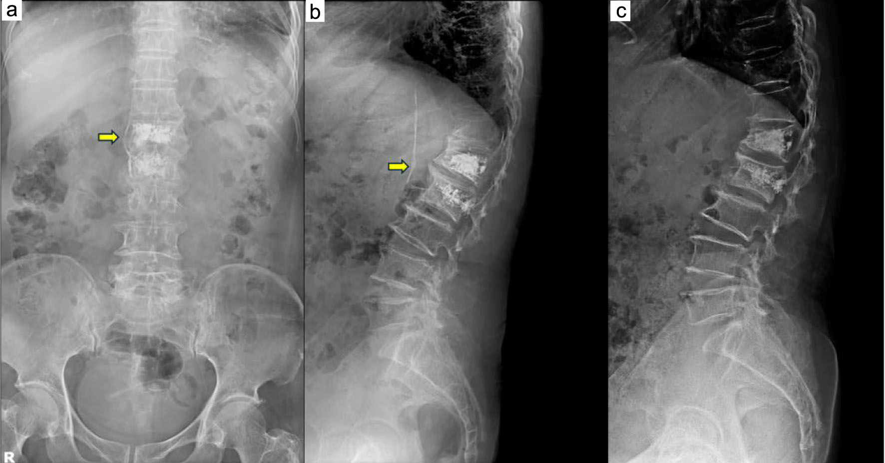
↓  Figure 2. Postoperative and follow-up lumbar radiographs. (a, b) Postoperative anteroposterior and lateral radiographs showing venous leakage of the bone cement following percutaneous vertebroplasty at L2. (c) Follow-up lumbar radiograph obtained 2 weeks later, showing that the previously observed venous cement leakage was no longer visible.
Figure 2.
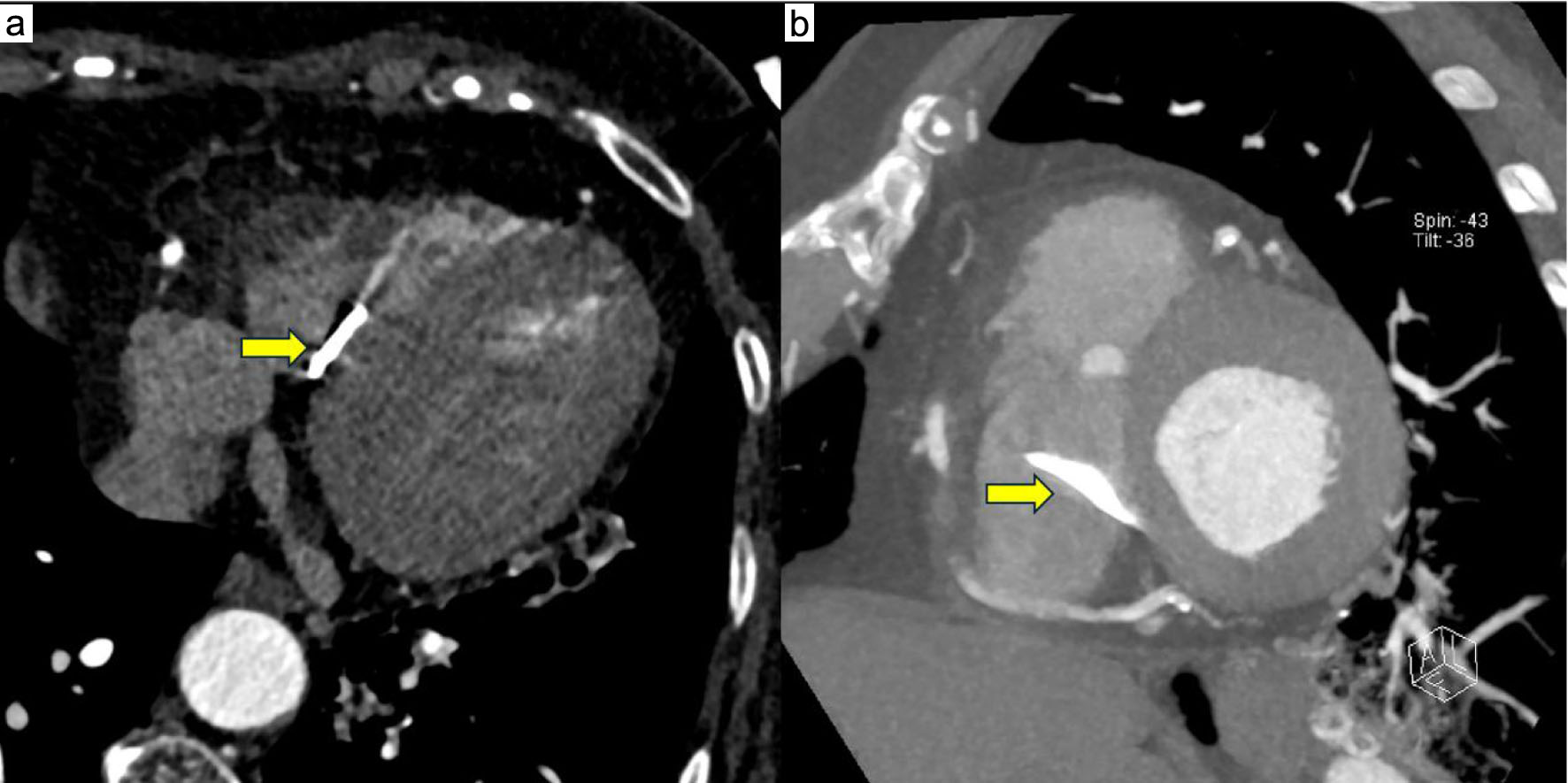
↓  Figure 3. Cardiac CT scans. (a, b) Axial and reconstructed views showing a linear, cane-shaped hyperdense foreign body extending from the RA into the RV. CT: computed tomography; RA: right atrium; RV: right ventricle.
Figure 3.
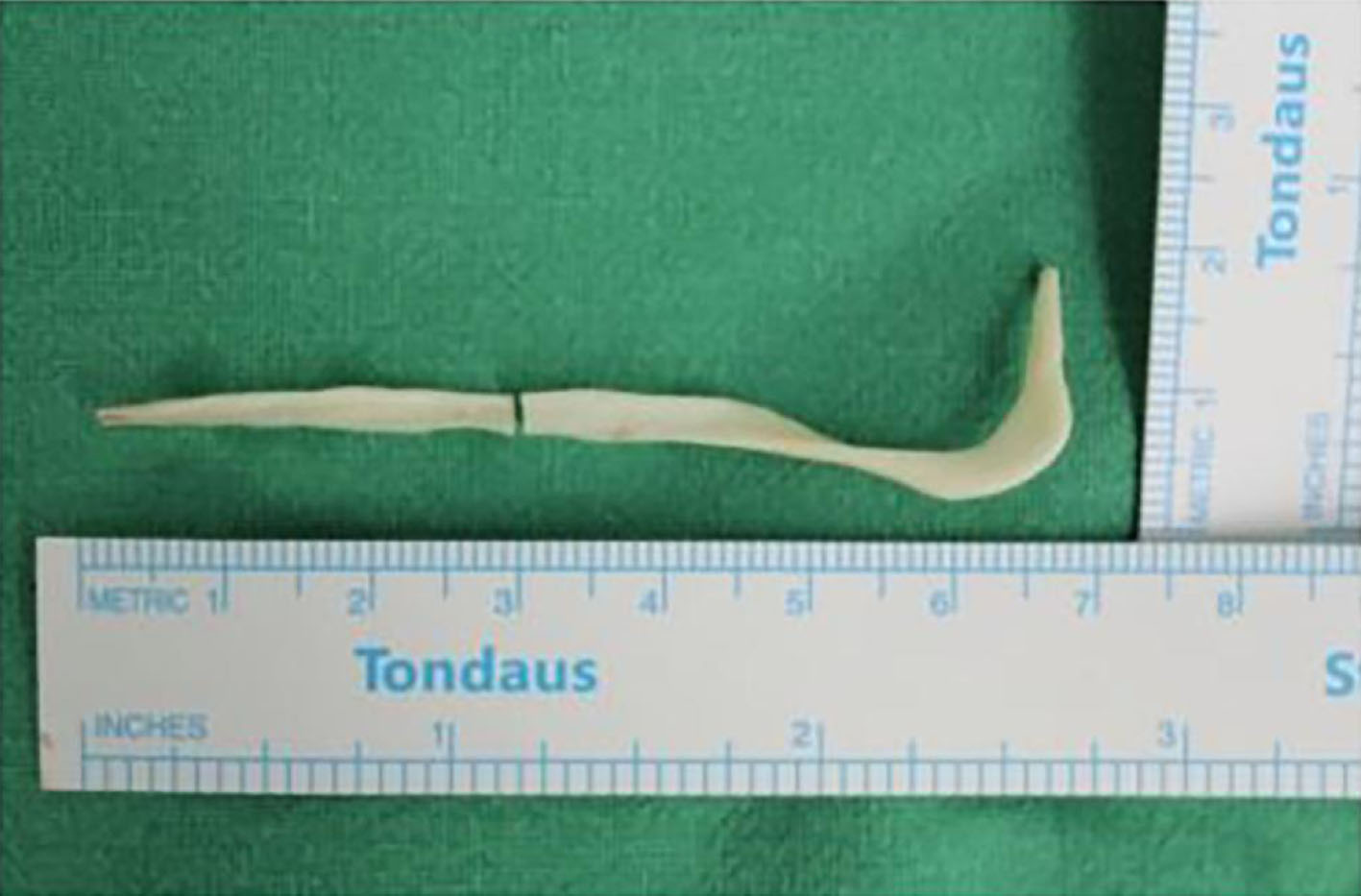
↓  Figure 4. Intraoperative photograph of the removed bone cement. The extracted cane-shaped bone cement after successful surgical removal via open-heart surgery.
Figure 4.