| Journal of Medical Cases, ISSN 1923-4155 print, 1923-4163 online, Open Access |
| Article copyright, the authors; Journal compilation copyright, J Med Cases and Elmer Press Inc |
| Journal website https://jmc.elmerpub.com |
Case Report
Volume 000, Number 000, August 2026, pages 000-000

Hyperviscosity Syndrome as a Rare Presentation of Immunoglobulin G Kappa Smoldering Multiple Myeloma Successfully Treated With Daratumumab
Muralidhar Idamakantia, d  , Rani Indrani Bijjama , Alexei Bakhirevb, Manoj Kumarc
aAdult Internal Medicine Services (AIMS), Presbyterian Healthcare Services (PHS), Albuquerque, NM 87106, USA
bPathology Associates of Albuquerque (PAA), Albuquerque, NM 87106, USA
cDepartment of Hematology/Oncology, Presbyterian Healthcare Services (PHS), Albuquerque, NM 87106, USA
dCorresponding Author: Muralidhar Idamakanti, Adult Internal Medicine Services (AIMS), Presbyterian Healthcare Services (PHS), Albuquerque, NM 87106, USA
Manuscript submitted June 30, 2026, accepted August 7, 2026, published online August 25, 2026
Short title: Daratumumab Treatment of HVS in IgG SMM
doi: https://doi.org/10.14740/jmc5398
| Abstract | ▴Top |
Hyperviscosity syndrome (HVS) is an oncologic emergency most commonly associated with Waldenstrom macroglobulinemia and immunoglobulin (Ig)M paraproteinemia. Clinically, significant HVS in smoldering multiple myeloma (SMM), particularly IgG-associated disease without overt CRAB (hypercalcemia, renal insufficiency, anemia, and bone lesions) criteria, is exceedingly rare. We report a unique case of IgG kappa smoldering myeloma complicated by symptomatic HVS, manifesting with retinal hemorrhage, epistaxis, and gross hematuria, despite the absence of overt end-organ myeloma-defining events. A 70-year-old patient with Sjogren syndrome and longstanding monoclonal gammopathy showed gradual progression from high-risk monoclonal gammopathy of undetermined significance (MGUS) to smoldering myeloma over several years. Bone marrow biopsy showed 10% plasma cells, with no high-risk cytogenetic abnormalities on limited fluorescence in situ hybridization (FISH) testing. Positron emission tomography/computed tomography (PET/CT) imaging repeatedly showed no fluorodeoxyglucose (FDG)-avid osseous disease. In the setting of HVS, serum viscosity peaked at 5.7 centipoise, with concomitant IgG elevation to 5,463 mg/dL and an M-protein of 3.01 g/dL. Given symptomatic hyperviscosity, treatment with daratumumab was initiated despite the absence of SLiM (≥ 60% clonal plasma cells, light chain ratio ≥ 100, and magnetic resonance imaging (MRI) focal lesions)-CRAB criteria for overt multiple myeloma. Following therapy, serum viscosity rapidly improved from 5.7 to 2.2, with corresponding reductions in IgG and M-protein levels and resolution of bleeding manifestations. This case highlights that symptomatic hyperviscosity may occur in IgG smoldering myeloma at a relatively modest plasma cell burden and may itself represent a clinically meaningful indication for early therapeutic intervention.
Keywords: Hyperviscosity syndrome; Smoldering multiple myeloma; Waldenström macroglobulinemia; paraproteinemia; hematuria; retinal hemorrhage; epistaxis; Daratumumab
| Introduction | ▴Top |
Hyperviscosity syndrome (HVS) is a hematologic emergency caused by increased resistance to blood flow from elevated serum proteins or cellular components [1]. The syndrome classically presents with a triad of mucosal bleeding, visual abnormalities, and neurologic manifestations due to impaired microcirculatory perfusion [2]. Common clinical manifestations include epistaxis, retinal hemorrhage, blurred vision, headache, dizziness, ataxia, and altered mental status [3].
HVS is most commonly associated with Waldenstrom macroglobulinemia, owing to the intravascular distribution and pentameric structure of immunoglobulin (Ig)M paraproteins, which substantially increase serum viscosity even at relatively low concentrations [4]. In contrast, hyperviscosity associated with IgG monoclonal gammopathies is considerably less common because IgG molecules are smaller and more widely distributed into the extravascular compartment [5, 6].
Smoldering multiple myeloma (SMM) is an intermediate asymptomatic plasma cell neoplasm characterized by serum monoclonal protein ≥ 3 g/dL and/or 10–60% clonal bone marrow plasma cells in the absence of myeloma-defining SLiM-CRAB (≥ 60% clonal bone marrow plasma cells, light chain ratio ≥ 100, MRI focal lesions, hypercalcemia, renal insufficiency, anemia, and bone lesions) criteria [7]. Although patients with high-risk SMM carry a substantial risk of progression to symptomatic multiple myeloma, they typically lack end-organ manifestations at diagnosis [8]. HVS occurring in SMM is exceedingly rare, particularly in IgG-associated disease without extensive marrow involvement or overt myeloma-defining organ damage [9].
Recent advances in risk stratification and early intervention strategies for high-risk SMM have led to increasing interest in plasma cell-directed therapy before progression to overt multiple myeloma [10]. Daratumumab, an anti-CD38 monoclonal antibody, has demonstrated promising efficacy in delaying disease progression in high-risk SMM, including in the phase III AQUILA trial, which evaluated daratumumab versus active monitoring in high-risk smoldering multiple myeloma [11].
We report a rare case of IgG kappa smoldering myeloma complicated by clinically significant HVS, manifesting as retinal hemorrhage, epistaxis, and gross hematuria despite absence of overt CRAB criteria. The patient demonstrated marked improvement in serum viscosity and clinical manifestations following initiation of daratumumab therapy.
| Case Report | ▴Top |
This is a 70-year-old male with a history of Sjogren syndrome, who was initially evaluated in June 2018 for elevated globulin levels and found to have an IgG kappa monoclonal gammopathy, with an M-protein of 1.18 g/dL and a free light chain ratio of 3.39. Over subsequent years, serial laboratory monitoring demonstrated a gradual progression of paraproteinemia, with rising M-protein levels and worsening free light chain ratio (Tables 1, 2).
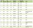 Click to view | Table 1. Chronologic Disease Progression |
Click to view | Table 2. Selected Hematologic and Myeloma Laboratory Data |
Bone marrow biopsy performed in August 2020 demonstrated 6% plasma cells, consistent with high-risk monoclonal gammopathy of undetermined significance (MGUS) (Fig. 1). Peripheral blood smear showed rouleaux formation without any evidence of agglutination (Fig. 2). Skeletal imaging, including radiographs and serial positron emission tomography/computed tomography (PET/CT) studies, repeatedly showed no lytic lesions or fluorodeoxyglucose (FDG)-avid osseous disease.
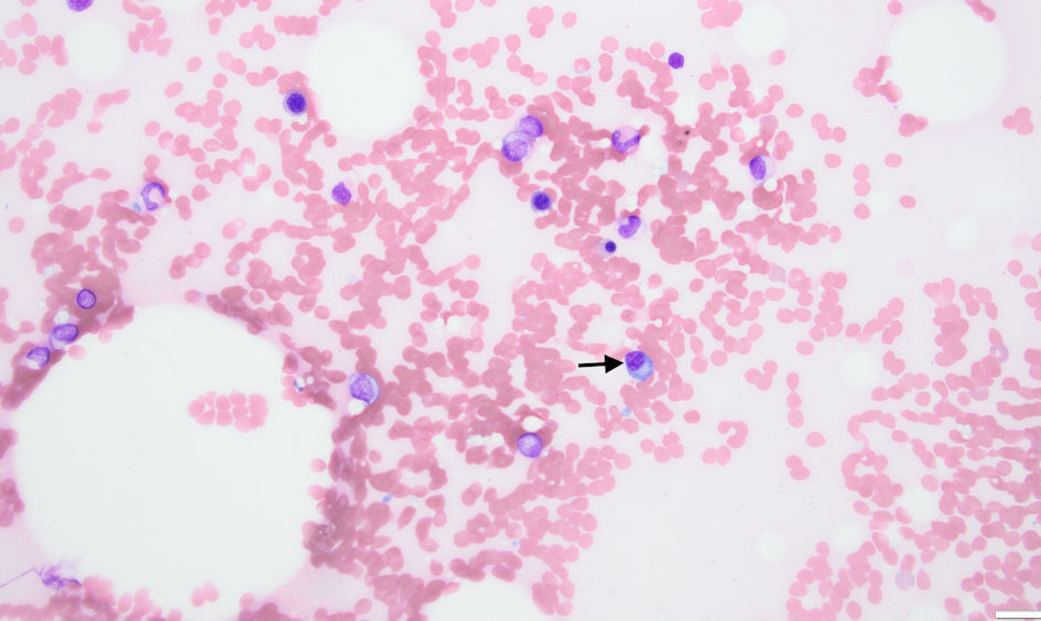 Click for large image | Figure 1. Bone marrow biopsy (with Wright-Giemsa staining) at initial diagnosis of high-risk MGUS demonstrating approximately 6% plasma cells (black arrow), consistent with high-risk MGUS. MGUS: monoclonal gammopathy of undetermined significance. |
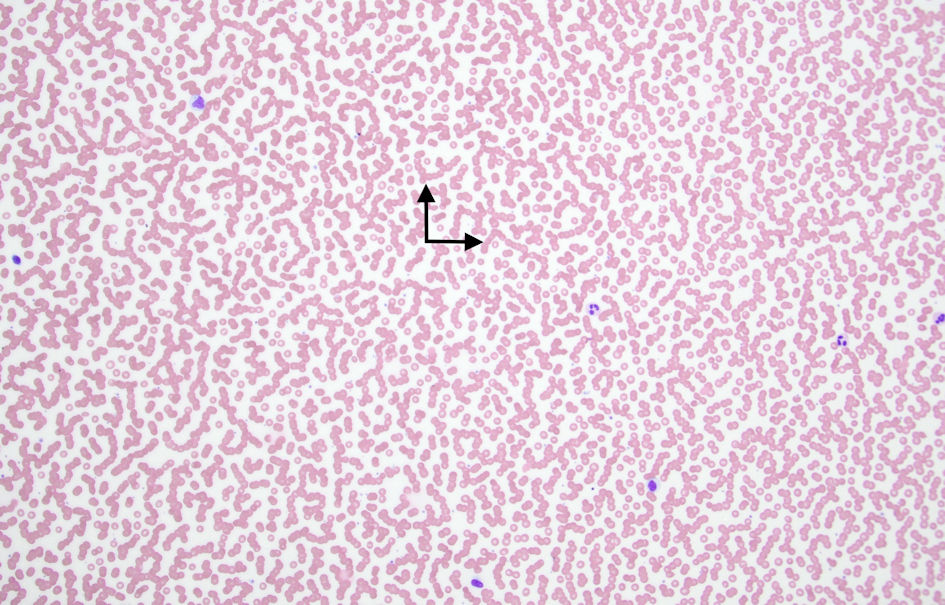 Click for large image | Figure 2. Peripheral blood smear (with Wright-Giemsa staining) demonstrating well-formed rouleaux (Black arrows). |
By June 2025, a repeat bone marrow biopsy demonstrated progression to SMM, with approximately 10% plasma cells and 2% monoclonal kappa-restricted plasma cells by flow cytometry. The cells expressed CD138, CD38, and CD56 (Fig. 3). The peripheral blood smear showed rouleaux formation and red blood cell agglutination, suggesting a significant rheologic disturbance associated with paraproteinemia (Fig. 4). Fluorescence in situ hybridization (FISH) studies were negative for tumor protein p53 (TP53) deletion, 1q gain, and 1p loss, though additional testing was limited by insufficient specimen.
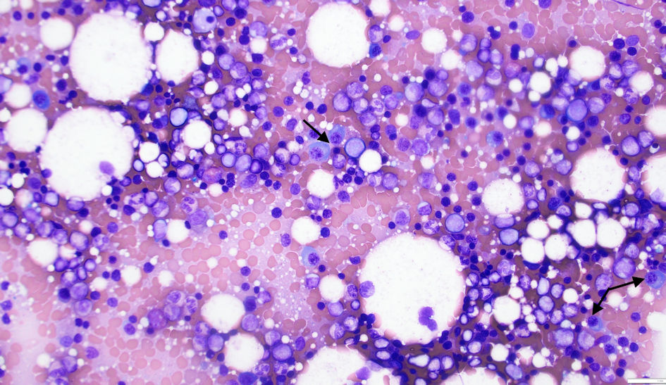 Click for large image | Figure 3. Bone marrow biopsy (with Wright-Giemsa staining) showing approximately 10% plasma cells (black arrows), meeting diagnostic criteria, and demonstrating progression to smoldering multiple myeloma. |
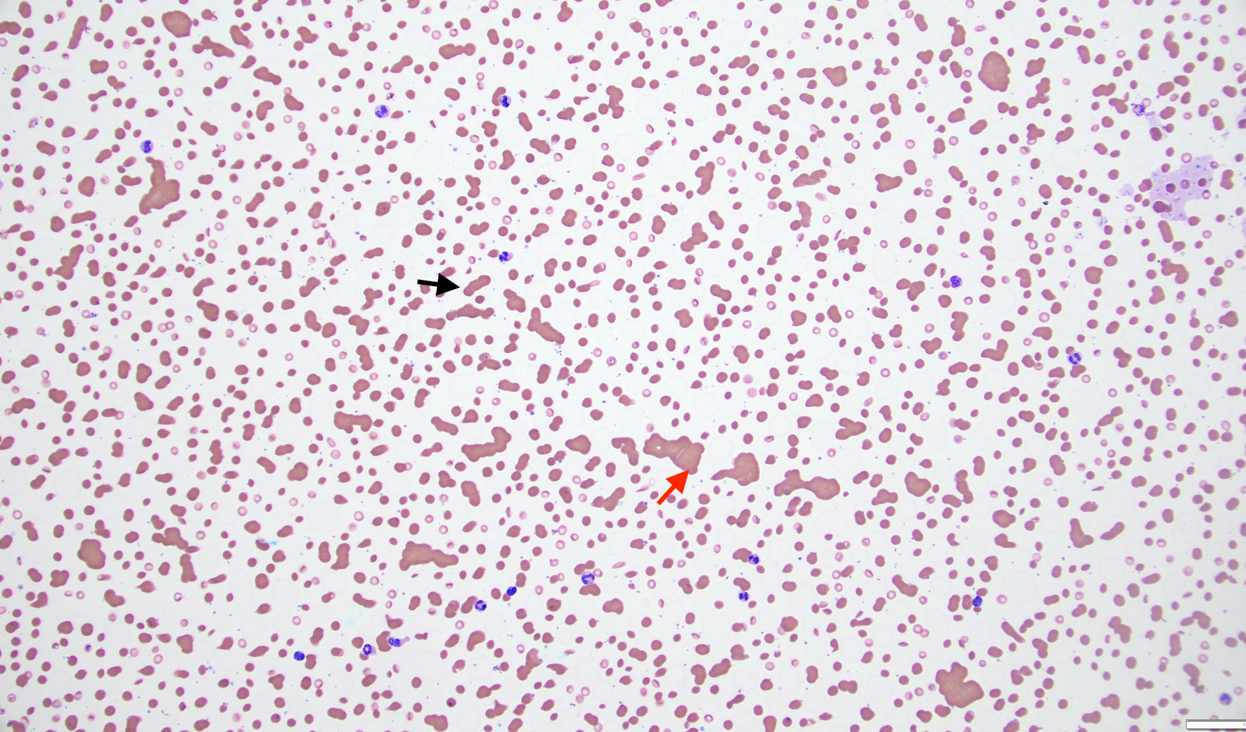 Click for large image | Figure 4. Peripheral blood smear (with Wright-Giemsa staining) demonstrating rouleaux formation (black arrows) and red blood cell agglutination (red arrow) associated with elevated monoclonal immunoglobulin levels and hyperviscosity syndrome. |
Despite the absence of classic CRAB criteria, the patient developed progressive signs suggestive of HVS. In January 2026, the patient presented to the emergency department with gross hematuria and lower abdominal discomfort, necessitating Foley catheter placement and continuous bladder irrigation. Imaging revealed prostatomegaly and bladder wall thickening without evidence of urinary tract malignancy or obstructive pathology. Coagulation studies, including prothrombin time (PT), partial thromboplastin time (PTT), fibrinogen, and factor X activity, were within normal limits. Subsequently, the patient developed retinal hemorrhage and recurrent epistaxis. Further evaluations in February 2026 revealed markedly elevated serum viscosity of 5.7 centipoise. Laboratory evaluation showed an IgG elevation to 5,463 mg/dL, with M-protein at 3.01 g/dL and total protein at 10.9 g/dL (Tables 1–3). PET/CT imaging remained negative for active myelomatous lesions.
Click to view | Table 3. Serum Viscosity Trend Following Daratumumab Therapy |
Given symptomatic hyperviscosity attributed to progressive smoldering myeloma, treatment was recommended despite the absence of overt multiple myeloma-defining events to prevent recurrence of HVS and related complications. The patient had previously declined therapy and pursued a holistic approach but agreed to initiate daratumumab in April 2026 after extensive counseling. After treatment initiation, the patient experienced rapid biochemical improvement. Serum viscosity decreased from 5.7 to 2.2 over approximately 3 months, accompanied by reductions in IgG from 5,463 mg/dL to 2,653 mg/dL and in M-protein from 3.01 g/dL to 1.35 g/dL (Table 3, Fig. 5). Cytopenias improved significantly, with no recurrence of bleeding or HVS episodes and no treatment-limiting toxicities reported (Tables 1, 3). These findings were confirmed by repeat eye and physical examinations.
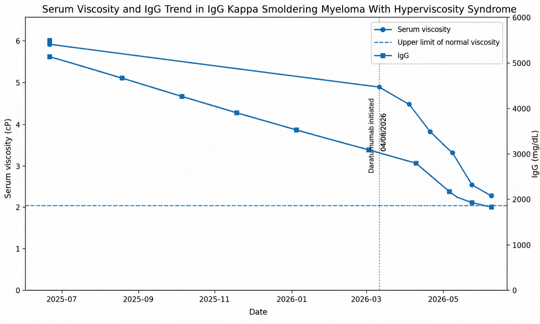 Click for large image | Figure 5. Trends in serum viscosity and IgG levels following initiation of daratumumab therapy. Trends consistent with a significant hematologic response and resolution of hyperviscosity-related symptoms. Ig: |
| Discussion | ▴Top |
HVS is an uncommon but potentially life-threatening complication of plasma cell dyscrasias characterized by impaired blood flow due to increased serum viscosity from circulating paraproteins [1]. The classic clinical triad includes mucosal bleeding, visual disturbances, and neurologic manifestations [2]. HVS is most frequently associated with Waldenstrom macroglobulinemia because IgM is a large pentameric immunoglobulin predominantly confined to the intravascular compartment [4]. Common causes of HVS are included in Table 4. In contrast, symptomatic HVS secondary to IgG paraproteinemia is rare and generally requires substantially higher immunoglobulin concentrations [5].
Click to view | Table 4. Common Causes of Hyperviscosity Syndrome |
SMM is defined by the presence of serum monoclonal protein ≥ 3 g/dL and/or 10–60% clonal plasma cells in the bone marrow in the absence of myeloma-defining SLiM-CRAB features [7] (Table 5). While SMM is associated with risk of progression to overt multiple myeloma, clinically significant HVS occurring before transformation is exceedingly uncommon [8].
Click to view | Table 5. Myeloma-Defining CRAB Criteria |
Our patient developed multiple manifestations consistent with symptomatic HVS, including retinal hemorrhage, recurrent epistaxis, and gross hematuria, in the setting of elevated serum viscosity of 5.7 centipoise. Importantly, alternative causes of bleeding were not identified. Coagulation parameters, including PT, PTT, fibrinogen, and factor X activity, were within normal limits, and extensive urologic imaging failed to demonstrate structural pathology accounting for recurrent hematuria.
Ocular manifestations are among the most characteristic findings of HVS and are often considered highly suggestive of clinically significant disease [12]. Retinal venous engorgement, flame hemorrhages, cotton wool spots, and papilledema may result from impaired retinal microcirculation [13, 14]. The retinal hemorrhage observed in this patient strongly supported clinically significant hyperviscosity rather than an isolated laboratory abnormality.
An additional notable feature of this case is the relatively modest marrow plasma cell burden despite severe rheologic complications. Bone marrow biopsy demonstrated approximately 10% plasma cells, barely meeting diagnostic criteria for smoldering myeloma, yet the patient developed marked symptomatic hyperviscosity. This finding suggests that factors beyond tumor burden alone, including immunoglobulin subtype, polymerization tendencies, molecular interactions, and intravascular distribution, may substantially influence serum viscosity [5].
Peripheral blood smear findings showed rouleaux formation and red blood cell agglutination, both classic hematologic manifestations of significant paraproteinemia [15, 16]. Rouleaux formation occurs when monoclonal proteins reduce the zeta potential between erythrocytes, promoting aggregation and impairing microvascular flow [15, 16].
Management should be individualized based on symptom severity, progression risk, comorbidities, and treatment goals. Urgent plasmapheresis is traditionally considered first-line therapy for acute, severe HVS. However, plasmapheresis provides only a rapid, temporary reduction in paraprotein levels; durable control requires therapy directed at the underlying plasma-cell clone [11, 17]. The AQUILA trial demonstrated improved progression-free survival with daratumumab monotherapy in patients with high-risk smoldering myeloma [11]. In high-risk smoldering myeloma, alternatives to daratumumab include lenalidomide alone or lenalidomide plus dexamethasone, both of which have shown delayed progression in randomized trials. More intensive regimens based on proteasome inhibitors or multidrug combinations remain investigational and may carry greater toxicity [17].
However, symptomatic hyperviscosity in smoldering myeloma remains poorly described in the literature and may be an underrecognized indication for early therapeutic intervention. Daratumumab, a monoclonal antibody targeting CD38, has demonstrated substantial efficacy in plasma cell dyscrasias through direct cytotoxicity, immune modulation, and depletion of clonal plasma cells [18]. Daratumumab monotherapy was selected in this case for its anti-plasma-cell activity, favorable tolerability, and evidence supporting early treatment in high-risk smoldering myeloma [17]. After initiating daratumumab, our patient experienced rapid improvement in serum viscosity from 5.7 to 2.2 centipoise, with corresponding reductions in IgG and M-protein levels. Importantly, retinal hemorrhage, epistaxis, and hematuria resolved with therapy, strongly supporting a causal relationship between paraproteinemia and hyperviscosity in this case.
Cases of HVS associated with IgG smoldering myeloma remain exceedingly rare in the literature. Most reported cases of hyperviscosity in plasma cell disorders involve overt multiple myeloma, particularly IgM/IgA-associated disease, due to increased polymerization [6]. This case therefore underscores the importance of maintaining clinical suspicion for HVS even in non-IgM plasma cell disorders without overt CRAB features or extensive marrow involvement. It also demonstrates that symptomatic hyperviscosity may warrant early therapeutic intervention in otherwise asymptomatic smoldering myeloma, and that timely initiation of plasma cell-directed therapy may prevent irreversible ophthalmologic and/or neurologic complications.
Conclusions
This case describes a rare presentation of IgG kappa SMM complicated by clinically significant HVS, manifesting as retinal hemorrhage, epistaxis, and gross hematuria. Despite the absence of overt CRAB features and a relatively low marrow plasma cell burden, the patient developed severe, symptomatic hyperviscosity, with serum viscosity reaching 5.7 centipoise. Daratumumab therapy led to rapid improvement in serum viscosity and clinical symptoms. This case highlights the importance of recognizing HVS in IgG-associated intermediate plasma cell disorders and suggests that symptomatic hyperviscosity may warrant treatment initiation in selected patients.
Learning points
Clinically significant HVS can occur in IgG kappa SMM despite the absence of SLiM-CRAB criteria. Retinal hemorrhage, epistaxis, hematuria, or rouleaux formation should prompt evaluation for hyperviscosity. This case highlights symptomatic hyperviscosity as a potential indication for early plasma cell-directed therapy, with daratumumab producing rapid clinical and serologic improvement (Fig. 6).
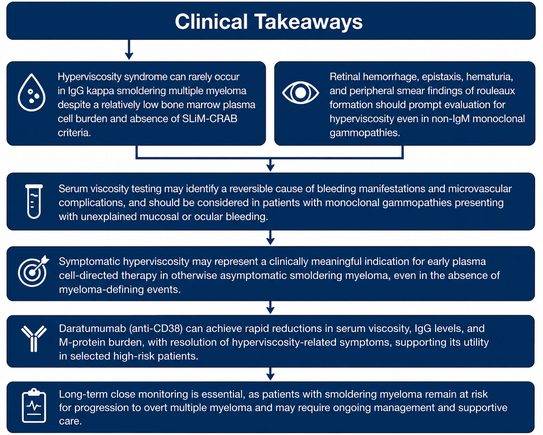 Click for large image | Figure 6. Learning points. |
Acknowledgments
The authors thank all the investigators whose work contributed to the understanding of hyperviscosity and to the treatment of SMM.
Financial Disclosure
The authors declare that they have no financial relationship with any commercial entity that is interested in the subject of this manuscript.
Conflict of Interest
The authors declare no conflicts of interest.
Informed Consent
This manuscript does not use patient identifiers or patient photographs. The patient’s treating oncologist, who is a co-author of this report, obtained verbal consent from the patient.
Author Contributions
All the authors participated actively in various sections of this manuscript prior to submission. Dr. Idamakanti (primary author) started the case report, designed the article and actively wrote and edited multiple sections of the manuscript, including abstract, case report, discussion, and conclusions, along with a literature review pertinent to the above sections. Dr. Kumar (co-author, corresponding oncologist) identified the rarity of the case and participated in the manuscript design, editing, and literature review. Dr. Bijjam (co-author) participated in manuscript editing, correcting grammar, literature review, and helped with obtaining MRI images. Dr. Bakhirev (co-author) provided histopathology slides and input on pathogenesis.
Data Availability
The authors declare that the data supporting the findings of this case report are available within the article. All data analyzed during this study were obtained from previously published studies and are available in the public domain.
Abbreviations
HVS: hyperviscosity syndrome; SMM: smoldering multiple myeloma; MGUS: monoclonal gammopathy of undetermined significance; IgG: immunoglobulin G; IgM: immunoglobulin M; CRAB: hypercalcemia, renal insufficiency, anemia, and bone lesions; SLiM: ≥ 60% clonal plasma cells, light chain ratio ≥ 100, and magnetic resonance imaging focal lesions; PET/CT: positron emission tomography/computed tomography; FDG: fluorodeoxyglucose; FISH: fluorescence in situ hybridization; TP53: tumor protein p53; CD: cluster of differentiation; PT: prothrombin time; PTT: partial thromboplastin time; WBC: white blood cell; cP: centipoise; IMWG: International Myeloma Working Group; anti-CD38: anti-cluster of differentiation 38 monoclonal antibody
| References | ▴Top |
- Stone MJ, Bogen SA. Evidence-based focused review of management of hyperviscosity syndrome. Blood. 2012;119(10):2205-2208.
doi pubmed - Gertz MA. Acute hyperviscosity: syndromes and management. Blood. 2018;132(13):1379-1385.
doi pubmed - Mehta J, Singhal S. Hyperviscosity syndrome in plasma cell dyscrasias. Semin Thromb Hemost. 2003;29(5):467-471.
doi pubmed - Bey T, Qdaisat A. Hyperviscosity syndrome: an oncologic emergency. Emerg Med Clin North Am. 2025;43(3):445-452.
doi pubmed - Dumas G, Merceron S, Zafrani L, Canet E, Lemiale V, Kouatchet A, Azoulay E. [Hyperviscosity syndrome]. Rev Med Interne. 2015;36(9):588-595.
doi pubmed - Halaseh RM, Kaur S, Khanna R, Conkling P, Sarna N, Magee A. Hyperviscosity syndrome in IgA multiple myeloma. QJM. 2022;115(1):30-31.
doi pubmed - Rajkumar SV, Dimopoulos MA, Palumbo A, Blade J, Merlini G, Mateos MV, Kumar S, et al. International Myeloma Working Group updated criteria for the diagnosis of multiple myeloma. Lancet Oncol. 2014;15(12):e538-548.
doi pubmed - Lakshman A, Rajkumar SV, Buadi FK, Binder M, Gertz MA, Lacy MQ, Dispenzieri A, et al. Risk stratification of smoldering multiple myeloma incorporating revised IMWG diagnostic criteria. Blood Cancer J. 2018;8(6):59.
doi pubmed - Kwaan HC. Hyperviscosity in plasma cell dyscrasias. Clin Hemorheol Microcirc. 2013;55(1):75-83.
doi pubmed - Mateos MV, Hernandez MT, Giraldo P, de la Rubia J, de Arriba F, Lopez Corral L, Rosinol L, et al. Lenalidomide plus dexamethasone for high-risk smoldering multiple myeloma. N Engl J Med. 2013;369(5):438-447.
doi pubmed - Dimopoulos MA, Voorhees PM, Schjesvold F, Cohen YC, Hungria V, Sandhu I, Lindsay J, et al. Daratumumab or active monitoring for high-risk smoldering multiple myeloma. N Engl J Med. 2025;392(18):1777-1788.
doi pubmed - Menke MN, Feke GT, McMeel JW, Treon SP. Ophthalmologic techniques to assess the severity of hyperviscosity syndrome and the effect of plasmapheresis in patients with Waldenstrom's macroglobulinemia. Clin Lymphoma Myeloma. 2009;9(1):100-103.
doi pubmed - Lai CC, Chang CH. Hyperviscosity-related retinopathy and serous macular detachment in Waldenstrom's macroglobulinemia: A mortal case in 5 years. Eur J Ophthalmol. 2022;32(4):NP109-NP114.
doi pubmed - Watson JA, Olson DJ, Zhang AY. Hyperviscosity Retinopathy Due to Waldenstrom macroglobulinemia: a case report and literature review. J Vitreoretin Dis. 2021;5(6):520-524.
doi pubmed - Van Camp B, Reynaert P, Broodtaerts L. Studies on the origin of the precursor cells in multiple myeloma, Waldenstrom's macroglobulinaemia and benign monoclonal gammopathy. I. Cytoplasmic isotype and idiotype distribution in peripheral blood and bone marrow. Clin Exp Immunol. 1981;44(1):82-89.
pubmed - Girard LP, Soekojo CY, Ooi M, Poon LM, Chng WJ, de Mel S. Immunoglobulin M paraproteinaemias. Cancers (Basel). 2020;12(6):1688.
doi pubmed - Schwartz J, Padmanabhan A, Aqui N, Balogun RA, Connelly-Smith L, Delaney M, Dunbar NM, et al. Guidelines on the use of therapeutic apheresis in clinical practice-evidence-based approach from the Writing Committee of the American Society for Apheresis: the seventh special issue. J Clin Apher. 2016;31(3):149-162.
doi pubmed - van de Donk NW, Janmaat ML, Mutis T, Lammerts van Bueren JJ, Ahmadi T, Sasser AK, Lokhorst HM, et al. Monoclonal antibodies targeting CD38 in hematological malignancies and beyond. Immunol Rev. 2016;270(1):95-112.
doi pubmed
This article is distributed under the terms of the Creative Commons Attribution 4.0 International License (CC BY 4.0), which permits unrestricted use, distribution, and reproduction in any medium, including commercial use, provided the original work is properly cited.
Journal of Medical Cases is published by Elmer Press Inc.