| Journal of Medical Cases, ISSN 1923-4155 print, 1923-4163 online, Open Access |
| Article copyright, the authors; Journal compilation copyright, J Med Cases and Elmer Press Inc |
| Journal website https://jmc.elmerpub.com |
Case Report
Volume 17, Number 11, November 2026, pages 628-636

Reversible Thyroid Nodule in Van Wyk–Grumbach Syndrome: An Unusual Case Report
Laila A. AlBishi
Department of Pediatrics, Faculty of Medicine, University of Tabuk, PO Box 71491, Tabuk, Saudi Arabia
Manuscript submitted June 21, 2026, accepted September 18, 2026, published online October 2, 2026
Short title: Thyroid Nodule in Van Wyk–Grumbach
doi: https://doi.org/10.14740/jmc5386
| Abstract | ▴Top |
Van Wyk–Grumbach syndrome (VWGS) is a rare complication of longstanding, untreated primary hypothyroidism in children. It is characterized by isosexual precocious puberty, delayed bone age, and multicystic ovarian enlargement despite the presence of severe hypothyroidism. Although thyroid enlargement is common, a discrete thyroid nodule has not previously been documented in this syndrome. This study presents a 6-year-old Saudi girl who exhibited a 2-day history of vaginal hemorrhage and a history of breast development for 6 months. The physical examination demonstrated a palpable right thyroid nodule. The laboratory evaluation revealed a severe primary hypothyroidism, high thyroid autoantibodies, elevated prolactin levels, and prepubertal levels of basal gonadotropins. A thyroid ultrasound demonstrated a well-defined, hypoechoic nodule. Further testing revealed that the child had a delayed bone age, multi-cystic ovaries, and pituitary hyperplasia. After levothyroxine treatment, the patient’s thyroid hormone levels normalized. In addition, there was progressive shrinkage of the thyroid nodule, pituitary hypertrophy, and ovarian cysts, along with significant improvement in linear growth. This case adds to the previously unreported association of a reversible thyroid nodule with VWGS and highlights the importance of early diagnosis and appropriate management of severe hypothyroidism to prevent unnecessary or invasive diagnostic tests.
Keywords: Van Wyk–Grumbach syndrome; Thyroid nodule; Juvenile hypothyroidism; Precocious puberty; Autoimmune thyroiditis
| Introduction | ▴Top |
Van Wyk–Grumbach syndrome (VWGS), first described in 1960, is a rare clinical manifestation of longstanding juvenile hypothyroidism characterized by the paradoxical occurrence of isosexual precocious puberty despite severe thyroid hormone deficiency [1]. The syndrome classically manifests breast development and vaginal bleeding in girls or testicular enlargement without virilization in boys, accompanied by delayed bone age and multi-cystic gonadal enlargement [2]. These manifestations result from chronic elevation of thyroid-stimulating hormone (TSH), which exhibits structural homology with follicle-stimulating hormone (FSH) and can activate FSH receptors at supraphysiologic concentrations, leading to gonadal hyperstimulation [3]. Concomitant hyperprolactinemia, resulting from unopposed thyrotropin-releasing hormone (TRH) stimulation of lactotrophs, further enhances ovarian sensitivity to gonadotropins and contributes to the clinical phenotype [4].
Although VWGS has been well-characterized with respect to its reproductive and systemic manifestations, focal structural changes within the thyroid gland, specifically, discrete nodules, have received limited attention. Most reports focus on diffuse thyroid enlargement (goiter) as a consequence of chronic TSH stimulation, consistent with the pathophysiology of autoimmune thyroiditis or iodine deficiency [5, 6]. However, there are no prior reports describing discrete thyroid nodules as a clinical manifestation of VWGS. In this context, it is particularly pertinent that elevated TSH levels over extended periods may promote the growth of thyroid tissue and ultimately lead to follicular hyperplasia and nodule formation in various clinical contexts [7]. The present case report describes a 6-year-old girl with VWGS who presented with a palpable thyroid nodule in addition to the classic features of the syndrome. To our knowledge, no prior case of VWGS has reported a discrete, reversible thyroid nodule. This is an important observation as it suggests that long-term high levels of TSH may cause localized structural changes in the thyroid in severely hypothyroid children. It also emphasizes the importance of conducting a comprehensive structural evaluation of the thyroid by physically examining the child and using ultrasound when children present with VWGS.
This case also features the broader clinical significance of recognizing VWGS as a reversible cause of precocious puberty and pituitary enlargement. Misdiagnosis may result in unnecessary invasive investigations, including pituitary surgery, and inappropriate hormonal interventions [8]. Early recognition and treatment with levothyroxine leads to gradual but complete resolution of all endocrine abnormalities, including regression of ovarian cysts, pituitary hyperplasia [9], and possibly regression of a discrete thyroid nodule.
| Case Report | ▴Top |
Investigations
A 6-year-old girl from Saudi Arabia came to the pediatric endocrine clinic with a 2-day history of vaginal bleeding. Her parents reported that she had breast enlargement on both sides over the previous 6 months, with no growth underarm or pubic hair. They also noticed that she had become less social and demonstrated decreased concentration at her school. These variations were corroborated by the schoolteacher. A full-body checkup revealed no issues, including headaches, changes in vision, unusual movements, or behavioral shifts. There was no history of local trauma, abnormal bleeding from other sites, dysuria, or foreign-object insertion.
Two years before this child’s presentation, the patient had been assessed for poor appetite but never returned for follow-up. At that time, her height was 97 cm, her weight was 16 kg (10th and 25th centiles, respectively), and she had no abnormal findings recorded. The patient’s family history was significant for consanguinity, as well as her mother’s history of maternal autoimmune thyroiditis, for which she is currently on treatment. The patient is the second of her family’s children delivered vaginally at term, with an uneventful outcome. The child had met all developmental milestones and was up to date on vaccinations.
Physical examination of the patient revealed a dull-looking face with puffiness around both eyes and cheek fullness. Figure 1 displays growth measurement data for this patient. Her height of 101.3 cm is below the third percentile, corresponding to a −2.5 standard deviation score (SDS), while her weight of 26 kg places her at the 90th percentile for her age, and her BMI is 25.49 kg/m2. Her mother’s and father’s heights were 152.4 and 158.2 cm, respectively; her calculated target height is 148.8 cm. Assessment for puberty revealed a girl in Tanner stage B3P1 (B for breast development; P for pubic hair), but no axillary hair, no virilization, and no galactorrhea. On neck examination, there was a palpable right thyroid mass approximately 1.2 × 1.5 × 1.0 cm in size. It was smooth, firm in consistency, and non-tender. There was no cervical lymphadenopathy. The physical findings were consistent with a benign condition, but could not be ruled out for cancer by physical exam alone (a further ultrasound demonstrated more on the thyroid nodule). Further signs were negative for café au lait spots, bone deformities, and papilledema.
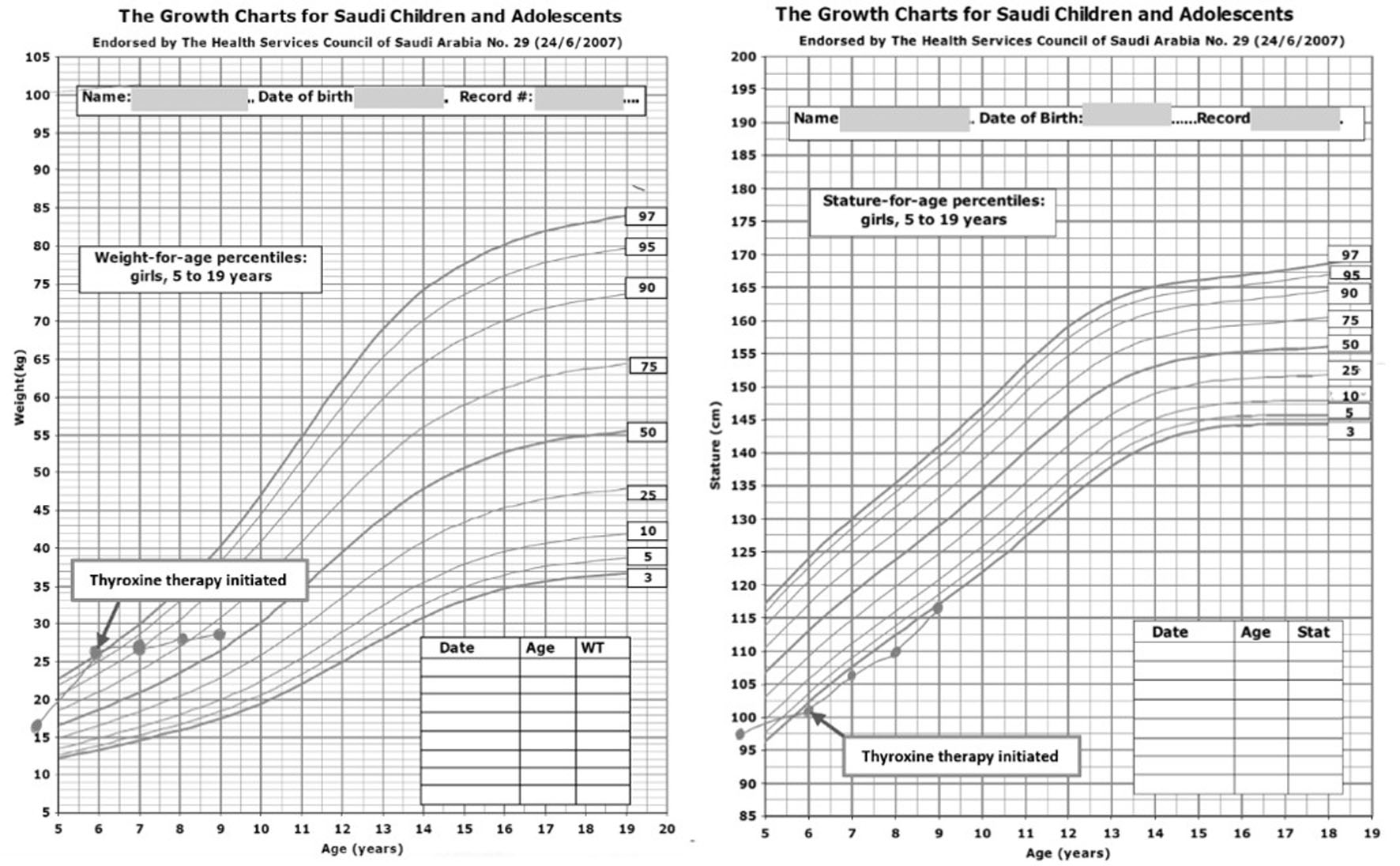 Click for large image | Figure 1. Anthropometric measurements before and after levothyroxine therapy. Weight-for-age and height-for-age percentile charts for Saudi girls illustrating the patient’s growth trajectory at presentation. The plotted points indicate improvements in growth parameters before and after the initiation of levothyroxine therapy (arrow). |
Diagnosis
Laboratory results demonstrated that there was significant primary hypothyroidism based on very high TSH (> 75 mIU/L), low free thyroxine (FT4, 9.1 pmol/L), and low free triiodothyronine (FT3, 3.4 pmol/L). The lab data indicated significant glandular failure over time. The presence of high autoantibody activity confirmed autoimmune thyroiditis.
The prolactin (PRL) level was very high at 173 ng/mL and therefore consistent with TRH stimulating secretion. Gonadal hormone levels were prepubertal with luteinizing hormone (LH) of < 0.21 IU/L and follicle-stimulating hormone (FSH) of 7.01 U/L. Estradiol levels were higher than expected (82 pg/mL), suggesting ovarian hyperstimulation. Both progesterone and dehydroepiandrosterone sulfate (DHEA-S) were within the normal limits. This makes precocious puberty due to an adrenal gland abnormality unlikely.
Macrocytic hyperchromic anemia was identified via complete blood count (CBC). Anemia in association with hypothyroidism can be seen in approximately 55% of patients (with no identifiable nutritional deficits) [10]. Levels of insulin-like growth factor-1 (IGF-1) and insulin-like growth factor-binding protein-3 (IGFBP-3) were determined to be in the normal range for screening of short stature. All laboratory testing results, including thyroid function tests, autoantibodies, and CBC, are listed in Table 1. A thyroid ultrasound confirmed diffusely enlarged thyroid lobes with heterogeneous hypoechoic echotexture and increased vascularity, consistent with autoimmune thyroiditis. In the right thyroid lobe, there was a nodule measuring 1.2 × 1.5 × 1.0 cm (estimated volume 0.94 mL) with well-defined margins and no microcalcifications, no spread beyond the thyroid, and no significant vascularity. Accordingly, it was rated as American College of Radiology Thyroid Imaging Reporting and Data System category 3 (ACR TI-RADS 3) (mildly suspicious with a low chance of cancer) (Fig. 2a, b). A bone age X-ray, read using the Greulich-Pyle method, was 4.5 years, reported as delayed for the chronological age. This moderate delay suggests that the patient still has good potential for linear growth once thyroid hormone treatment is started. Pelvic ultrasound revealed bilateral multicystic ovaries. The right ovary measured 2.3 × 3.6 cm, and the left ovary measured 2.7 × 3.2 cm indicating a bilateral ovarian enlargement. Multiple peripheral follicles created a characteristic “necklace” appearance, reflecting chronic FSH stimulation in the absence of ovulation. The uterus was prepubertal in size and configuration (Fig. 2c, d). Pituitary magnetic resonance imaging (MRI) demonstrated a homogeneously enhancing sellar mass measuring 20 × 15 × 13 mm with extension into the suprasellar cistern and grade 1 cavernous sinus extension (Knosp classification), without optic chiasm compression. The pituitary stalk was midline with normal enhancement, and the posterior pituitary bright spot was preserved. These findings were consistent with pituitary hyperplasia secondary to chronic hypothyroidism rather than a true adenoma (Fig. 3a, b).
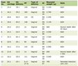 Click to view | Table 1. Baseline and Longitudinal Follow-Up Laboratory Parameters in Patients Diagnosed With Van Wyk–Grumbach Syndrome |
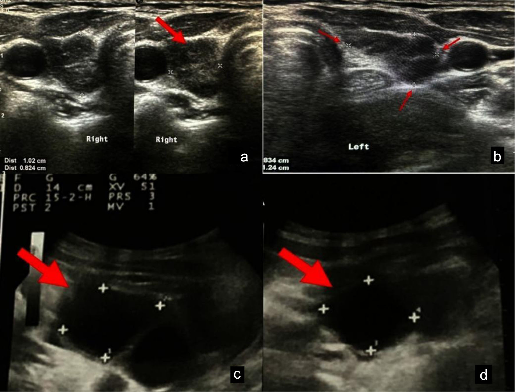 Click for large image | Figure 2. Thyroid and pelvic ultrasound imaging. (a, b) Thyroid ultrasound demonstrates a hypoechoic nodule in the right lobe. (c, d) Pelvic ultrasound showing bilateral multi-cystic ovaries (arrows) consistent with Van Wyk-Grumbach syndrome and a prepubertal uterus in size and configuration. |
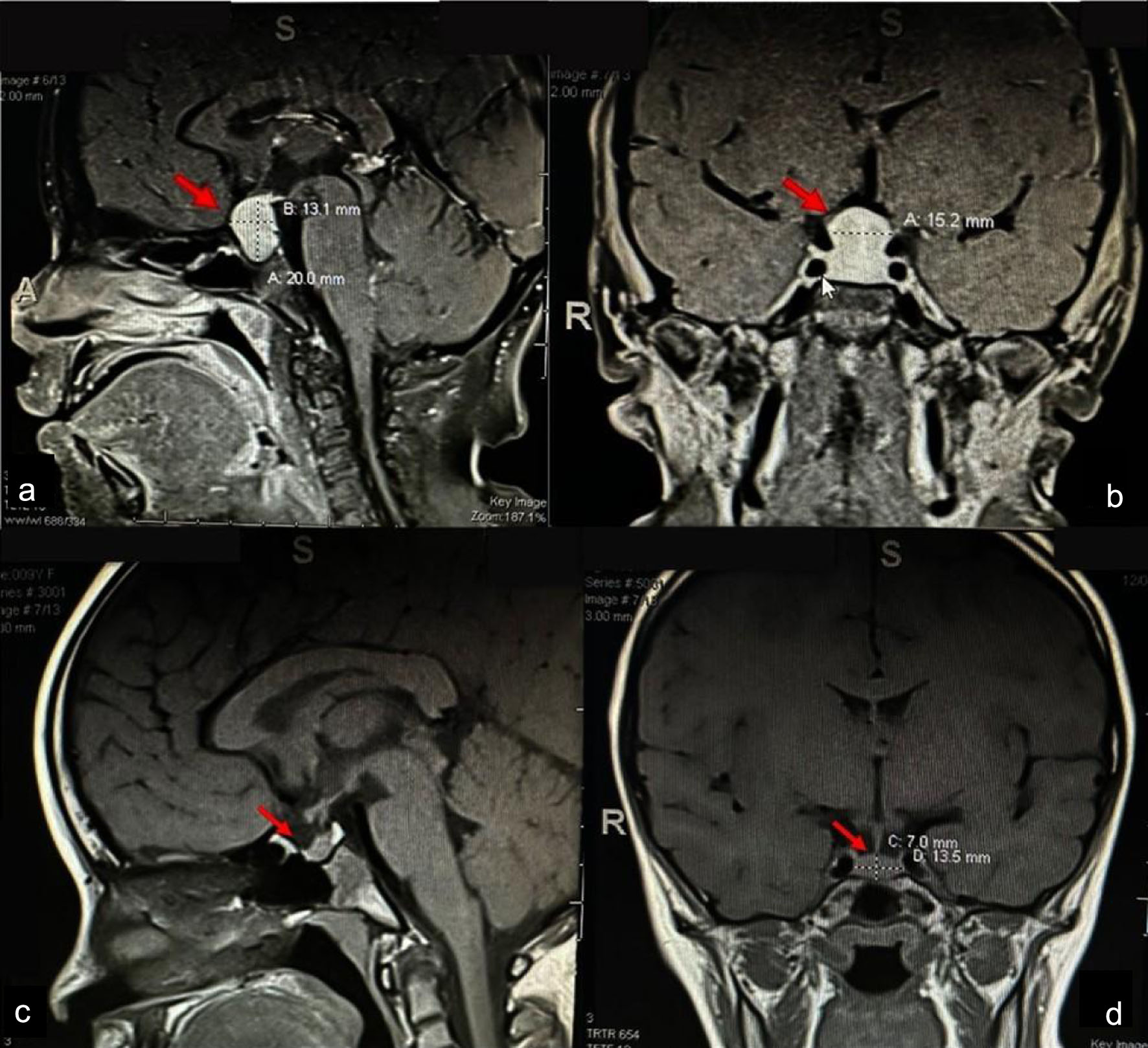 Click for large image | Figure 3. Pituitary magnetic resonance imaging. (a, b) Baseline sagittal and coronal T1-weighted post-contrast images showing pituitary hyperplasia with suprasellar extension (arrows). (c, d) Follow-up imaging at 9 months demonstrated a marked reduction in pituitary size after levothyroxine therapy. |
Treatment
The patient was initiated on levothyroxine replacement therapy at 100 µg/day, with thyroid function tests monitored and the dose titrated accordingly. The dose was selected based on pediatric guidelines for severe hypothyroidism, which recommend higher initial doses to achieve more rapid normalization of thyroid function. Monthly thyroid function tests were monitored to guide dose titration, with adjustments made to maintain TSH within the target range of 0.5–2.0 mIU/L to optimize regression of the thyroid nodule and pituitary hyperplasia [11]. No additional hormonal interventions were administered. Specifically, gonadotropin-releasing hormone (GnRH) agonists were not indicated, as the precocious puberty in VWGS is peripheral (gonadotropin-independent) and resolves with thyroid hormone replacement alone. Similarly, no surgical intervention was pursued for the pituitary mass or thyroid nodule, as both were expected to undergo progressive regression with normalization of thyroid function.
Follow-up and outcomes
Vaginal bleeding resolved completely within 1 week of initiating levothyroxine therapy, likely reflecting withdrawal of estrogen support and endometrial shedding. This rapid response provided early reassurance to both the patient and her family. During 3 months of follow-up, thyroid function tests normalized: TSH decreased from > 75 to 4.2 mIU/L and FT4 increased from 9.1 to 18.3 pmol/L (Fig. 4; Table 1). Prolactin levels also dropped, and TSH normalized to 28 ng/mL by month 3. This trend confirmed that hyperprolactinemia had arisen from chronic TRH overstimulation.
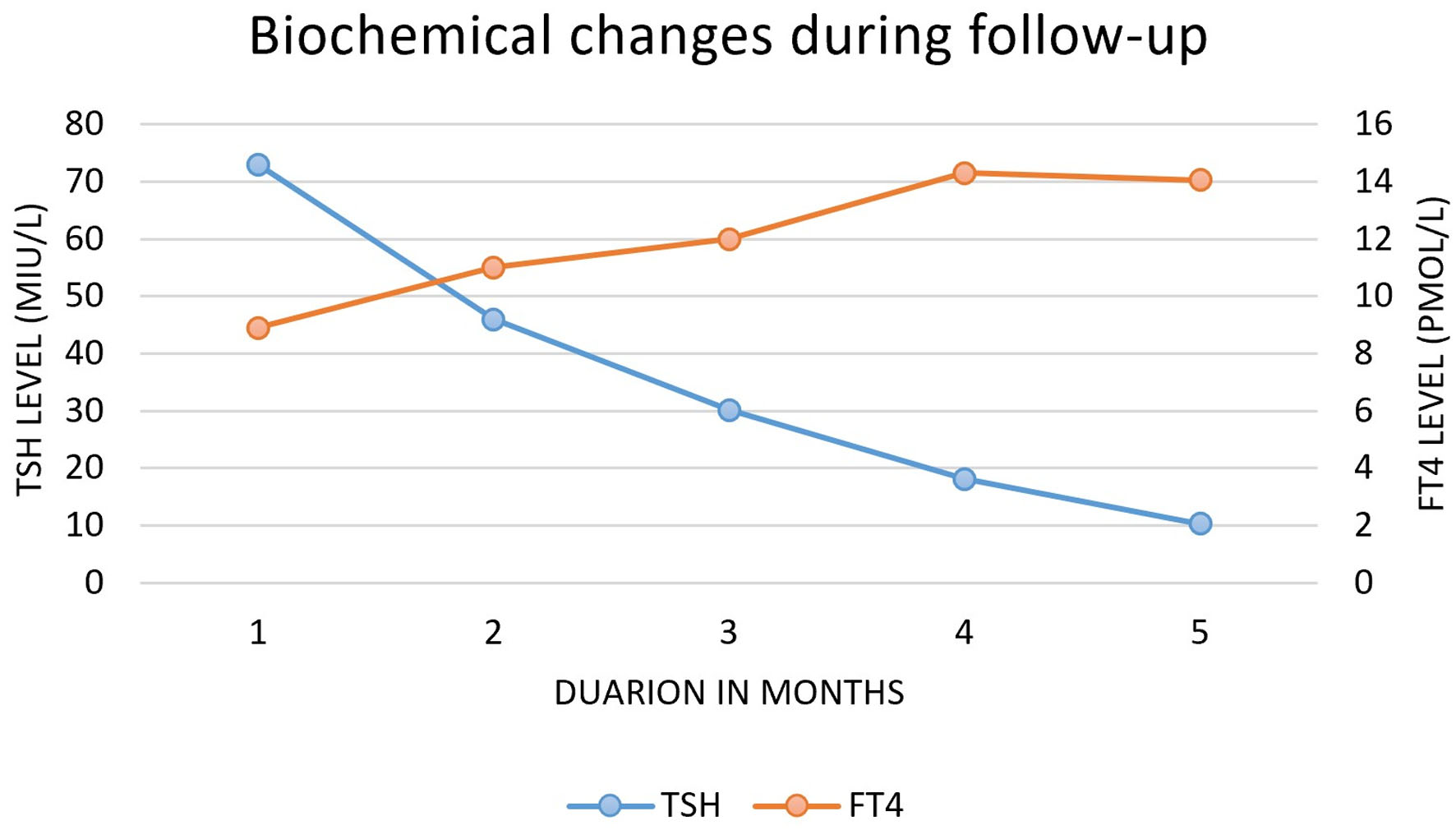 Click for large image | Figure 4. Temporal changes in thyroid function tests during levothyroxine therapy. Serial measurements of thyroid-stimulating hormone (TSH) and free thyroxine (FT4) during follow-up after initiation of levothyroxine therapy. The graph demonstrates progressive normalization of thyroid function, with a marked decline in TSH levels accompanied by a corresponding increase in FT4 concentrations over the follow-up period. |
Follow-up thyroid ultrasounds showed a steady decrease in the size of the thyroid nodule. After 6 months of levothyroxine therapy, the nodule had shrunk to 0.7 × 0.9 × 0.6 cm (volume 0.20 mL), representing a 79% reduction in volume. The close temporal and quantitative correlation between TSH reduction and nodule volume regression provides strong support for a TSH-driven, reversible mechanism rather than a true neoplastic process.
Follow-up pelvic ultrasound at 9 months showed complete resolution of the previously noted multicystic ovarian changes, with normalization of ovarian volumes and return to normal prepubertal appearance of less than 2 cm. Repeat pituitary magnetic resonance imaging (MRI) at 6 months demonstrated a marked reduction in pituitary size from 20 × 15 × 13 to 12 × 10 × 8 mm (volume 0.5 mL; 75% reduction), with resolution of suprasellar extension and restoration of normal pituitary configuration (Fig. 3c, d). This dramatic regression confirmed the diagnosis of reversible pituitary hyperplasia rather than a true adenoma.
Anthropometric parameters improved over the follow-up period. At 9 months, the patient’s height was 106.8 cm (−2.0 SDS), representing a gain of 5.5 cm, indicating substantial catch-up growth (the normal growth velocity for a 6-year-old child is approximately 5–6 cm per year). Weight remained stable at 26.5 kg. Breast development showed gradual regression, with Tanner stage decreasing from B3 to B2 by 9 months. No further episodes of vaginal bleeding occurred. The patient’s school performance and social engagement improved markedly, as reported by parents and teachers.
At the most recent follow-up (12 months post-treatment initiation), the patient remains on levothyroxine 50 µg daily. Thyroid function is stable (TSH 1.8 mIU/L, FT4 16.2 pmol/L). Long-term monitoring is planned to assess final adult height, pubertal progression, and thyroid nodule status.
| Discussion | ▴Top |
This case documents the first report of a discrete reversible thyroid nodule with VWGS, thereby expanding the recognized clinical spectrum of this rare disorder. The patient presented all classic features of VWGS including severe hypothyroidism, isosexual precocious puberty, multi-cystic ovarian enlargement, delayed bone age, and pituitary hyperplasia, along with a novel finding of a palpable thyroid nodule. Most literature on VWGS focuses on reproductive and systemic effects, with less attention to structural changes in the thyroid. However, biochemical evidence for thyroid dysfunction has been well documented in all previous published cases of VWGS. Structural imaging has usually been limited to diffuse thyroid diseases (such as autoimmune thyroiditis) or glandular enlargement. Diffuse goiter is frequently documented and represents an indication of either autoimmune thyroiditis or iodine deficiency [12]. A cohort of 30 children with VWGS did not routinely perform thyroid ultrasound, and no nodules were described [13]. Other case reports and series have documented diffuse heterogeneous echotexture and increased vascularity consistent with thyroiditis, but without discrete nodular lesions [14]. Consequently, whether the lack of documented nodules in VWGS represents genuine biological rarity, insufficient recognition, or reporting bias remains unclear. This knowledge gap possesses clinical significance because, in the absence of systematic data, clinicians lack evidence-based guidance for managing a child with VWGS who presents with a thyroid nodule. Focal thyroid nodules have not been demonstrated in most of the literature to date (Table 2) [9, 12, 13, 15–33].
Click to view | Table 2. Reported Thyroid Morphological and Structural Abnormalities Documented in Published Pediatric Cases of Van Wyk–Grumbach Syndrome |
Our case has reassuring sonographic features of a thyroid nodule with low-risk stratification (ACR TI-RADS 3), which favors a benign hyperplastic nodule [15] that showed regression following levothyroxine therapy; however, they do not rule out malignancy, as thyroid nodules in pediatrics carry a higher risk of malignancy than in adults. Although data on the well-documented regression of other TSH-dependent manifestations in VWGS, including ovarian cysts and pituitary hyperplasia [23], are available, the absence of histological confirmation for the thyroid nodule does not definitively establish a causal relationship.
Importantly, future prospective studies of VWGS may evaluate thyroid ultrasound both at diagnosis and after achieving euthyroidism to establish the true prevalence of discrete nodules and their natural history. In the setting of severe hypothyroidism, nodules with low-risk sonographic features may represent TSH-dependent changes that regress with levothyroxine therapy; therefore, careful serial monitoring before invasive evaluation may be a reasonable approach in selected cases.
Recognition of the peripheral nature of precocious puberty in VWGS is essential to avoid unnecessary GnRH agonist therapy. Treatment should instead focus exclusively on thyroid hormone replacement therapy, accompanied by monitoring for the potential development of central precocious puberty [17]. Similarly, pituitary hyperplasia should not prompt surgery or biopsy, as it typically resolves with thyroid hormone replacement alone. A long-term follow-up is required to monitor growth, pubertal progression, thyroid function, and structural changes [12].
This report is limited by its single-patient report, which limits the ability to draw broader conclusions about the occurrence and clinical relevance of thyroid nodules in VWGS. Fine-needle aspiration was not performed because the nodule had low-risk sonographic features (ACR TI-RADS 3) [34] and demonstrated interval regression after levothyroxine therapy. We cannot definitively rule out a coexisting benign neoplasm that regressed coincidentally, although this scenario remains unlikely given the close temporal relationship between TSH reduction and nodule volume regression. In addition, the duration of follow-up was relatively short, preventing assessment of long-term outcomes, including possible recurrence of the thyroid lesion and its impact on growth and pubertal development. Larger studies with extended follow-up are needed to clarify further the relationship between severe hypothyroidism and potentially reversible thyroid nodular changes in this syndrome.
To our knowledge, there are no reports that describe discrete thyroid nodules in patients with VWGS. Thus, the current case report is more of a hypothesis-generating report than a documentation of VWGS. Future case reports are needed to determine whether thyroid nodules occur more frequently in patients with severe, longstanding hypothyroidism than has been reported.
Learning points
VWGS involved the following diagnostic features in children: isosexual puberty, an advanced bone age, and clinical evidence of longstanding hypothyroidism.
Severe primary hypothyroidism does not simply result in enlarged or diffusely enlarged thyroid but may include thyroid nodular changes.
Elevated TSH levels over time may cause focal thyroid nodular hyperplasia, contributing to thyroid nodules.
Thyroxine replacement therapy may regress not only pituitary hyperplasia and ovarian cysts, but also thyroid nodules, thereby preventing unnecessary invasive surgical procedures.
Thorough clinical evaluation and ultrasound imaging are required when evaluating children with VWGS.
Acknowledgments
None to declare.
Financial Disclosure
None to declare.
Conflict of Interest
None to declare.
Informed Consent
Written informed consent to publish this case report has been obtained from the patient’s legal representative.
Author Contributions
LAA was responsible for the care of patients, for the collection and analysis of data, for the literature search, for writing the first draft of the article, and for approval of the final version of the article.
Data Availability
The data from this report can be obtained from the corresponding author upon request. Confidentiality of patient data will be maintained.
| References | ▴Top |
- Wyk J, Grumbach M. Syndrome of precocious menstruation and galactorrhea in juvenile hypothyroidism: an example of hormonal overlap in pituitary feedback. Journal of Pediatrics. 1960; 57(3):416-435.
- Fernandez-Gonzalez SM, Perez Vila MM, Prado-Carro AM. Precocious puberty in hypothyroidism: mini-review of Van Wyk-Grumbach syndrome. J Endocr Soc. 2023;7(12):bvad135.
doi pubmed - Ryan GL, Feng X, d'Alva CB, Zhang M, Van Voorhis BJ, Pinto EM, Kubias AE, et al. Evaluating the roles of follicle-stimulating hormone receptor polymorphisms in gonadal hyperstimulation associated with severe juvenile primary hypothyroidism. J Clin Endocrinol Metab. 2007;92(6):2312-2317.
doi pubmed - Dutta D, Maisnam I, Ghosh S, Mukhopadhyay P, Mukhopadhyay S, Chowdhury S. Panhypopituitarism with empty sella a sequel of pituitary hyperplasia due to chronic primary hypothyroidism. Indian J Endocrinol Metab. 2012;16(Suppl 2):S282-284.
doi pubmed - Zimmermann MB, Boelaert K. Iodine deficiency and thyroid disorders. Lancet Diabetes Endocrinol. 2015;3(4):286-295.
doi pubmed - Caturegli P, De Remigis A, Rose NR. Hashimoto thyroiditis: clinical and diagnostic criteria. Autoimmun Rev. 2014;13(4-5):391-397.
doi pubmed - Haymart MR, Repplinger DJ, Leverson GE, Elson DF, Sippel RS, Jaume JC, Chen H. Higher serum thyroid stimulating hormone level in thyroid nodule patients is associated with greater risks of differentiated thyroid cancer and advanced tumor stage. J Clin Endocrinol Metab. 2008;93(3):809-814.
doi pubmed - Eom KS, See-Sung C, Kim JD, Kim JM, Kim TY. Primary hypothyroidism mimicking a pituitary macroadenoma: regression after thyroid hormone replacement therapy. Pediatr Radiol. 2009;39(2):164-167.
doi pubmed - Baranowski E, Hogler W. An unusual presentation of acquired hypothyroidism: the Van Wyk-Grumbach syndrome. Eur J Endocrinol. 2012;166(3):537-542.
doi pubmed - Eram F, Quamri MA, Zaidi A, Sonam. A Comprehensive review of anaemia associated with hypothyroidism. New Emir Med J. 2024;05:e02506882282237.
doi - Hietamaki J, Karkinen J, Iivonen AP, Vaaralahti K, Tarkkanen A, Almusa H, Huopio H, et al. Presentation and diagnosis of childhood-onset combined pituitary hormone deficiency: a single center experience from over 30 years. EClinicalMedicine. 2022;51:101556.
doi pubmed - Cao Y, Zhao X, Huang W, Xi Y, Wu H. Case Report: Van Wyk-Grumbach syndrome presenting as vaginal bleeding: diagnostic value of pituitary and pelvic imaging. Front Pediatr. 2025;13:1736671.
doi pubmed - Kusuma Boddu S, Ayyavoo A, Hebbal Nagarajappa V, Kalenahalli KV, Muruda S, Palany R. Van Wyk Grumbach syndrome and ovarian hyperstimulation in juvenile primary hypothyroidism: lessons from a 30-case cohort. J Endocr Soc. 2023;7(6):bvad042.
doi pubmed - Nicolescu CR, Bazus L, Stephan JL. Severe acquired hypothyroidism and Van Wyk-Grumbach syndrome in two children. Case Rep Pediatr. 2024;2024:8919177.
doi pubmed - Sanjeevaiah AR, Sanjay S, Deepak T, Sharada A, Srikanta SS. Precocious puberty and large multicystic ovaries in young girls with primary hypothyroidism. Endocr Pract. 2007;13(6):652-655.
doi pubmed - Patni N, Cervantes LF, Diaz A. Elevated alpha-fetoprotein levels in Van Wyk-Grumbach syndrome: a case report and review of literature. J Pediatr Endocrinol Metab. 2012;25(7-8):761-767.
doi pubmed - Bhagwat NM, Dalwadi PP, Joshi AS, Pawal PS, Varthakavi PK. Asymmetrical Ovarian Enlargement: Caught Timely Before the Cut! J Pediatr Adolesc Gynecol. 2015;28(3):e83-85.
doi pubmed - Kocova M, Zdraveska N, Kacarska R, Kochova E. Diagnostic approach in children with unusual symptoms of acquired hypothyroidism. When to look for pituitary hyperplasia? J Pediatr Endocrinol Metab. 2016;29(3):297-303.
doi pubmed - Leonardi A, Penta L, Cofini M, Lanciotti L, Principi N, Esposito S. Rhabdomyolysis in a young girl with Van Wyk-Grumbach syndrome due to severe hashimoto thyroiditis. Int J Environ Res Public Health. 2018;15(4):704.
doi pubmed - Reddy P, Tiwari K, Kulkarni A, Parikh K, Khubchandani R. Van Wyk Grumbach syndrome: a rare consequence of hypothyroidism. Indian J Pediatr. 2018;85(11):1028-1030.
doi pubmed - Lee SJ, Moon JE, Lee GM, Cho MH, Ko CW. An Alport syndrome boy with Van Wyk-Grumbach syndrome induced by prolonged untreated congenital hypothyroidism. Ann Pediatr Endocrinol Metab. 2020;25(2):132-136.
doi pubmed - Gupta J, Lin-Su K. Van Wyk-Grumbach syndrome in a female pediatric patient with trisomy 21: a case report. Int J Pediatr Endocrinol. 2020;2020:2.
doi pubmed - Riaz M, Ibrahim MN, Laghari TM, Hanif MI, Raza J. Van Wyk Grumbach syndrome. J Coll Physicians Surg Pak. 2020;30(12):1332-1334.
doi pubmed - Kandasamy D, Malik R, Sharma R, Jana M. Case 308: Van Wyk-Grumbach syndrome. Radiology. 2022;305(3):746-750.
doi pubmed - Sherwani P, Nandolia KK, Joshi K, Lourembam R. Imaging in Van Wyk Grumbach syndrome: An uncommon presentation of hypothyroidism. SA J Radiol. 2023;27(1):2572.
doi pubmed - Waddankeri S, Waddankeri M, Waddankeri S, Arora K. The clinical septet of Van Wyk-Grumbach syndrome: a case series from a tertiary care centre in Kalyana Karnataka, India. touchREV Endocrinol. 2023;19(1):98-102.
doi pubmed - Marzouk A, Mezzi R, Zelfani S, Jelaila N, Ayeb S, Bouaziz A. Pediatric psoriasis associated with Van Wyk Grumbach syndrome: a case report. Tunis Med. 2023;101(10):780-782.
pubmed - Rathod S, Jajoo S, Shanoo A, Dande A, Kaplish D. Solving the dilemma: Van Wyk-Grumbach syndrome. Cureus. 2024;16(5):e61382.
doi pubmed - Baioumi A, Kolenova A, Avatapalle HB. An unusual cause of bleeding in primary hypothyroidism. Clin Pediatr Endocrinol. 2024;33(2):71-75.
doi pubmed - Mukhopadhyay I, Roy B, Varma A, Singh S. Diverse nature of ovarian tumours: Van Wyk-Grumbach syndrome. BMJ Case Rep. 2024;17(12):e261639.
doi pubmed - Ganta AG, Desta WT. Challenge faced in diagnosis and treatment of congenital hypothyroidism presenting with isosexual puberty and bilateral ovarian mass in low and middle income country: a case report. BMC Endocr Disord. 2025;25(1):234.
doi pubmed - Sivasubramanian D, Senthilkumar V, Aravind S, Rajasekar AR, Prasaanth S, Sanil S. Van Wyk-Grumbach Syndrome with bilateral inguinal hernia: A case report. Int J Surg Case Rep. 2025;127:110975.
doi pubmed - Gil M, Martino F, Besseghine G, Rojas Ortiz MV, Rojas S, Rodriguez ME, Arcari A, et al. Peripheral precocious puberty secondary to severe hypothyroidism. Arch Argent Pediatr. 2025;123(3):e202410456.
doi pubmed - Tessler FN, Middleton WD, Grant EG, Hoang JK, Berland LL, Teefey SA, Cronan JJ, et al. ACR Thyroid Imaging, Reporting and Data System (TI-RADS): White Paper of the ACR TI-RADS Committee. J Am Coll Radiol. 2017;14(5):587-595.
doi pubmed
This article is distributed under the terms of the Creative Commons Attribution 4.0 International License (CC BY 4.0), which permits unrestricted use, distribution, and reproduction in any medium, including commercial use, provided the original work is properly cited.
Journal of Medical Cases is published by Elmer Press Inc.