| Journal of Medical Cases, ISSN 1923-4155 print, 1923-4163 online, Open Access |
| Article copyright, the authors; Journal compilation copyright, J Med Cases and Elmer Press Inc |
| Journal website https://jmc.elmerpub.com |
Case Report
Volume 17, Number 9, September 2026, pages 498-502

Postoperative Adjuvant Treatment Decision for Early Epstein–Barr Virus-Associated Gastric Cancer
Xiao Feng Hea, c, Bo Yuan Huangb, c, Jing Jing Tenga, Xiao Jun Yangb, d
aFirst Clinical Medical College, Gansu University of Chinese Medicine, Lanzhou 730000, Gansu, China
bDepartment of Hepatobiliary Surgery, Gansu Provincial Hospital, Lanzhou 730000, Gansu, China
cThe authors contributed equally to this report.
dCorresponding Author: Xiao Jun Yang, Department of Hepatobiliary Surgery, Gansu Provincial Hospital, Lanzhou 730000, Gansu, China
Manuscript submitted April 23, 2026, accepted June 18, 2026, published online July 28, 2026
Short title: Adjuvant Treatment for Early EBVaGC
doi: https://doi.org/10.14740/jmc5353
| Abstract | ▴Top |
Epstein–Barr virus-associated gastric cancer (EBVaGC) is a relatively rare clinical subtype. Despite its generally favorable prognosis, preoperative diagnosis remains challenging, and no established consensus exists regarding postoperative adjuvant treatment strategies for early-stage patients with high-risk pathological factors. This study reports a female case of early-stage EBVaGC of the lymphoepithelioma-like carcinoma subtype complicated with perineural invasion (a high-risk factor), and systematically explores the etiology, pathological features, key diagnostic points, individualized adjuvant therapy options, and prognosis of EBVaGC. The aim is to improve clinicians’ understanding of this gastric cancer subtype, reduce its misdiagnosis rate, and accelerate the development of standardized diagnostic and treatment pathways. A case of a 60-year-old female patient admitted with “abdominal pain and distension for more than 1 year” was reported, and postoperative pathology confirmed EBVaGC. The clinical manifestations and diagnostic and treatment process were analyzed in combination with a review of relevant literature from recent years. Preoperative gastroscopic biopsy revealed poorly differentiated adenocarcinoma of the gastric mucosa. The patient underwent laparoscopic radical subtotal gastrectomy with lymph node dissection. Postoperative pathology confirmed EBVaGC. After symptomatic treatment, the patient improved and was discharged from the hospital. One month after surgery, no disease progression was observed and the SOX regimen was administered. The patient tolerated the treatment well, and the next cycle of chemotherapy is currently planned. EBVaGC lacks specific clinical manifestations, and laboratory findings may be within normal ranges at the time of onset. Reliance solely on conventional gastroscopy and histopathological biopsy can easily lead to misdiagnosis and inappropriate treatment. Epstein–Barr virus–encoded RNA in situ hybridization is the core diagnostic modality for definitive diagnosis. This subtype generally has a favorable prognosis. However, for patients with high-risk pathological factors such as perineural invasion and tumor invasion into the muscularis propria, standardized postoperative individualized adjuvant therapy should be administered based on tumor stage and molecular profiles.
Keywords: Epstein–Barr virus-associated gastric cancer; Lymphoepithelioma-like carcinoma; EBER in situ hybridization; Adjuvant therapy
| Introduction | ▴Top |
The global incidence and mortality risks of gastric cancer remain high, and precise subtyping of its molecular subtypes is of great significance for clinical treatment decision making and prognosis assessment. Epstein–Barr virus-associated gastric carcinoma (EBVaGC), as one of the four major molecular subtypes of gastric cancer defined by The Cancer Genome Atlas Research Network in 2014, accounts for approximately 8–10% of all gastric cancers, with a higher incidence rate in East Asia. The occurrence of this type of gastric cancer is closely related to EBV infection of gastric mucosal epithelial cells. However, for patients with relatively small tumors and negative surgical margins, whether postoperative adjuvant therapy is needed and what the optimal adjuvant treatment strategy should be—particularly the value of targeted therapy and immunotherapy—remain under investigation. Through an in-depth analysis of an early-stage typical EBVaGC case and a review of relevant literature, this article aims to provide a reference for the clinical diagnosis and treatment of this disease.
| Case Report | ▴Top |
A 60-year-old female patient presented with no significant past medical history or relevant family history. The patient presented with abdominal pain and distension for more than 1 year without obvious triggers, accompanied by nausea and acid reflux. The symptoms were initially ignored. After symptom aggravation, she underwent gastroscopy and biopsy at a local hospital, which demonstrated malignant transformation of a gastric ulcer (gastric mucosa) with poorly differentiated adenocarcinoma, without further subtyping. Physical examination revealed no abnormalities except for tenderness in the upper abdomen. After admission, serum tumor marker panel (six gastric cancer markers) was completed, and all results were within normal ranges. In addition, contrast-enhanced abdominopelvic computed tomography (CT) showed localized thickening and enhancement of the gastric antrum, highly suggestive of a neoplastic lesion (Fig. 1).
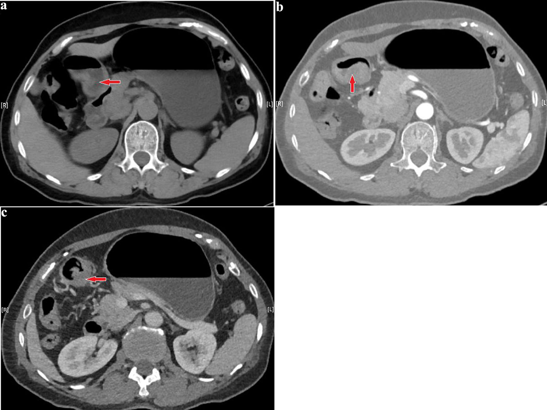 Click for large image | Figure 1. (a) Preoperative computed tomography (CT) shows localized thickening along the greater curvature of the gastric antrum with protruding into the gastric lumen; the maximum thickness is approximately 17 mm, and the size is approximately 12 × 34 mm. (b, c) Contrast-enhanced scans demonstrate marked heterogeneous enhancement. Red arrows indicate the location of the lesion. No enlarged retroperitoneal lymph nodes are observed. |
After evaluation, there were no obvious contraindications to surgery. Therefore, laparoscopic subtotal gastrectomy with gastrojejunostomy plus abdominal lymph node dissection was performed under general anesthesia. Intraoperative blood loss was approximately 100 mL, and the procedure was uneventful. Postoperatively, the patient received symptomatic treatments including fasting, gastrointestinal decompression, anti-infection, and nutritional support, after which she improved and was discharged. Histopathological examination of the resected specimens (stomach, omentum, and lymph nodes), with supplementary Epstein–Barr virus–encoded RNA (EBER) in situ hybridization and molecular biomarker testing, revealed the following findings: (gastric angle) small round cell malignant tumor. Immunohistochemistry confirmed EBVaGC of ulcerative type, lymphoepithelioma-like carcinoma. The tumor invaded the muscularis propria (T2). Perineural invasion was present, with no definite lymphovascular invasion. No cancer metastasis was found in any of the submitted lymph nodes (including those from the greater curvature, lesser curvature, and other groups) (0/26). All resection margins were R0 (Fig. 2). Immunohistochemistry results: tumor cells tested positive for EBER in situ hybridization (+), confirming EBV association. Immunophenotype: CKP (+), P53 (wild-type expression pattern), P63 (–), P40 (–), Her-2 (score 0, negative), Claudin18.2 (3+, strong positivity in 70% of tumor cells). Mismatch repair protein testing: MLH1 (+), MSH2 (+), MSH6 (+), PMS2 (+), indicating proficient mismatch repair (pMMR).
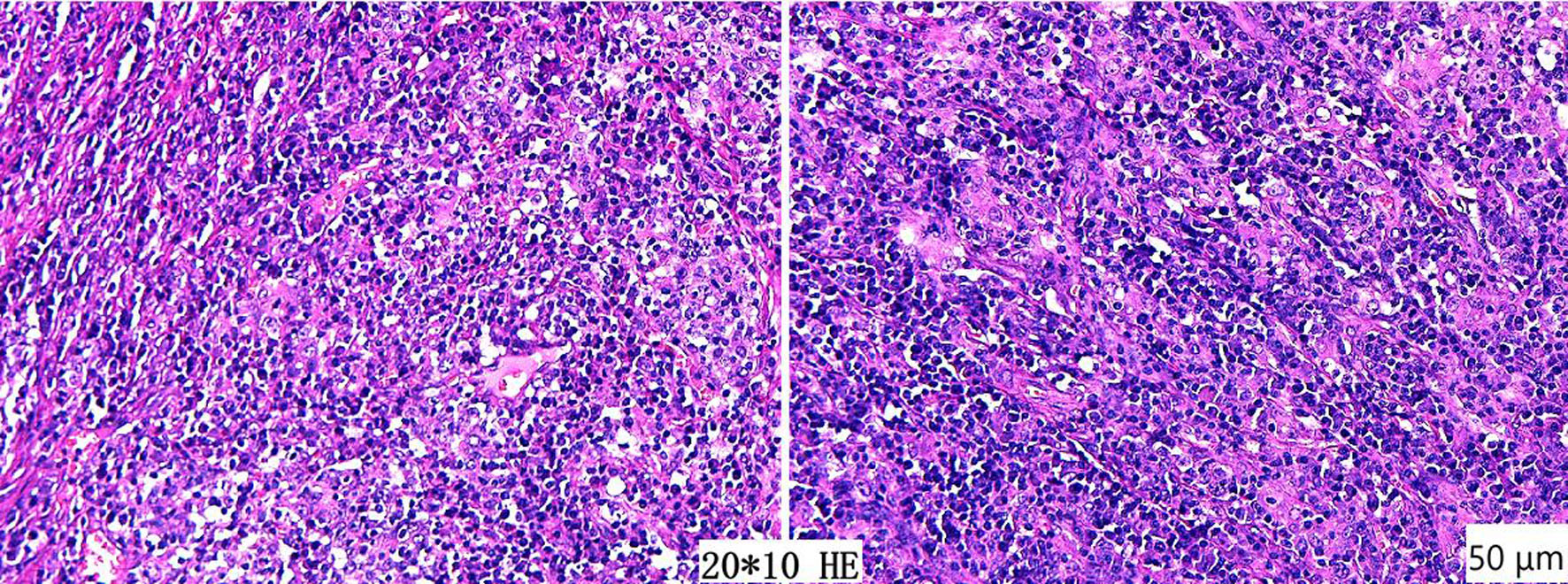 Click for large image | Figure 2. Postoperative pathological findings show a small round cell malignant tumor at the gastric angle, consistent with ulcerative-type lymphoepithelioma-like carcinoma of Epstein–Barr virus-associated gastric cancer. All resection margins are negative, and no lymph node metastasis is observed (hematoxylin and eosin staining, original magnification × 200; scale bar = 50 µm). |
One month later, the patient returned for follow-up. Serum thymidine kinase 1 and contrast-enhanced CT of the chest and abdomen were both normal, showing no tumor recurrence or metastasis and no lymphadenopathy (Fig. 3). The overall disease control was stable. Based on the immunohistochemistry results, the first cycle of the SOX regimen was administered (oxaliplatin 200 mg intravenously; S-1 50 mg orally twice daily on days 1–14). The treatment course went smoothly, and the patient tolerated it well without any toxicities or adverse reactions. Our department is currently conducting active follow-up, and the second cycle is planned to be given after 21 days.
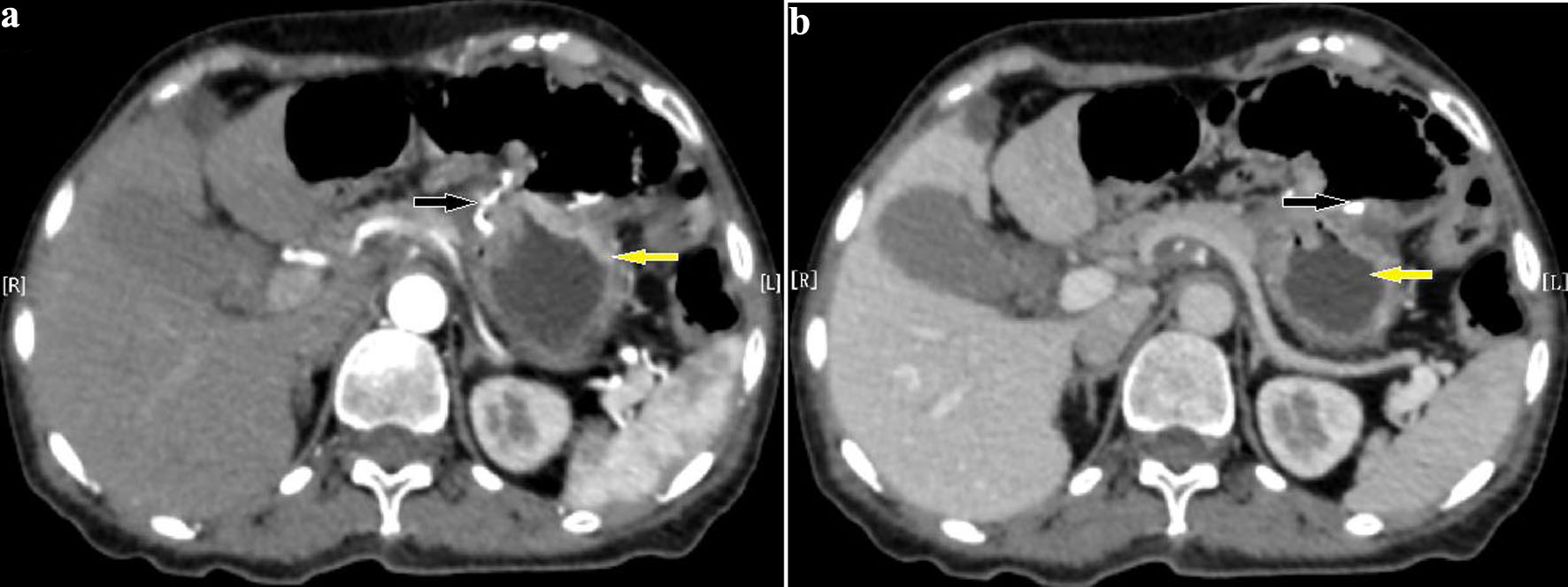 Click for large image | Figure 3. (a, b) Pre-first-cycle chemotherapy computed tomography (CT) after surgery shows postoperative changes in the distal stomach. The nodular protrusion below the cardia is related to the surgical procedure. Yellow arrows indicate the remnant stomach, and black arrows indicate the gastrojejunal anastomosis. No obviously enlarged retroperitoneal lymph nodes are observed. |
| Discussion | ▴Top |
Epidemiological data show that the prevalence of EBVaGC in Asian populations ranges from approximately 2% to 10% [1]. This type of gastric cancer is not exclusive to males; females also have a risk of developing the disease, and there is no clear age limit for onset. Of note, serum tumor marker levels in EBVaGC are mostly within the normal range at the time of diagnosis. The tumor morphology is predominantly ulcerative, and the depth of tumor invasion is often limited to T1–T2 stages, with TNM staging frequently classified as stage I–II [2, 3]. Related studies have confirmed that EBV can infect the inflamed mucosal epithelial cells of Hp-associated gastritis via the ephrin A2 receptor, and the two synergistically promote epithelial-mesenchymal transition (EMT), thereby increasing the risk of carcinogenesis [4]. EBVaGC has distinct clinicopathological characteristics. The core histological feature is carcinoma with lymphoid stroma (CLS) or similar changes, among which the classic CLS subtype (i.e., lymphoepithelioma-like carcinoma (LELC)) accounts for up to 75% [5]. Pathologically, it presents as poorly differentiated adenocarcinoma accompanied by extensive diffuse lymphocytic infiltration. Gastroscopy combined with biopsy is the mainstay of gastric cancer diagnosis, but the definitive diagnosis of EBVaGC must rely on EBER in situ hybridization, which is also the gold standard for distinguishing this subtype from EBV-negative adenocarcinoma, microsatellite instability-type gastric cancer, and other subtypes.
The core of gastric cancer treatment lies in accurate disease assessment and dynamic evaluation of patient status. For early-stage and some advanced-stage patients, surgical resection is the main treatment modality. Studies have shown that the surgical approaches for EBVaGC are primarily total gastrectomy and distal gastrectomy, with the key determinants being tumor location and size, and there is no need to extend the extent of lymph node dissection [6]. While ensuring the curative effect of tumor resection, this approach maximizes the preservation of postoperative physiological function of the digestive tract and improves long-term quality of life. Chemotherapy serves as the mainstay of perioperative adjuvant treatment. However, programmed cell death protein 1 (PD-1) immunotherapy and Claudin18.2-targeted therapy are of great value for patients who are intolerant to chemotherapy or have inoperable advanced tumors. Amplification of the 9p24.1 locus drives high expression of JAK2, PD-L1, and PD-L2. The JAK2/STAT1/IRF-1 pathway further upregulates PD-L1, creating a typical “hot tumor” immune microenvironment [3]. This allows immune checkpoint inhibitors, including anti-PD-L1 antibodies, to work effectively. It also explains why EBVaGC patients tend to have a relatively favorable prognosis. EBVaGC often shows high expression of PD-L1, significantly higher than that in non-LELC subtypes, which represents an adaptive resistance of the tumor in response to immune pressure [7]. Moreover, the PD-L1 Combined Positive Score (CPS) can directly influence the efficacy of immunotherapy, with markedly different objective response rates (ORRs) depending on the score. EBV positivity is an independent predictor of immunotherapy efficacy in pMMR gastric cancer patients. The ORR in EBV+/pMMR patients reaches 54.5%, which is comparable to that in deficient mismatch repair (dMMR) patients [8], providing a clinical reference for adjuvant immunotherapy after gastric cancer surgery. Claudin18.2 is a tight junction protein with restricted expression in normal tissues, but it shows extremely high expression rates in EBVaGC, making it a characteristic molecular signature. Meanwhile, the PI3KCA mutation rate in this subtype reaches 80%, which can activate related pathways involved in tumor proliferation and drug resistance. The latest guidelines also recommend a monoclonal antibody targeting Claudin18.2 in combination with chemotherapy as a grade I recommendation for advanced gastric cancer that is Claudin18.2-positive and HER2-negative.
The overall prognosis of EBVaGC is significantly better than that of EBV-negative gastric cancer (EBVnGC). In particular, patients with the LELC subtype and without lymph node metastasis have longer disease-free survival and overall survival. Moreover, the disease control rate of first-line chemotherapy with fluoropyrimidines plus platinum reaches as high as 90.3–100%, with significant survival benefits [3, 4]. An extremely low lymph node metastasis rate is a core characteristic of EBVaGC, which is closely related to the immune barrier formed by activated T cells in the tumor microenvironment. High CD3+ T cell infiltration is an independent protective prognostic factor for EBVaGC, and its infiltration density is significantly higher in EBVaGC than in EBVnGC [1]. In contrast, elevated carcinoembryonic antigen (CEA) levels and pTNM stage III are independent risk factors affecting overall survival [6]. During postoperative follow-up, plasma EBV-DNA load and endoscopy can sensitively reflect recurrence and dynamically monitor chemotherapy efficacy, with better sensitivity than routine examinations. They should be incorporated into the long-term follow-up protocol [3, 9].
Based on the early detection, small lesion size, and relevant pathological features of this patient, the necessity of postoperative chemotherapy in adjuvant treatment decisions remains somewhat controversial. Although conventional postoperative adjuvant chemotherapy regimens for stage II gastric cancer (such as XELOX or SOX) offer survival benefits, considering that EBVaGC generally has a favorable prognosis and is a “hot” tumor, whether it can derive maximum benefit from traditional cytotoxic chemotherapy, or whether immunotherapy-based regimens could play a role in the adjuvant setting, remains to be further elucidated. Following departmental discussion, we ultimately adopted the SOX regimen. This decision was based on: first, the high sensitivity of EBVaGC to fluoropyrimidine plus platinum combinations, with a considerable disease control rate [4], which also aligns with the treatment consensus for pT2 stage with high-risk factors (perineural invasion); second, this regimen can eliminate microscopic residual disease while maximally preserving the function of immune cells in the tumor microenvironment. Taking into account the patient’s financial situation and family wishes, this treatment plan was finally determined. If recurrence or metastasis occurs in the future, CLDN18.2-targeted therapy combined with chemotherapy could be a priority option.
Conclusion
This report presents a rare case with early-stage EBVaGC (LELC) confirmed by postoperative pathology, exhibiting a molecular phenotype of EBER positivity, HER-2 negativity, high Claudin18.2 expression, and pMMR status, with a postoperative tumor stage of pT2N0M0. This case intuitively demonstrates the unique pathomorphological and molecular characteristics of EBVaGC. Deepening the understanding of this gastric cancer subtype is an important foundation for achieving precise diagnosis and evidence-based prognostic assessment. Systematic testing of key molecular biomarkers including EBER, PD-L1, and CLDN18.2 in gastric cancer patients can effectively compensate for the limited diagnostic capacity in primary healthcare settings, reduce the misdiagnosis rate of EBVaGC, and provide precise guidance for the selection of targeted therapy and immunotherapy regimens in the event of disease recurrence or metastasis. This represents a core strategy for delivering individualized patient care. Future prospective clinical studies are still needed to optimize the adjuvant treatment model for EBVaGC, with the aim of further improving patients’ quality of life and long-term survival outcomes on the premise of achieving radical tumor control.
Acknowledgments
The authors have no relevant acknowledgments to declare.
Financial Disclosure
This study was supported by the Lanzhou Municipal Science and Technology Plan Project (Project Title: Study on the Molecular Mechanism of STIL in Regulating Proliferation and Apoptosis of Hepatocellular Carcinoma Cells; Grant No. 2023-ZD-26).
Conflict of Interest
The authors declare no conflict of interest.
Informed Consent
Written informed consent was obtained from the patient for the publication of this case report.
Author Contributions
Xiao Feng He: case collection, analysis, and manuscript preparation; Bo Yuan Huang: conception and design of study; acquisition of data; and data analysis and interpretation; Jing Jing Teng: acquisition of data; data analysis and interpretation; Xiao Jun Yang: approval of final version of manuscript.
Data Availability
Any inquiries regarding supporting data availability of this study should be directed to the corresponding author.
| References | ▴Top |
- Jia X, Guo T, Li Z, Zhang M, Feng Y, Dong B, Li Z, et al. Clinicopathological and immunomicroenvironment characteristics of Epstein-Barr virus-associated gastric cancer in a Chinese population. Front Oncol. 2020;10:586752.
doi pubmed - Li G, Zhou Z, Wang Z, Wang Z. Assessing Epstein-Barr virus in gastric cancer: clinicopathological features and prognostic implications. Infect Agent Cancer. 2023;18(1):11.
doi pubmed - Yang J, Liu Z, Zeng B, Hu G, Gan R. Epstein-Barr virus-associated gastric cancer: A distinct subtype. Cancer Lett. 2020;495:191-199.
doi pubmed - Corallo S, Lasagna A, Filippi B, Alaimo D, Tortorella A, Serra F, Vanoli A, et al. Unlocking the potential: Epstein-Barr Virus (EBV) in gastric cancer and future treatment prospects, a literature review. Pathogens. 2024;13(9).
doi pubmed - Yanai H, Chihara D, Harano M, Sakaguchi E, Murakami T, Nishikawa J. Endoscopic and pathologic motifs for the clinical diagnosis of Epstein-Barr virus-associated gastric cancer. DEN Open. 2021;1(1):e7.
doi pubmed - Gu L, Zhang Q, Hu Y, Xiang Z, Ji S, Yu W, Liu X, et al. Demographic/clinicopathological characteristics and prognosis of resectable Epstein-Barr virus-associated gastric cancer: a nested case-control study from an Eastern China. Front Oncol. 2025;15:1602091.
doi pubmed - Wei XL, Liu QW, Liu FR, Yuan SS, Li XF, Li JN, Yang AL, et al. The clinicopathological significance and predictive value for immunotherapy of programmed death ligand-1 expression in Epstein-Barr virus-associated gastric cancer. Oncoimmunology. 2021;10(1):1938381.
doi pubmed - Bai Y, Xie T, Wang Z, Tong S, Zhao X, Zhao F, Cai J, et al. Efficacy and predictive biomarkers of immunotherapy in Epstein-Barr virus-associated gastric cancer. J Immunother Cancer. 2022;10(3).
doi pubmed - Yanai H, Fujiwara J, Toyama E, Okuda H, Miura O, Kaino S, Nishikawa J. Endoscopic diagnosis and therapy for Epstein-Barr virus-associated gastric cancer. Microorganisms. 2023;11(11):2619.
doi pubmed
This article is distributed under the terms of the Creative Commons Attribution 4.0 International License (CC BY 4.0), which permits unrestricted use, distribution, and reproduction in any medium, including commercial use, provided the original work is properly cited.
Journal of Medical Cases is published by Elmer Press Inc.