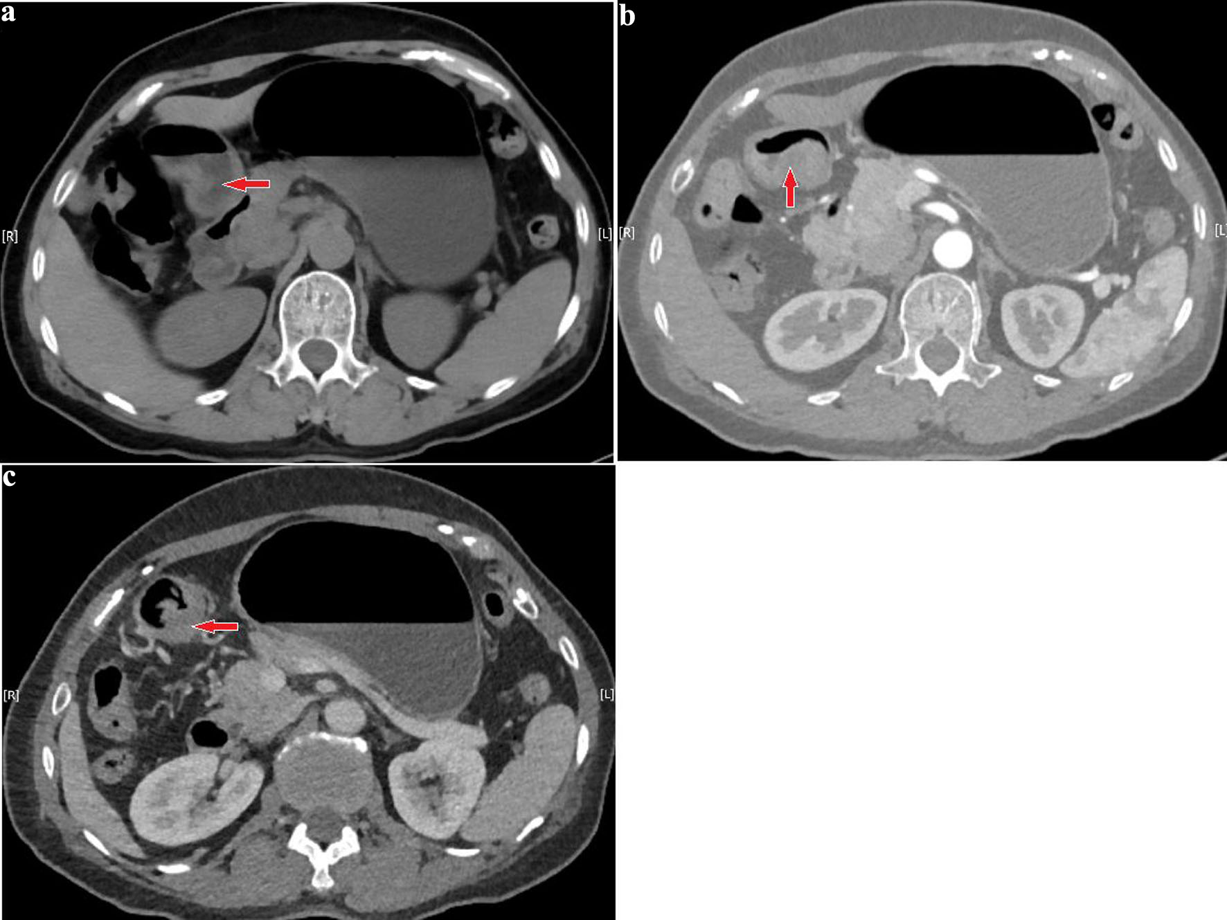
↓ Figure 1. (a) Preoperative computed tomography (CT) shows localized thickening along the greater curvature of the gastric antrum with protruding into the gastric lumen; the maximum thickness is approximately 17 mm, and the size is approximately 12 × 34 mm. (b, c) Contrast-enhanced scans demonstrate marked heterogeneous enhancement. Red arrows indicate the location of the lesion. No enlarged retroperitoneal lymph nodes are observed.
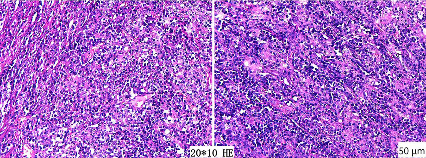
↓ Figure 2. Postoperative pathological findings show a small round cell malignant tumor at the gastric angle, consistent with ulcerative-type lymphoepithelioma-like carcinoma of Epstein–Barr virus-associated gastric cancer. All resection margins are negative, and no lymph node metastasis is observed (hematoxylin and eosin staining, original magnification × 200; scale bar = 50 µm).
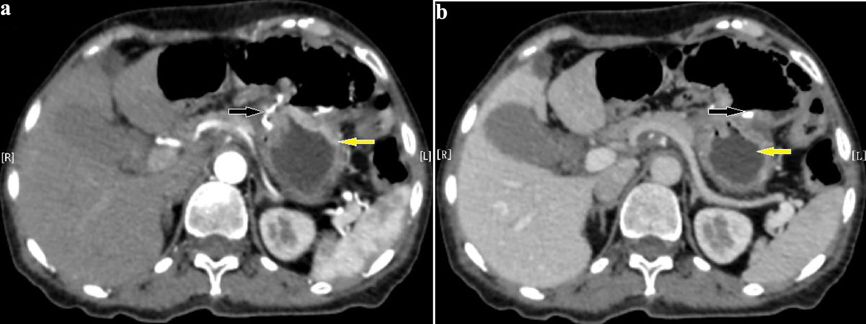
↓ Figure 3. (a, b) Pre-first-cycle chemotherapy computed tomography (CT) after surgery shows postoperative changes in the distal stomach. The nodular protrusion below the cardia is related to the surgical procedure. Yellow arrows indicate the remnant stomach, and black arrows indicate the gastrojejunal anastomosis. No obviously enlarged retroperitoneal lymph nodes are observed.