| Journal of Medical Cases, ISSN 1923-4155 print, 1923-4163 online, Open Access |
| Article copyright, the authors; Journal compilation copyright, J Med Cases and Elmer Press Inc |
| Journal website https://jmc.elmerpub.com |
Case Report
Volume 17, Number 9, September 2026, pages 464-469

The Hidden Link: Cryoglobulinemic Membranoproliferative Glomerulonephritis Secondary to Monoclonal Gammopathy of Renal Significance
Samar Mehyara, e, Amanda Gomesb, Harneet K. Ghummanc, Avneet K. Ghummanc, Noor Eweisa, Syedehmaryam Pishvaa, Jerry Kenmoed
aDepartment of Internal Medicine, McLaren Bay Region Hospital, Bay City, MI, USA
bDepartment of Nephrology, McLaren Bay Region Hospital, Bay City, MI, USA
cDepartment of Internal Medicine, McLaren Health Care/Michigan State University, Flint City, MI, USA
dHematology and Medical Oncology Department, McLaren Greater Lansing, Lansing City, MI, USA
eCorresponding Author: Samar Mehyar, Department of Internal Medicine, McLaren Bay Region Hospital, Bay City, MI, USA
Manuscript submitted March 2, 2026, accepted May 25, 2026, published online July 28, 2026
Short title: Cryoglobulinemic MPGN Secondary to MGRS
doi: https://doi.org/10.14740/jmc5318
| Abstract | ▴Top |
Membranoproliferative glomerulonephritis (MPGN) is a histopathological pattern that represents a spectrum of glomerular injury diseases. MPGN is usually associated with infections, autoimmune diseases and monoclonal gammopathies. Monoclonal gammopathy of renal significance (MGRS) is any proliferative B-cell or plasma-cell disease that causes abnormal buildup of monoclonal proteins in the kidneys. It is usually underdiagnosed due to its rarity. We present a case of a 74-year-old woman referred to nephrology with worsening creatinine levels, proteinuria and microscopic hematuria. Laboratory evaluation showed low C3 levels and elevated serum free light chain ratio. Kidney biopsy showed immune deposits consistent with cryoglobulinemic glomerulonephritis secondary to MGRS. Patient was treated with prednisone and mycophenolate mofetil, resulting in improvement in the creatinine functions, proteinuria, and hematuria.
Keywords: Membranoproliferative; Glomerulonephritis; MPGN; MGRS; MGUS
| Introduction | ▴Top |
Membranoproliferative glomerulonephritis (MPGN) is a glomerular injury that is characterized by endocapillary proliferation and mesangial deposits of immune complexes [1]. MPGN usually presents as nephritic syndrome. MPGN is subclassified as immune complex MPGN (IC-MPGN) and complement 3 glomerulopathy (C3G). IC-MPGN can occur secondary to infections, autoimmune diseases, paraproteinemia and malignancy [2]. Cryoglobulinemic MPGN is most associated with infections, specifically hepatitis B and C virus [3, 4].
MPGN is a rare glomerular disease and one of the less common causes of nephrotic syndrome. Its incidence has decreased over the decades, and it currently accounts for around two cases per million population per year. It has variable and unusual clinical presentations from being asymptomatic to hematuria. It can be primary (idiopathic), and secondary to various causes such as infections, autoimmune diseases, and tumors [5]. Cryoglobulinemia is a rare medical condition, it is characterized by immunoglobulins that deposit at lower temperatures in the small and medium-sized blood vessels causing endothelial injury and organ damage [6]. We present a case of a 74-year-old woman who was initially diagnosed with monoclonal gammopathy of unknown significance (MGUS) after initial serology and bone marrow biopsy. Her creatinine levels continued to spike up until a computed tomography (CT)-guided biopsy of the kidney confirmed a mixed cryoglobulinemic glomerulonephritis. This is a unique case, as the patient was known to have an immunoglobulin (Ig)G kappa paraprotein and was initially diagnosed with MGUS following bone marrow biopsy. MGUS is considered a premalignant condition that can evolve to multiple myeloma (MM) in some patients. Moreover, MGUS is common in patients with chronic kidney disease (CKD) and is more prevalent in older patients with CKD. The diagnosis of monoclonal gammopathy of renal significance (MGRS) by kidney biopsy is important for guiding treatment and improving renal prognosis.
| Case Report | ▴Top |
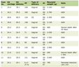
A74-year-old woman with past medical history of stable CKD stage III attributed to hypertension, nonischemic cardiomyopathy, and paroxysmal atrial fibrillation was referred to the nephrology because of worsening creatinine. She was initially found to have microscopic hematuria and proteinuria during a previous hospital stay for Clostridium difficile infection. In March 2021, during a workup for post herpetic neuralgia by neurology, the patient was found to have an IgG kappa paraprotein; however, she was not evaluated by hematology at that time. The patient was referred to nephrology in June 2022 with microscopic hematuria, proteinuria, and worsening creatinine levels (from 1 to 1.7 mg/dL). At that time, proteinuria was quantified at 0.4 mg/g, urine microscopy showed persistent microscopic hematuria with blood urine of 250 (Tables 1, 2). Serum protein electrophoresis (SPEP) showed elevated kappa free light chain 10.37 mg/L (normal range is 3.3–19.4 mg/L). Lambda free light chain was 1.75 mg/L (normal range is 5.7–26.3 mg/L), kappa/lambda free light chain ratio was 5.93 (normal range is 0.26–1.65). Serum immunoelectrophoresis showed IgM kappa level of 0.3 g/dL (normal range is 3.3–19.4 mg/L), and IgG Kappa level of 0.45 g/dL (normal range is 3.3–19.4 mg/L) (Table 3). Urine protein electrophoresis (UPEP) was negative. Results were remarkable for a low C3 level of 77 mg/dL. Further laboratory workup was negative for cryoglobulin, antinuclear antibodies (ANA), myeloperoxidase–proteinase 3 (MPO/PR3), anti–glomerular basement membrane (anti-GBM) antibodies, C4, hepatitis B and C, human immunodeficiency virus (HIV), anti-Ro, and anti-La levels (Table 4). Patient was referred to oncology for further evaluation and bone marrow biopsy.
 Click to view | Table 1. CBC, CMP and Iron Studies |
Click to view | Table 2. Urine Studies |
Click to view | Table 3. Protein Electrophoresis |
Click to view | Table 4. Autoimmune Workup |
Bone marrow biopsy showed a normocellular to slightly hypocellular marrow (10–20% cellular for age) with trilineage hematopoiesis. A single small lymphoid aggregate was noted. No large clusters of plasma cells were identified. CD138 highlighted approximately 1–2% plasma cells. Flow cytometry demonstrated reticulin fibrosis with occasional lymphoid aggregates in the bone marrow. Congo red staining was negative for amyloidosis. The bone scan was unremarkable. A second bone marrow aspirate and biopsy done a couple of months later showed a 10–20% cellular, small kappa restricted clonal B-cell population by flow, and CG 46,XX. MM fluorescence in situ hybridization (FISH) panel was normal. There was no increase in blasts (more than 1%), and the plasma cells count was 1%, with no definite increase in plasma cells or reticulin fibrosis.
The patient underwent a CT-guided left kidney biopsy, which showed a membranoproliferative pattern of glomerular injury with masked immune deposits overlying glomerulonephritis and kappa and lesser IgG and lambda light chains (Figs. 1–6), most consistent with mixed cryoglobulinemic glomerulonephritis. Initially, the patient was monitored conservatively; however, in the spring of 2024, the patient’s urine protein-to-creatinine ratio (UPC) increased to 1.4 g, prompting initiation of intervention. The patient was started on oral prednisone 40 mg daily with tapering, mycophenolate mofetil (MMF) and prophylaxis for Pneumocystis jirovecii pneumonia (PJP), resulting in improvement in proteinuria and serum creatinine. Patient has successfully remained on MMF with a stable serum creatinine level of 1.4 mg/dL. The UPC has decreased to 0.2 mg/g, with resolution of microscopic hematuria.
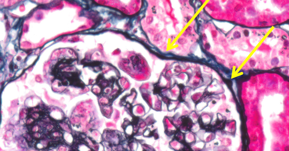 Click for large image | Figure 1. Intraluminal cryoglobulin deposits (yellow arrows). |
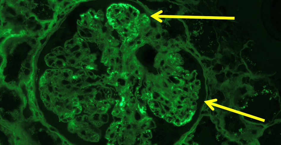 Click for large image | Figure 2. IgG immunofluorescence (yellow arrows) indicating cryoglobulinemic glomerulonephritis. Ig: immunoglobulin. |
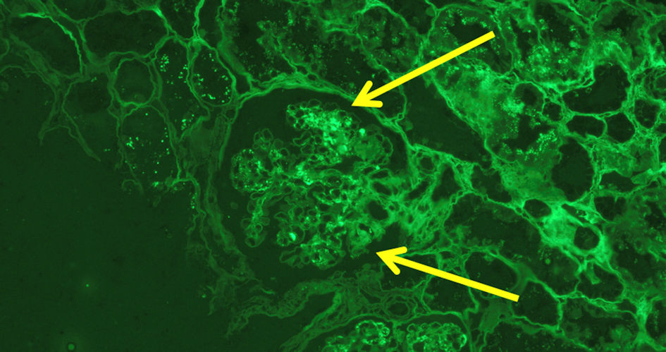 Click for large image | Figure 3. Immunofluorescence staining showing IgM deposition (yellow arrows). Ig: immunoglobulin. |
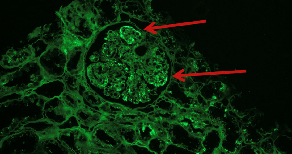 Click for large image | Figure 4. Immunofluorescence stain showing kappa light chain deposits (red arrows). |
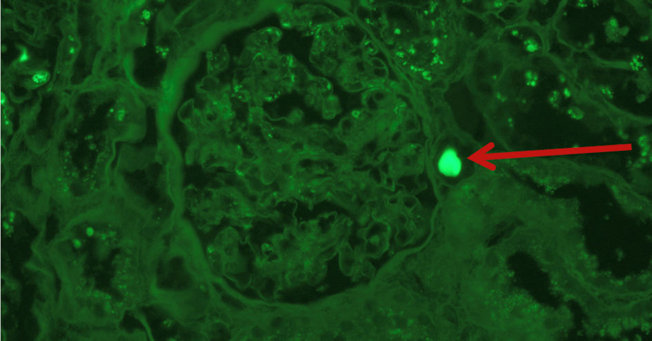 Click for large image | Figure 5. Immunofluorescence stain kappa deposits (red arrow). |
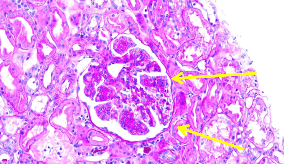 Click for large image | Figure 6. MPGN pattern on periodic acid Schiff (yellow arrows). |
| Discussion | ▴Top |
This case involves a woman presented with worsening proteinuria, microscopic hematuria, and increasing creatinine levels. Laboratory evaluation showed a low C3 level and negative results for hepatitis B and C, autoimmune and cryoglobulin levels. SPEP revealed increase in the kappa, lambda, and kappa/lambda ratio. Bone marrow biopsy findings were suggestive of MGUS. A repeated kidney biopsy was performed to confirm the diagnosis of MGRS and guide appropriate treatment. Patient ultimately required immunosuppression for MPGN with prednisone followed by a taper and MMF, with subsequent stabilization of the renal function and proteinuria and resolution of microscopic hematuria.
MPGN is a rare condition characterized by mesangial hypercellularity and subendothelial deposition of the complements or immune complexes causing glomerular wall thickening [5]. It accounts for about 7–10% of all glomerulonephritis cases [5]. MPGN in older patients is commonly associated with cryoglobulinemia and hepatitis infections [7].
Recent classification of MPGN is based on immunofluorescence examination findings. It can be subdivided into immune complex-mediated subtype, complement-mediated subtype, and a subtype without immune complexes or complements. Immune complex-mediated subtype is associated with chronic antigenemia with or without the presence of complement levels. It is mainly caused by infections, most commonly hepatitis B and C, autoimmune diseases such as systemic lupus erythematosus (SLE) and Sjogren’s, monoclonal gammopathy, or idiopathic [8].
According to the 2021–2022 International Kidney and Monoclonal Gammopathy Research Group (IKMG) consensus, MGRS is defined as any proliferative B-cell or plasma-cell disorder that produces nephrotoxic monoclonal immunoglobulins [9]. MGRS can be caused by a variety of kidney diseases including MGUS, amyloidosis and proliferative glomerulonephritis with monoclonal immunoglobulin deposits (PGNMID). A renal biopsy should always be done whenever MGRS is suspected [10].
MGUS has a prevalence of approximately 3% in people over 50 years of age, 5% in those over 70, and 8% in those over 80 years of age. As CKD is more common in patients over 60 years of age, patients with MGUS can also coincidentally have CKD. In old patients with MGUS and renal manifestations such as elevated creatinine, hematuria or proteinuria, performing renal biopsy is crucial to avoid missing a diagnosis of MGRS kidney disorders, which is more likely to occur in old patients [10].
MGRS does not meet the criteria for overt MM but is associated with significant mortality, depending on the severity of the monoclonal immunoglobulin deposition in the kidneys and other systemic organs. Renal failure is a common presenting symptom, with or without microscopic hematuria [11]. Current treatment of MGRS associated with MPGN depends on the symptoms and the degree of B-cell proliferation. In patients who are asymptomatic or have mild symptoms and low-grade proliferation, observation is recommended. However, patients with progressive symptoms may require further treatment [11].
MPGN is usually caused by infections such hepatitis C virus, autoimmune diseases, and less commonly, MGRS. It is common in older patients with CKD [11, 12]. The prevalence of MPGN in patients with MGUS is underrecognized and understudied due to rarity of the disease, and more research is needed to better understand the association between both. Kidney biopsy is crucial whenever MGRS is suspected, it should be undertaken in patients with a paraprotein, elevated creatinine levels, hematuria or proteinuria. This is important to assess severity and determine treatment, especially in older patients with negative serological workup for infectious and autoimmune diseases. This case shows the importance of immunosuppressive therapy over clinical observation in symptomatic patients for the management of the disease.
Conclusions
MPGN is a pattern of glomerular injury that is characterized by subendothelial and mesangial deposition of immune complexes. It is usually caused by infections such as hepatitis C virus infection, autoimmune disease, and less commonly from MGRS. Kidney biopsy is important to confirm the diagnosis, assess its severity, and determine the type of treatment. Further studies are still needed to acknowledge the prevalence of MPGN in patients with MGUS to start the appropriate treatment in a timely manner.
Acknowledgments
The authors would like to thank the multidisciplinary care team involved in the patient’s management, including nephrology, hematology, oncology, and primary care.
Financial Disclosure
This research received no specific grant from any funding agency in the public, commercial, or not-for-profit sectors.
Conflict of Interest
The authors declare that they have no conflict of interest related to this case report.
Informed Consent
Written informed consent was obtained from the patient for publication of this case report and any accompanying images.
Author Contributions
Samar Mehyar, MD: primary author, manuscript drafting, and clinical documentation. Amanda Gomes, MD: case conceptualization, manuscript revision, editing, and clinical input and supervision. Harneet K. Ghumman, MD: manuscript revision and editing. Avneet K. Ghumman, MD: manuscript revision and editing. Syedehmaryam Pishva, MD: manuscript revision and clinical oversight. Noor Eweis, MD: manuscript revision and clinical oversight. Jerry Kenmoe: supervision, final review, and clinical input. All authors read and approved the final version of the manuscript.
Data Availability
All data supporting the findings of this case report are included within the manuscript. Additional information is available from the corresponding author upon reasonable request.
| References | ▴Top |
- Fogo AB, Lusco MA, Najafian B, Alpers CE. AJKD atlas of renal pathology: membranoproliferative glomerulonephritis. Am J Kidney Dis. 2015;66(3):e19-20.
doi pubmed - Bomback AS, Charu V, Fakhouri F. Challenges in the diagnosis and management of immune complex-mediated membranoproliferative glomerulonephritis and complement 3 glomerulopathy. Kidney Int Rep. 2025;10(1):17-28.
doi pubmed - Zhang Y, Meng X, Wang J, Hou Q, Xu Y, Sun J, Wu S, et al. A case of renal damage induced by cryoglobulinemia associated with Monoclonal Gammopathy of Renal Significance. BMC Nephrol. 2025;26(1):410.
doi pubmed - Bhandari J, Awais M, Aeddula NR. Cryoglobulinemia [Internet]. PubMed. Treasure Island (FL): StatPearls Publishing; 2020. Available from: https://www.ncbi.nlm.nih.gov/books/NBK557606/.
- Nakagawa N, Kimura T, Sakate R, Isaka Y, Narita I. Demographics and treatment of patients with primary membranoproliferative glomerulonephritis in Japan using a national registry of clinical personal records. Clin Exp Nephrol. 2023;27(11):928-935.
doi pubmed - Desbois AC, Cacoub P, Saadoun D. Cryoglobulinemia: an update in 2019. Joint Bone Spine. 2019;86(6):707-713.
doi pubmed - Bernardes TP, Mastroianni-Kirsztajn G. Membranoproliferative glomerulonephritis: current histopathological classification, clinical profile, and kidney outcomes. J Bras Nefrol. 2023;45(1):45-50.
doi pubmed - KDIGO 2021 clinical practice guideline for the management of glomerular diseases [Internet]. 2021. Available from: https://kdigo.org/wp-content/uploads/2017/02/KDIGO-Glomerular-Diseases-Guideline-2021-English.pdf.
- Fermand JP, Bridoux F, Kyle RA, Kastritis E, Weiss BM, Cook MA, Drayson MT, et al. How I treat monoclonal gammopathy of renal significance (MGRS). Blood. 2013;122(22):3583-3590.
doi pubmed - Bridoux F, Leung N, Hutchison CA, Touchard G, Sethi S, Fermand JP, Picken MM, et al. Diagnosis of monoclonal gammopathy of renal significance. Kidney Int. 2015;87(4):698-711.
doi pubmed - Chowdhury R, Gupta S. Monoclonal gammopathies and CKD progression: an unfinished story. Clin J Am Soc Nephrol. 2024;19(3):280-282.
doi pubmed - Wang CH, Hogan JJ. Monoclonal gammopathies and kidney disease: searching for significance. Clin J Am Soc Nephrol. 2018;13(12):1781-1782.
doi pubmed
This article is distributed under the terms of the Creative Commons Attribution 4.0 International License (CC BY 4.0), which permits unrestricted use, distribution, and reproduction in any medium, including commercial use, provided the original work is properly cited.
Journal of Medical Cases is published by Elmer Press Inc.