| Journal of Medical Cases, ISSN 1923-4155 print, 1923-4163 online, Open Access |
| Article copyright, the authors; Journal compilation copyright, J Med Cases and Elmer Press Inc |
| Journal website https://jmc.elmerpub.com |
Case Report
Volume 17, Number 9, September 2026, pages 453-463

When Virchow’s Triad Goes on Overdrive: Catastrophic Multi-System Thrombosis in the Setting of New-Onset Heart Failure in a Previously Healthy Man
Bhanu C. Ramachandrana, Yolande Mbomeb, Tanuj Guptab, Karan Bhanushalib, John Sabub, c, d
aDepartment of Internal Medicine, SUNY Downstate Health Sciences University, Brooklyn, NY, USA
bDepartment of Cardiology, SUNY Downstate Health Sciences University, Brooklyn, NY, USA
cKings County Hospital, Brooklyn, NY, USA
dCorresponding Author: John Sabu, Department of Cardiology, SUNY Downstate Health Sciences University, Brooklyn, NY, USA
Manuscript submitted April 28, 2026, accepted July 8, 2026, published online July 28, 2026
Short title: Catastrophic Multi-System Thrombosis
doi: https://doi.org/10.14740/jmc5312
| Abstract | ▴Top |
Catastrophic multi-territory thromboembolism is an uncommon but life-threatening presentation that often signals an underlying hypercoagulable state or severe cardiac dysfunction. When all three components of Virchow’s triad converge simultaneously in a previously healthy individual, the clinical consequences can be devastating and the diagnostic workup both urgent and complex. We report a 58-year-old man with no prior medical history who presented with progressive dyspnea and was found to have bilateral pulmonary emboli, large mobile thrombi in both the left atrium and left ventricle, and extensive systemic arterial thrombosis culminating in left lower extremity ischemia. Imaging revealed an underlying heart failure with reduced ejection fraction with an estimated ejection fraction of 20% and concurrent atrial flutter. Comprehensive hypercoagulable workup including antiphospholipid antibody syndrome evaluation was negative. The patient required emergent left lower extremity embolectomy at the initial hospital, transfer to a tertiary center for repeat femoral-femoral bypass that subsequently thrombosed, and ultimately a left below-knee amputation. He was discharged on apixaban. Subsequent atrial fibrillation/flutter ablation achieved durable sinus rhythm. Genetic testing revealed a pathogenic TTN (titin) variant, providing a unifying etiology for his dilated cardiomyopathy. Serial echocardiography at 3 months demonstrated resolution of all intracardiac thrombi; repeat imaging at 6 months showed substantial left ventricular ejection fraction recovery to 42%. This case illustrates that new-onset heart failure with reduced ejection fraction with concurrent atrial flutter can serve as a sufficient and independent driver of catastrophic multi-territory thromboembolism without additional systemic hypercoagulable disease. It also demonstrates that with aggressive guideline-directed medical therapy, rhythm control, and sustained anticoagulation, meaningful ventricular recovery is achievable even after severe initial dysfunction. TTN-related cardiomyopathy should be considered in otherwise unexplained dilated cardiomyopathy, and genetic counseling should be offered to first-degree relatives.
Keywords: Thrombosis; Heart failure; Virchow’s triad; Myocardial stunning; TTN mutation; Atrial flutter ablation; Below-knee amputation; Cardiomyopathy
| Introduction | ▴Top |
Cardiac thrombi, particularly left ventricular (LV) thrombus, are typically associated with ischemic cardiomyopathy or severe LV systolic dysfunction, with a reported incidence of 2% to 36% in dilated cardiomyopathy (DCM) and up to 39% following anterior ST-segment elevation myocardial infarction [1, 2]. As described by Virchow’s triad, the pathogenesis of intracardiac thrombus formation involves the interplay of blood stasis from reduced ventricular function, endocardial injury, and a systemic hypercoagulable state [1, 3]. In heart failure (HF), low cardiac output and elevated right-sided filling pressures, compounded by decreased functional mobility, produce sluggish blood flow throughout the cardiovascular system, while cardiac dilatation and reduced myocardial contractility in both atria and ventricles further predispose to thrombus formation [3, 4]. Increases in thrombin generation, impaired fibrinolysis from neurohormonal activation, and endothelial dysfunction with diminished nitric oxide availability complete this prothrombotic milieu [3, 5]. Accordingly, the incidence of venous thromboembolism (VTE) in patients with acute decompensated HF not receiving thromboprophylaxis ranges from 4% to 26%, and LV thrombus, particularly when mobile or protuberant, carries up to a 22% risk of systemic embolization and a 37% risk of major adverse cardiovascular events [1, 6].
However, the simultaneous occurrence of large mobile thrombi in both the left ventricle and left atrium, bilateral pulmonary emboli, and extensive systemic arterial thrombosis as the initial presentation of previously unrecognized heart failure is exceedingly rare outside of advanced malignancy, antiphospholipid syndrome (APS), or catastrophic systemic inflammatory conditions [7, 8]. Patients at highest risk for embolization include those with atrial fibrillation or flutter, severe congestive heart failure, markedly dilated ventricles with severe systolic dysfunction, and mobile intracardiac thrombi [1, 9]. In a large national analysis of over 175,000 admissions with intracardiac thrombus, concurrent acute VTE and arterial thrombosis were among the strongest independent predictors of in-hospital mortality [10]. New-onset HF confers a twofold increased risk of VTE, with the highest risk concentrated in the first 30 days after diagnosis [11].
TTN-related DCM, caused by pathogenic variants in the gene encoding titin, accounts for approximately 18% to 25% of familial DCM and up to 27% of DCM cases identified in larger unselected cohorts, making it the single most common genetic cause of DCM [12]. TTN-related cardiomyopathy frequently presents with arrhythmia-mediated dysfunction, and its identification has important implications for surveillance of first-degree relatives and for prognostication [12, 13].
We present a case of catastrophic multi-territory thromboembolism as the sentinel presentation of TTN-related DCM with concurrent atrial flutter in a 58-year-old man with no prior medical history, detailing the complex acute management, prolonged post-acute course, and the remarkable degree of cardiac recovery achieved with sustained guideline-directed therapy, rhythm control, and anticoagulation.
| Case Report | ▴Top |
Medical history and initial presentation
A 58-year-old man with no known past medical history, no medications, and no family history of cardiac disease or thrombophilia presented to the emergency department with a 1-week history of progressive dyspnea, initially on exertion but progressing to rest. He reported fatigue, a non-productive cough, and left foot pain that began after dropping a heavy metal object on his left foot approximately 1 week prior. He denied chest pain, fever, or orthopnea, but noted awareness of a rapid heartbeat over the past week. The palpitations were intermittent with no diurnal variation or changes with activity. There was no history of recent immobilization, surgery, travel, or prior thromboembolic events.
Clinical findings
On arrival, the patient was tachypneic but maintained adequate oxygen saturation on room air. Vital signs revealed a heart rate in the 130s with an irregular rhythm (see full vitals in Table 1). Cardiopulmonary examination was notable for mild bibasilar crackles. Peripheral examination demonstrated mild tenderness over the left foot without significant edema or skin changes.
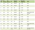 Click to view | Table 1. Initial Vitals |
Initial laboratory testing demonstrated an elevated high-sensitivity troponin (72 ng/L; repeat 64 ng/L), markedly elevated NT-proBNP (8,861 pg/mL), and elevated D-dimer (2,222 ng/mL). All labs obtained on admission are listed in Table 2. Chest radiography showed cardiomegaly with a right basilar opacity (Supplementary Material 1, jmc.elmerpub.com). The electrocardiogram revealed atrial flutter with 2:1 conduction (Fig. 1).
Click to view | Table 2. Admission Laboratory Values |
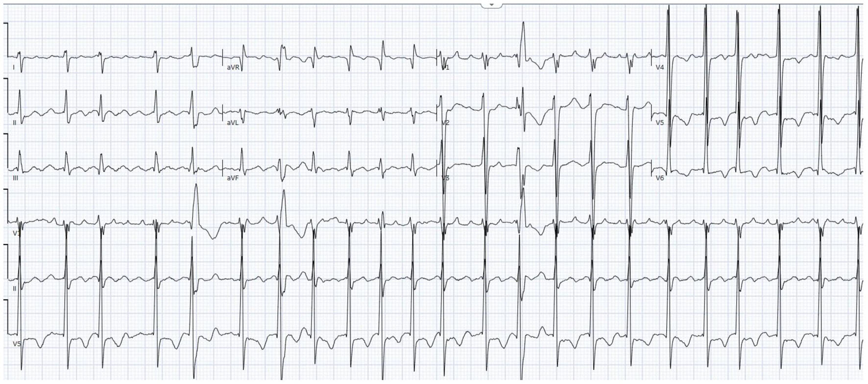 Click for large image | Figure 1. Twelve-lead electrocardiogram demonstrating 2:1 atrial flutter with classic sawtooth flutter waves, best visualized in the inferior leads (II, III, aVF), with a ventricular rate in the 130s. |
Diagnostic imaging
Computed tomography (CT) pulmonary angiography demonstrated filling defects in the proximal segmental and subsegmental pulmonary arteries of the right lower lobe and distal segmental and subsegmental arteries of the left lower lobe, consistent with bilateral pulmonary emboli (Fig. 2). Pleural-based wedge-shaped consolidations with internal air lucencies in the lateral right lower lobe were consistent with pulmonary infarction, and a moderate right-sided pleural effusion was present. There was no CT evidence of right heart strain. Lower extremity venous Doppler was negative for deep vein thrombosis.
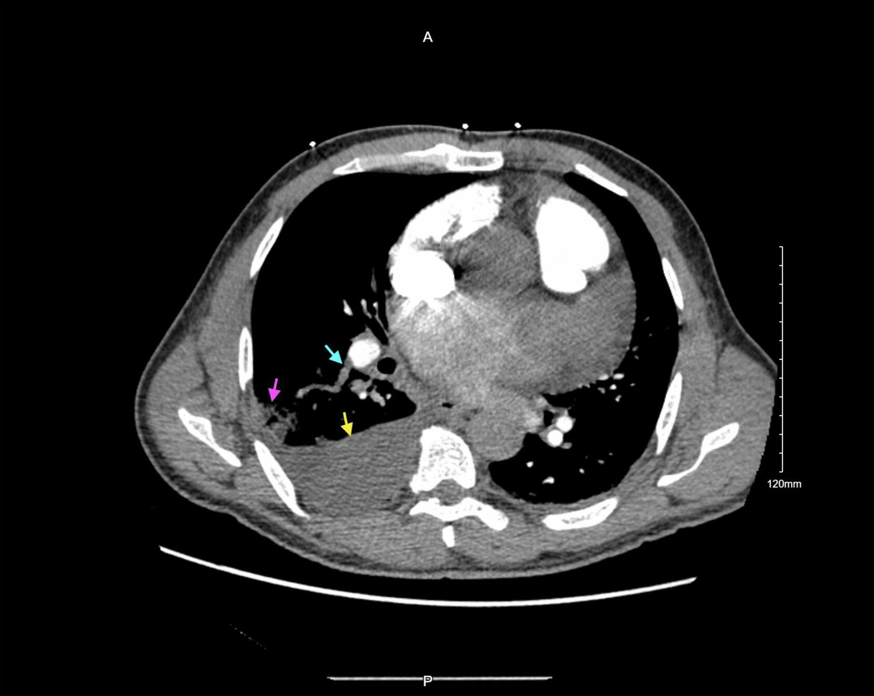 Click for large image | Figure 2. CT pulmonary angiography with contrast. Blue arrow: filling defect in the right lower lobe proximal segmental pulmonary artery, consistent with pulmonary embolism. Pink arrow: pleural-based wedge-shaped consolidation with internal air lucencies in the lateral right lower lobe, consistent with pulmonary infarction. Yellow arrow: right-sided pleural effusion. CT: computed tomography. |
Transthoracic echocardiography (TTE) demonstrated severely reduced LV systolic function with an estimated ejection fraction (EF) of 20%, a large mobile, heterogeneously echogenic LV apical thrombus (1.77 × 1.81 cm) with distinct borders separate from the underlying myocardium (Fig. 3), and a large mobile left atrial thrombus near the left atrial appendage (3.1 × 2.1 cm) (Fig. 4). The left and right atria were severely dilated with moderate right ventricular dilation. Pulmonary artery systolic pressure was elevated at 49 mm Hg, consistent with moderate pulmonary hypertension. Additional findings included a moderately dilated inferior vena cava and a small-to-moderate pericardial effusion. No patent foramen ovale or atrial septal defect was identified (see details of the TTE at presentation are in Table 3).
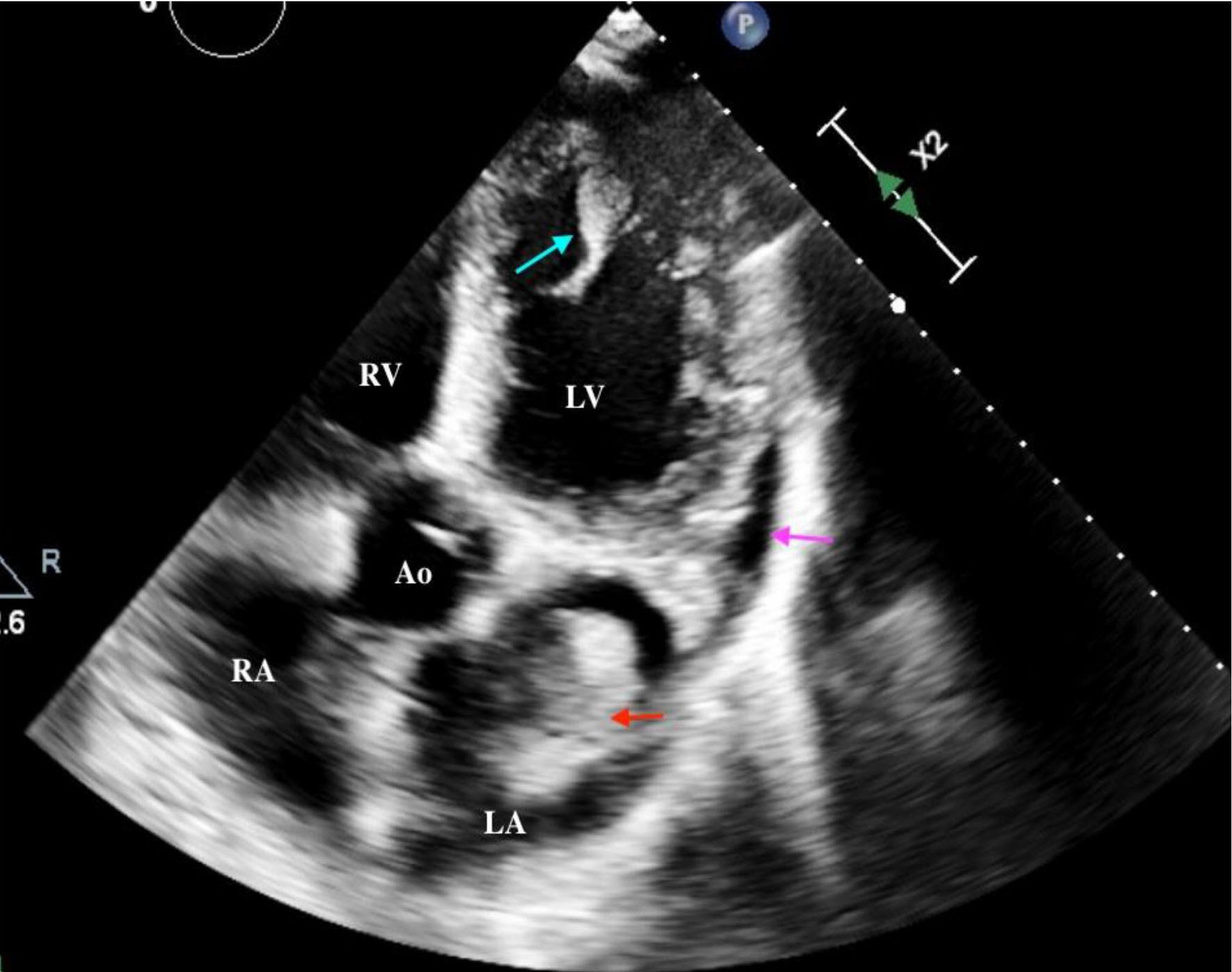 Click for large image | Figure 3. Apical five-chamber off-axis view demonstrating left atrial and left ventricular thrombus. Blue arrow: left ventricular apical thrombus (1.77 × 1.81 cm) with distinct borders; Red arrow: left atrial thrombus; Pink arrow: small-to-moderate pericardial effusion. Ao: aorta; LA: left atrium; LV: left ventricle; RA: right atrium; RV: right ventricle. |
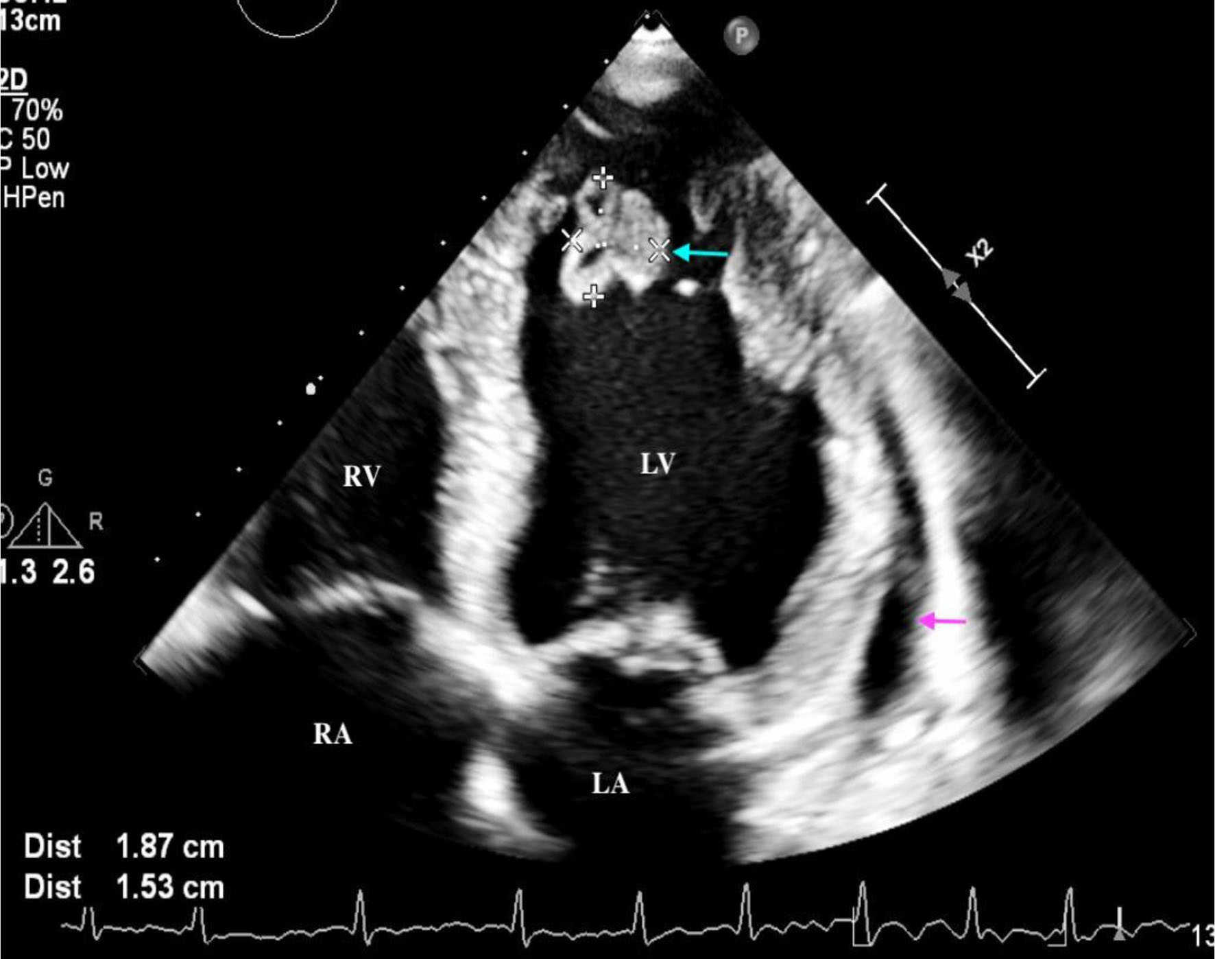 Click for large image | Figure 4. Apical four-chamber view on transthoracic echocardiography. Blue arrow: large mobile left atrial thrombus (3.1 × 2.1 cm) near the left atrial appendage, appearing as a mobile, heterogeneously echogenic mass. LA: left atrium; LV: left ventricle; RA: right atrium; RV: right ventricle. |
Click to view | Table 3. Transthoracic Echocardiographic Findings |
CT angiography of the abdomen and pelvis, performed given the high clinical suspicion for systemic arterial embolization, suspicion for malignancy in the context of large mobile intracardiac thrombi and the patient’s left foot pain, revealed extensive arterial thrombosis involving the left common iliac artery (Fig. 5), left internal iliac artery and its branches, and the left external iliac artery extending to the common femoral artery.
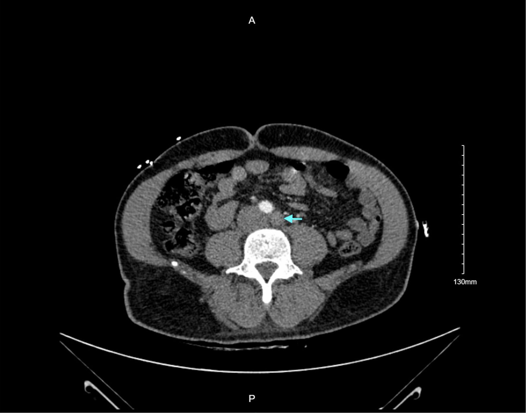 Click for large image | Figure 5. CT angiography of the abdomen and pelvis. Blue arrow: filling defect within the left common iliac artery representing acute arterial thrombus, part of extensive arterial thrombosis extending through the left iliac system to the common femoral artery. CT: computed tomography. |
Management and hospital course
Initial surgical intervention
The patient was taken emergently to the operating room for left lower limb embolectomy for acute limb ischemia, with arteriogram and four-compartment fasciotomy. He was subsequently admitted to the cardiac critical care unit. Rate control was initiated with metoprolol, and intravenous amiodarone was started for rhythm management. Full-dose anticoagulation with unfractionated heparin was maintained from a surgical standpoint. Repeat echocardiography 2 days post-embolectomy demonstrated unchanged findings.
Hypercoagulable and hematologic workup
Hematology was consulted for comprehensive evaluation. Anticardiolipin antibodies (IgG and IgM), anti-beta-2-glycoprotein I antibodies, and lupus anticoagulant were all negative. Additional testing demonstrated normal ADAMTS13 activity, negative CALR and MPL exon mutations, normal antithrombin III, and negative JAK2 V617F mutation (Table 4). Inflammatory workup including procalcitonin was normal; blood and sputum cultures showed no growth. A urinary tract infection was identified and treated with ceftriaxone. Workup for connective tissue disease, nephrotic syndrome, and non-bacterial thrombotic endocarditis was unrevealing (Table 4).
Click to view | Table 4. Differential Diagnosis of Unprovoked Multi-Territory Thromboembolism, Rationale for Consideration, and Result of Evaluation in This Patient |
Repeat imaging confirmed acute-on-chronic left lower extremity limb ischemia. The patient underwent femoral-femoral bypass, left below-knee popliteal artery exposure with popliteal and tibial Fogarty thrombectomy, left posterior tibial thrombectomy, angiography, and left lower extremity four-compartment fasciotomy. Doppler signal was restored to the left popliteal artery but remained absent distally. Despite maximal surgical revascularization, irreversible distal ischemia necessitated a left below-knee amputation (BKA), performed approximately 4 weeks after initial presentation. The perioperative course required vasopressor support. Postoperatively, the patient was maintained on a heparin infusion, transitioned to oral amiodarone for atrial fibrillation/flutter management, and continued on guideline-directed medical therapy (GDMT) for heart failure with reduced ejection fraction (HFrEF) including sacubitril/valsartan, empagliflozin, and spironolactone. The patient was discharged on apixaban (5 mg twice daily), the selected long-term anticoagulant given the combination of residual intracardiac thrombus, atrial flutter, and prior systemic embolism, with plan for indefinite anticoagulation pending cardiac recovery and thrombus resolution.
Following discharge, the patient subsequently underwent catheter ablation for recurrent atrial fibrillation and flutter. Ablation achieved durable restoration of sinus rhythm with complete resolution of atrial fibrillation and flutter on subsequent monitoring.
Genetic cardiomyopathy panel testing identified a pathogenic TTN (titin gene) variant (Table 4), providing a unifying molecular diagnosis for his DCM. Given the autosomal dominant inheritance pattern of TTN-related DCM with reduced penetrance, genetic counseling and cascade screening were recommended for first-degree relatives.
TTE at 3 months demonstrated complete resolution of both the LV and left atrial thrombi, with the patient remaining in sinus rhythm on continued apixaban and GDMT. Echocardiography at 6 months showed substantial recovery of LVEF to 42%, along with normalization of left atrial and ventricular dimensions compared with the initial presentation, consistent with reverse cardiac remodeling in the setting of sustained rhythm control, GDMT, and relief of the tachycardia-mediated cardiomyopathy component.
| Discussion | ▴Top |
This case illustrates several intersecting themes of contemporary cardiology and nephrology: the convergence of Virchow’s triad in the setting of unrecognized HFrEF, the catastrophic clinical consequences of delayed cardiac diagnosis, the therapeutic challenges of multi-territory thromboembolism requiring iterative surgical and pharmacologic management, the emerging role of genetic testing in DCM, and the meaningful cardiac recovery achievable when the underlying substrate is systematically addressed.
The primacy of hemodynamic dysfunction in driving multi-territory thrombosis
The simultaneous occurrence of LV and left atrial thrombi, bilateral pulmonary emboli, and extensive systemic arterial thrombosis as the inaugural presentation of an unrecognized cardiomyopathy is exceedingly rare. Outside of APS, advanced malignancy, and catastrophic systemic inflammatory states, most reported cases of multi-territory thromboembolism have an identifiable hypercoagulable driver [7, 8]. In the present case, however, comprehensive hematologic evaluation, including antiphospholipid antibody testing, myeloproliferative neoplasm markers (JAK2, CALR, MPL), ADAMTS13 activity, and antithrombin III, was entirely negative. The profound hemodynamic derangement from newly diagnosed severe systolic HF with an EF of 20% in the setting of concurrent atrial flutter was, ultimately, the most plausible and parsimonious unifying explanation.
As described by Virchow’s triad, the pathogenesis of intracardiac thrombus formation results from the interplay of blood stasis, endocardial injury, and systemic hypercoagulability [3]. In HF, each of these components is simultaneously activated: low cardiac output and elevated filling pressures produce stagnant blood flow; cardiac dilatation stretches and functionally injures the endocardium; and neurohormonal activation, including elevated thrombin generation and impaired fibrinolysis via increased plasminogen activator inhibitor-1, generates a systemic prothrombotic milieu [3, 5]. The addition of atrial flutter, which creates the atrial mechanical standstill and blood stasis conducive to left atrial and appendage thrombus formation, amplified each component of this triad, particularly the stasis element, to a degree sufficient to produce simultaneous thrombus formation across multiple vascular territories.
The importance of APS exclusion and its caveats
APS is arguably the single most important diagnosis to exclude in any patient presenting with unprovoked multi-territory thromboembolism [14, 15]. Exclusion was pursued rigorously in this patient, with lupus anticoagulant, anticardiolipin antibodies, and anti-beta-2-glycoprotein I antibodies all returning negative. However, two critical caveats merit emphasis. First, APS diagnosis requires persistent positivity on two occasions at least 12 weeks apart, and single-point negative testing in the acute setting does not definitively exclude the condition. Second, heparin therapy can produce false-positive lupus anticoagulant results [14, 16]. Repeat testing off anticoagulation at 12-week interval should be considered if clinical suspicion remains elevated, particularly if thrombosis recurs despite adequate anticoagulation. In this patient, the sustained absence of recurrent thromboembolism on long-term apixaban, combined with the clear cardiac etiology, argues against a concurrent APS phenotype.
The TTN mutation: a unifying molecular diagnosis with clinical implications
The identification of a pathogenic TTN variant by genetic cardiomyopathy panel testing provides the most likely molecular etiology for this patient’s DCM. Titin is the largest protein in the human body, functioning as a molecular spring within the sarcomere that governs passive myocardial stiffness and mechanosensation. Truncating variants in TTN (TTNtv) are the single most common identified genetic cause of DCM, identified in approximately 18% to 25% of familial DCM cases and 5% to 6% of sporadic DCM [12, 13]. TTN-related DCM frequently co-segregates with atrial fibrillation and flutter, a feature of the clinical phenotype that is mechanistically distinct from secondary arrhythmia: TTN variants impair cardiomyocyte mechanical homeostasis and may promote both atrial and ventricular remodeling through sarcomeric dysfunction, explaining why arrhythmia management is particularly important in this genetic subgroup [12].
From a clinical management standpoint, the TTN diagnosis has immediate implications for family members. TTN-related DCM follows autosomal dominant inheritance with variable and often age-dependent penetrance; unaffected first-degree relatives harbor the pathogenic variant at a 50% prior probability and may develop DCM over time, particularly in response to cardiac stressors such as pregnancy, viral infection, or tachyarrhythmia. Cascade genetic screening of first-degree relatives, followed by serial echocardiographic surveillance of variant carriers, is now standard practice per international DCM guidelines and the 2022 American Heart Association/American College of Cardiology (AHA/ACC) guideline for the diagnosis and treatment of heart failure [17, 18].
The surgical course of this patient exemplifies the iterative and often refractory nature of embolic arterial occlusion arising from intracardiac sources when the underlying cardiac disease and atrial arrhythmia remain untreated. Initial embolectomy achieved partial limb salvage, but graft thrombosis reflected ongoing embolization from persistent intracardiac thrombi in the setting of uncontrolled atrial flutter and ongoing HFrEF. The decision to proceed with femoral-femoral bypass recognized the extent of proximal arterial occlusion, yet graft failure in the context of ongoing thromboembolism necessitated BKA as the definitive procedure. This sequence underscores a fundamental principle: surgical revascularization for cardioembolism cannot succeed in durable fashion until the cardiac source is effectively treated. Anticoagulation, cardiac rhythm control, and optimization of ventricular function are not ancillary interventions in this setting but co-equal components of the treatment strategy alongside surgical arterial management.
Anticoagulation: strategy and duration
The AHA scientific statement on LV thrombus management recommends anticoagulation with a vitamin K antagonist (VKA) or direct oral anticoagulant (DOAC) for at least 3 to 6 months in patients with LV thrombus in the setting of DCM, with consideration of indefinite therapy in those whose LV systolic function does not improve beyond an EF of 35% or who have persistent apical akinesis [1]. In the present case, the choice of apixaban at discharge was appropriate given the multiplicity of indications: persistent intracardiac thrombus at discharge, atrial flutter (which carries an embolic risk comparable to atrial fibrillation and warrants anticoagulation per the same risk-stratification principles), and prior systemic embolism [19].
The subsequent resolution of intracardiac thrombi at 3-month follow-up echocardiography and EF recovery to 42% at 6 months raises the important clinical question of optimal anticoagulation duration. Per AHA guidance, discontinuation of anticoagulation may be considered when LVEF improves beyond 35% and thrombus has resolved, though this must be balanced against the patient’s prior catastrophic embolic history and the atrial flutter/fibrillation burden [1]. Following successful ablation with durable sinus rhythm restoration, ongoing shared decision-making regarding anticoagulation duration, incorporating the patient’s bleeding risk, functional recovery trajectory, and preference, is warranted. Notably, there is no evidence that surgery for persistent LV thrombus offers net benefit over medical therapy, reinforcing that anticoagulation and GDMT are the primary therapeutic tools [1].
The role of atrial flutter/fibrillation ablation in cardiac recovery
The contribution of uncontrolled atrial flutter to this patient’s severe initial ventricular dysfunction may have been substantial. Tachycardia-mediated cardiomyopathy, a reversible form of HF resulting from sustained rapid ventricular rates impairing myocardial perfusion and energetics, can occur with chronic atrial flutter and may account for a significant proportion of the observed ventricular dysfunction, particularly in patients without ischemic coronary disease [20]. The substantial LVEF improvement from 20% at presentation to 42% at 6 months, in a patient who underwent successful ablation with durable sinus rhythm restoration and who received optimized GDMT, is consistent with at least a partial tachycardia-mediated component to the initial cardiomyopathy, superimposed on the TTN-related structural substrate.
Catheter ablation for atrial flutter achieves durable sinus rhythm in 80% to 90% of patients and is now considered the preferred rhythm control strategy for typical isthmus-dependent atrial flutter over antiarrhythmic drug therapy given superior long-term efficacy and safety [21]. In patients with concurrent atrial fibrillation, as was present in this case, pulmonary vein isolation in addition to cavotricuspid isthmus ablation is generally recommended. The achievement of durable sinus rhythm in this patient was likely a critical contributor to ventricular recovery, alongside GDMT, by eliminating the rate-related cardiomyopathy component and reducing the ongoing thrombogenic atrial milieu.
Cardiac recovery: distinguishing TTN-related cardiomyopathy from reversible components
The improvement of LVEF from 20% to 42% at 6 months with resolution of all intracardiac thrombi represents a clinically significant and multifactorially determined recovery. Three mechanisms likely contributed in a non-additive fashion: (1) relief of tachycardia-mediated cardiomyopathy through successful ablation and rate control; (2) reverse remodeling from optimized neurohormonal blockade with the combination of sacubitril/valsartan (which has the strongest evidence for LV reverse remodeling among HF medications), an sodium–glucose cotransporter 2 inhibitor, and a mineralocorticoid receptor antagonist; and (3) alleviation of inflammatory and hemodynamic stressors from treated pulmonary embolism and restored forward cardiac output [18, 22]. Whether the residual ventricular dysfunction (EF 42%, below the normal threshold of 50% to 55%) represents the irreversible TTN-related structural substrate or an incompletely recovered reversible component remains to be determined by continued echocardiographic follow-up. Patients with TTN-related DCM who recover LVEF should remain on GDMT indefinitely given the propensity for relapse, particularly during physiological stressors, and should undergo continued genetic counseling and monitoring [12, 13].
In patients with acute pulmonary embolism without identifiable major reversible risk factors, occult cancer is diagnosed within the first year in 4% to 10% [23, 24]. Although this patient’s multi-territory thromboembolism ultimately had a clear hemodynamic and genetic explanation, age-appropriate cancer screening remains warranted as standard of care. Extended workup with positron emission tomography (PET)/CT for occult malignancy has not demonstrated a mortality benefit over standard cancer screening strategies in randomized trial data [23].
This case teaches several lessons applicable to clinicians across disciplines. First, new-onset HFrEF with concurrent atrial flutter can generate sufficient hemodynamic derangement to drive catastrophic multi-territory thromboembolism in the complete absence of other identifiable hypercoagulable states. Second, multi-territory thromboembolism demands simultaneous pursuit of cardiac optimization and surgical management: revascularization alone, without addressing the cardiac source of embolism, is predictably insufficient. Third, genetic evaluation should be considered early in unexplained DCM, as the identification of a pathogenic TTN variant has immediate implications for prognosis, management, and family screening. Fourth, meaningful ventricular recovery, including intracardiac thrombus resolution and LVEF improvement, is achievable even after severe initial dysfunction when rhythm is controlled and GDMT is optimized, offering a prognostically important and motivating finding for patients and clinicians alike. Fifth, the duration and agent of anticoagulation in this setting require individualized, dynamic reassessment as cardiac function evolves, integrating the patient’s atrial arrhythmia burden, LVEF trajectory, thrombus status, and bleeding risk.
Learning points
New-onset HFrEF with concurrent atrial flutter can generate sufficient hemodynamic derangement (blood stasis, endocardial injury, and a neurohormonal hypercoagulable milieu) to drive catastrophic multi-territory thromboembolism in the complete absence of other identifiable hypercoagulable disease.
Surgical revascularization for cardioembolism requires simultaneous cardiac source control: anticoagulation, rhythm management, and ventricular optimization are co-equal treatment priorities alongside arterial surgery and must proceed in parallel, not sequentially.
A pathogenic TTN (titin) variant should be considered in unexplained DCM. Its identification has immediate implications for prognosis, management, and cascade genetic screening of first-degree relatives, for whom echocardiographic surveillance is warranted.
APS evaluation in the acute setting must account for its limitations: a single negative result does not exclude the diagnosis (two positive tests ≥ 12 weeks apart are required), and heparin therapy may cause false-positive lupus anticoagulant results. Repeat testing off anticoagulation at 12 weeks should be considered if clinical suspicion persists.
Meaningful ventricular recovery, including complete intracardiac thrombus resolution and LVEF improvement from 20% to 42%, is achievable with GDMT, successful rhythm control, and sustained anticoagulation, even after catastrophic initial presentation.
Tachycardia-mediated cardiomyopathy from uncontrolled atrial flutter may contribute substantially to the degree of initial ventricular dysfunction; catheter ablation achieving durable sinus rhythm is an important reversible driver of recovery in addition to GDMT.
The choice and duration of anticoagulation in multi-territory cardioembolism require individualized, longitudinal reassessment as LVEF, atrial arrhythmia burden, thrombus status, and bleeding risk evolve over time.
| Supplementary Material | ▴Top |
Suppl 1. Chest X-ray at initial presentation revealing cardiomegaly (yellow arrow) and a hazy opacity in the right lower lung indicated by the red arrow (atelectasis vs. pneumonia).
Acknowledgments
None to declare.
Financial Disclosure
None to declare.
Conflict of Interest
None to declare.
Informed Consent
Verbal informed consent was obtained from the patient for publication of this case report.
Author Contributions
Bhanu C. Ramachandran: manuscript writing and revisions. Yolande Mbome: critical review. Tanuj Gupta and Karan Bhanushali: imaging curation and critical review. John Sabu: senior authorship, mentorship, and final approval.
Data Availability
The authors declare that data supporting the findings of this study are available within the article.
| References | ▴Top |
- Levine GN, McEvoy JW, Fang JC, Ibeh C, McCarthy CP, Misra A, Shah ZI, et al. Management of patients at risk for and with left ventricular thrombus: a scientific statement from the American Heart Association. Circulation. 2022;146(15):e205-e223.
doi pubmed - Camaj A, Fuster V, Giustino G, Bienstock SW, Sternheim D, Mehran R, Dangas GD, et al. Left ventricular thrombus following acute myocardial infarction: JACC State-of-the-Art review. J Am Coll Cardiol. 2022;79(10):1010-1022.
doi pubmed - Lin AY, Dinatolo E, Metra M, Sbolli M, Dasseni N, Butler J, Greenberg BH. Thromboembolism in heart failure patients in sinus rhythm: epidemiology, pathophysiology, clinical trials, and future direction. JACC Heart Fail. 2021;9(4):243-253.
doi pubmed - Lip GY, Gibbs CR. Does heart failure confer a hypercoagulable state? Virchow's triad revisited. J Am Coll Cardiol. 1999;33(5):1424-1426.
doi pubmed - Gurbel PA, Tantry US. Antiplatelet and anticoagulant agents in heart failure: current status and future perspectives. JACC Heart Fail. 2014;2(1):1-14.
doi pubmed - Shantsila E, Koziel M, Lip GY. Anticoagulation versus placebo for heart failure in sinus rhythm. Cochrane Database Syst Rev. 2021;5(5):CD003336.
doi pubmed - Hsu CH, Chao TH, Tsai WC, Li WT, Liu PY, Lin LJ, Chen JH, et al. Intracardiac thrombosis in multiple chambers and descending aorta manifested as systemic and pulmonary thromboembolism. Echocardiography. 2005;22(8):671-674.
doi pubmed - Meltzer RS, Visser CA, Fuster V. Intracardiac thrombi and systemic embolization. Ann Intern Med. 1986;104(5):689-698.
doi pubmed - Saric M, Armour AC, Arnaout MS, Chaudhry FA, Grimm RA, Kronzon I, Landeck BF, et al. Guidelines for the use of echocardiography in the evaluation of a cardiac source of embolism. J Am Soc Echocardiogr. 2016;29(1):1-42.
doi pubmed - Kwok CS, Abbas KS, Qureshi AI, Lip GYH. Cerebral infarction and in-hospital mortality for patients admitted to hospital with intracardiac thrombus: insights from the National Inpatient Sample. J Thromb Thrombolysis. 2023;56(2):342-350.
doi pubmed - Smilowitz NR, Zhao Q, Wang L, Shrestha S, Baser O, Berger JS. Risk of venous thromboembolism after new onset heart failure. Sci Rep. 2019;9(1):17415.
doi pubmed - Haggerty CM, Damrauer SM, Levin MG, Birtwell D, Carey DJ, Golden AM, Hartzel DN, et al. Genomics-first evaluation of heart disease associated with titin-truncating variants. Circulation. 2019;140(1):42-54.
doi pubmed - Tayal U, Prasad S, Cook SA. Genetics and genomics of dilated cardiomyopathy and systolic heart failure. Genome Med. 2017;9(1):20.
doi pubmed - Connors JM. Thrombophilia testing and venous thrombosis. N Engl J Med. 2017;377(12):1177-1187.
doi pubmed - Bauer KA. Long-term management of venous thromboembolism: a 61-year-old woman with unprovoked venous thromboembolism. JAMA. 2011;305(13):1336-1345.
doi pubmed - Garcia D, Erkan D. Diagnosis and management of the antiphospholipid syndrome. N Engl J Med. 2018;378(21):2010-2021.
doi pubmed - Arbelo E, Protonotarios A, Gimeno JR, Arbustini E, Barriales-Villa R, Basso C, Bezzina CR, et al. 2023 ESC Guidelines for the management of cardiomyopathies. Eur Heart J. 2023;44(37):3503-3626.
doi pubmed - Heidenreich PA, Bozkurt B, Aguilar D, Allen LA, Byun JJ, Colvin MM, Deswal A, et al. 2022 AHA/ACC/HFSA guideline for the management of heart failure: a report of the American College of Cardiology/American Heart Association Joint Committee on Clinical Practice Guidelines. J Am Coll Cardiol. 2022;79(17):e263-e421.
doi pubmed - Hindricks G, Potpara T, Dagres N, Arbelo E, Bax JJ, Blomstrom-Lundqvist C, Boriani G, et al. 2020 ESC Guidelines for the diagnosis and management of atrial fibrillation developed in collaboration with the European Association for Cardio-Thoracic Surgery (EACTS): the Task Force for the diagnosis and management of atrial fibrillation of the European Society of Cardiology (ESC) Developed with the special contribution of the European Heart Rhythm Association (EHRA) of the ESC. Eur Heart J. 2021;42(5):373-498.
doi pubmed - Gopinathannair R, Etheridge SP, Marchlinski FE, Spinale FG, Lakkireddy D, Olshansky B. Arrhythmia-induced cardiomyopathies: mechanisms, recognition, and management. J Am Coll Cardiol. 2015;66(15):1714-1728.
doi pubmed - Schmieder S, Ndrepepa G, Dong J, Zrenner B, Schreieck J, Schneider MA, Karch MR, et al. Acute and long-term results of radiofrequency ablation of common atrial flutter and the influence of the right atrial isthmus ablation on the occurrence of atrial fibrillation. Eur Heart J. 2003;24(10):956-962.
doi pubmed - Packer M, Anker SD, Butler J, Filippatos G, Pocock SJ, Carson P, Januzzi J, et al. Cardiovascular and renal outcomes with empagliflozin in heart failure. N Engl J Med. 2020;383(15):1413-1424.
doi pubmed - Carrier M, Lazo-Langner A, Shivakumar S, Tagalakis V, Zarychanski R, Solymoss S, Routhier N, et al. Screening for occult cancer in unprovoked venous thromboembolism. N Engl J Med. 2015;373(8):697-704.
doi pubmed - Creager MA, Barnes GD, Giri J, Mukherjee D, Jones WS, Burnett AE, Carman T, et al. 2026 AHA/ACC/ACCP/ACEP/CHEST/SCAI/SHM/SIR/SVM/SVN guideline for the evaluation and management of acute pulmonary embolism in adults: a report of the American College of Cardiology/American Heart Association Joint Committee on Clinical Practice Guidelines. J Am Coll Cardiol. 2026;87(13):1626-1710.
doi pubmed
This article is distributed under the terms of the Creative Commons Attribution 4.0 International License (CC BY 4.0), which permits unrestricted use, distribution, and reproduction in any medium, including commercial use, provided the original work is properly cited.
Journal of Medical Cases is published by Elmer Press Inc.