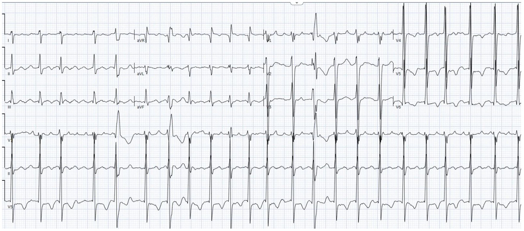
↓ Figure 1. Twelve-lead electrocardiogram demonstrating 2:1 atrial flutter with classic sawtooth flutter waves, best visualized in the inferior leads (II, III, aVF), with a ventricular rate in the 130s.
| Journal of Medical Cases, ISSN 1923-4155 print, 1923-4163 online, Open Access |
| Article copyright, the authors; Journal compilation copyright, J Med Cases and Elmer Press Inc |
| Journal website https://jmc.elmerpub.com |
Case Report
Volume 17, Number 9, September 2026, pages 453-463
When Virchow’s Triad Goes on Overdrive: Catastrophic Multi-System Thrombosis in the Setting of New-Onset Heart Failure in a Previously Healthy Man
Figures
Tables
| Parameter | Value |
|---|---|
| BSA: body surface area. | |
| Heart rate | 109 bpm |
| Blood pressure | 125/95 |
| Respiratory rate | 20 |
| Oxygen saturation | 100% on room air |
| Temperature | 97.7 °F |
| Weight/height/BSA | 168 lb/1.778 m/1.94 m2 |
| Laboratory parameter | Value | Reference range |
|---|---|---|
| CRP: C-reactive protein; eGFR: estimated glomerular filtration rate; NT-proBNP: N-terminal pro-B-type natriuretic peptide; RBC: red blood cell. | ||
| High-sensitivity troponin (initial) | 72 ng/L | < 14 ng/L |
| High-sensitivity troponin (repeat) | 64 ng/L | < 14 ng/L |
| NT-proBNP | 8,861 pg/mL | < 125 pg/mL |
| D-dimer | 2,222 ng/mL | < 500 ng/mL |
| Procalcitonin | 0.55 | < 0.10 ng/mL |
| ADAMTS13 activity | Normal | ≥ 67% |
| Anticardiolipin antibodies (IgG/IgM) | Negative | Negative |
| Anti-β2-glycoprotein I Antibodies | Negative | Negative |
| Lupus anticoagulant | Negative | Negative |
| Antithrombin III | Normal | 80–120% |
| CALR mutation | Negative | Negative |
| MPL exon mutation | Negative | Negative |
| Blood/sputum cultures | No growth | No growth |
| Urine culture | Positive (treated with ceftriaxone) | No growth |
| RBC count | 4.96 mill | Within normal limits |
| Platelet count | 267 | Within normal limits |
| Serum creatinine/eGFR | 1.0/82 | Within normal limits |
| Albumin | 3.2 | Low |
| CRP | 46.6 | Elevated |
| Protein C/protein S | Normal | - |
| Factor V Leiden/prothrombin G20210A | Negative for mutation | - |
| Parameter | Value |
|---|---|
| LA: left atrium; LAA: left atrial appendage; LV: left ventricular; LVDd/LVDs: LV end-diastolic/end-systolic diameter; LVEF: left ventricular ejection fraction; PASP: pulmonary artery systolic pressure; PFO/ASD: patent foramen ovale/atrial septal defect; TAPSE: tricuspid annular plane systolic excursion. | |
| LVEF | ∼20% (severely reduced) |
| LVDd | Not documented |
| LVDs | Not documented |
| Left atrial size | Severely dilated |
| Right atrial size | Severely dilated |
| Right ventricular size | Moderately dilated |
| TAPSE | Not documented |
| PASP | 49 mm Hg (moderate pulmonary hypertension) |
| Inferior vena cava | Moderately dilated |
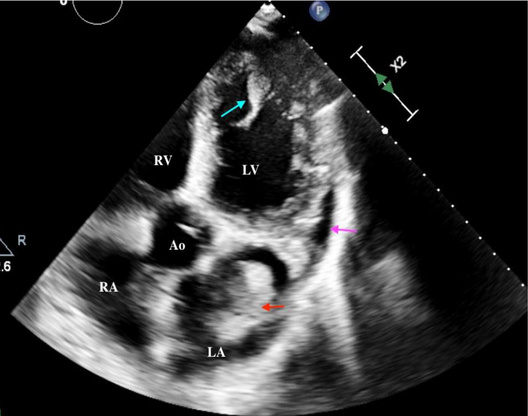
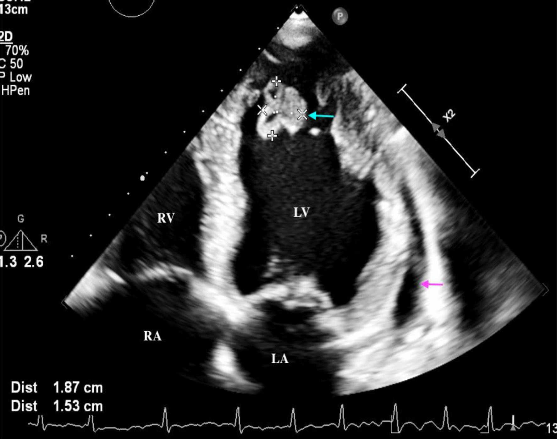
| Pericardial effusion | Small-to-moderate |
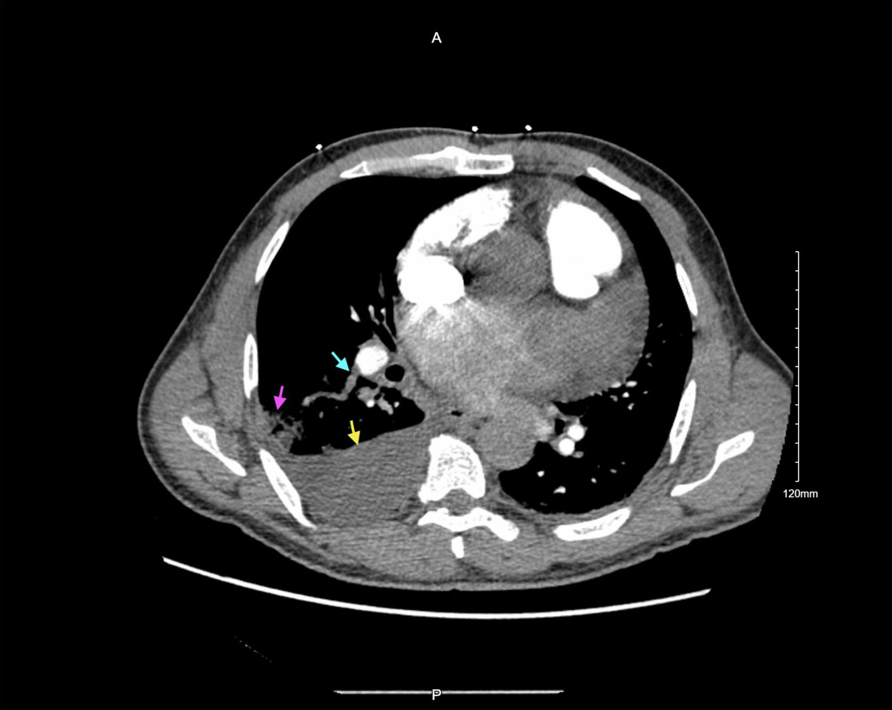
| LV apical thrombus | 1.77 × 1.81 cm, mobile |
| LA thrombus (near LAA) | 3.1 × 2.1 cm, mobile |
| PFO/ASD | Absent |
| Valvular pathology | Trace mitral and tricuspid regurgitation |
| Diagnosis | Key tests performed | Result/exclusion rationale |
|---|---|---|
| AHA/ACC: American Heart Association/American College of Cardiology; ANA: antinuclear antibody; APS: antiphospholipid syndrome; CT: computed tomography; ECG: electrocardiogram; EF: ejection fraction; HFrEF: heart failure with reduced ejection fraction; LA: left atrium; LV: left ventricular; MPN: myeloproliferative neoplasm; NBTE: non-bacterial thrombotic endocarditis; NT-proBNP: N-terminal pro-B-type natriuretic peptide; PE: pulmonary embolism; SLE: systemic lupus erythematosus; TTE: transthoracic echocardiography; TTP: thrombotic thrombocytopenic purpura; | ||
| APS | Lupus anticoagulant, anticardiolipin Ab (IgG/IgM), anti-β2-GP1 Ab | All negative (initial). Note: APS requires confirmation at ≥ 12 weeks; cannot be definitively excluded on single testing. Heparin therapy may produce false-positive LA results. |
| MPN | CALR mutation, MPL exon 10 mutation | Negative. JAK2 V617F negative. BCR-ABL negative |
| TTP | ADAMTS13 activity | Normal - TTP excluded. |
| Antithrombin III deficiency | Antithrombin III level | Normal - AT III deficiency excluded. |
| Protein C/protein S deficiency | Absent | Excluded |
| Factor V Leiden/prothrombin mutation | Negative | Excluded |
| Occult malignancy | Clinical exam, blood/sputum cultures, procalcitonin; CT imaging reviewed | No overt malignancy identified. Age-appropriate cancer screening recommended at follow-up per guidelines (2026 AHA/ACC PE guidelines). |
| NBTE | Echocardiography, blood cultures | No valvular vegetations or leaflet abnormalities on TTE; blood cultures negative. NBTE unlikely but TEE not performed. |
| Nephrotic syndrome | Urinalysis/urine protein | Negative for infection |
| Connective tissue disease (SLE) | Negative | ANA, anti-dsDNA negative. |
| Aortic source (mural thrombus/atheroma) | CT aorta reviewed as part of CT angiography | No aortic mural thrombus or complex atheroma identified. |
| Endocrine disorder (Cushing/hyperaldosteronism) | Cortisol was normal | No clinical features suggestive; excluded |
| Ischemic cardiomyopathy | Clinical assessment, ECG, echocardiography | Coronary angiography not performed. Diffuse LV dysfunction and absence of ischemic symptoms argue against ischemic etiology, but cannot be excluded. |
| New-onset HFrEF with atrial flutter (primary diagnosis) | Echocardiography, ECG, NT-proBNP, clinical course | EF 20%, atrial flutter with 2:1 conduction, markedly elevated NT-proBNP, all consistent. Most parsimonious unifying diagnosis. |