| Journal of Medical Cases, ISSN 1923-4155 print, 1923-4163 online, Open Access |
| Article copyright, the authors; Journal compilation copyright, J Med Cases and Elmer Press Inc |
| Journal website https://jmc.elmerpub.com |
Case Report
Volume 17, Number 12, December 2026, pages 685-690

A Rare Post–Hair Transplant Complication With Complete Recovery
Ekaterine Tcholadzea, b, Malvina Javakhadzeb, c, Lia Goliadzeb, Marine Kanashvilia, b, Megi Kobakhidzeb, Aleksandre Tavartkiladzec, Nia Iakobashvilid, Tinatin Kutubidzec
aTbilisi Medical Academy, Tbilisi, Georgia
bAcad. Vakhtang Bochorishvili Clinic, Tbilisi, Georgia
cTbilisi. Georgia. Infectious Diseases Department, Tbilisi State Medical University, Tbilisi, Georgia
dAversi Clinic, Tbilisi, Georgia
eCorresponding Author: Ekaterine Tcholadze, Tbilisi Medical Academy, Tbilisi, Georgia
Manuscript submitted February 6, 2026, accepted March 28, 2026, published online November 9, 2026
Short title: Hair Transplant Complication
doi: https://doi.org/10.14740/jmc5301
| Abstract | ▴Top |
Postoperative complications following hair transplantation are generally mild and self-limited; however, severe infectious or inflammatory reactions involving the periorbital or cervicofacial regions are exceedingly rare yet potentially vision- or life-threatening. Early recognition and prompt multidisciplinary management are essential, as delayed intervention may result in rapid soft-tissue progression, necrosis, and systemic instability. A 34-year-old man developed rapidly progressive cervicofacial edema 48 h after an uncomplicated hair transplantation procedure. The patient had no history of prior cranial surgery, trauma, or implanted prosthetic material. The swelling originated in the recipient area and rapidly extended to the forehead and bilateral periorbital regions, accompanied by severe pain, fever, hypotension, and hemodynamic instability. Laboratory evaluation demonstrated markedly elevated inflammatory markers (C-reactive protein 193 mg/L (reference < 5 mg/L), procalcitonin 51.43 ng/mL (reference < 0.05 ng/mL), lactate 9.68 mmol/L (reference 0.5–2.2 mmol/L)) with progressive leukopenia. Despite empirical broad-spectrum antibiotics, intensive care support, and multidisciplinary management, the patient’s condition deteriorated, with worsening edema, hemorrhagic–purulent bullae, bilateral pleural effusions, and atrial fibrillation with rapid ventricular response. Subsequent periorbital skin necrosis required surgical debridement. Later during hospitalization, neck soft-tissue involvement with multiple small fluid collections emerged. Gradual improvement followed escalation of antimicrobial therapy and corticosteroid administration; however, residual frontal muscle akinesia and right facial nerve paresis persisted. The patient eventually stabilized on long-term azathioprine therapy and achieved full clinical recovery. This case underscores that rapidly progressive cervicofacial edema, periorbital involvement, frontal muscle akinesia, and right facial nerve paresis after hair transplantation, although rare, may indicate a fulminant infectious or inflammatory process requiring urgent, multidisciplinary management. Early recognition, timely therapeutic escalation, and close monitoring are critical to preventing irreversible tissue damage and systemic complications. Increased awareness of such severe postoperative reactions can enhance clinical vigilance and improve patient outcomes.
Keywords: Hair transplantation; Soft-tissue infection; Periorbital necrosis; Facial nerve paresis; Complication
| Introduction | ▴Top |
Hair transplantation has become one of the most rapidly expanding procedures in contemporary aesthetic and reconstructive medicine. Despite its reputation as a generally safe intervention with low complication rates, emerging literature continues to emphasize that clinically significant postoperative events do occur. According to a recent large-scale review by Garg and Garg, postoperative complications—ranging from edema and folliculitis to rare cases of recipient-site necrosis—were identified in 2.8% of 2,896 patients undergoing hair transplantation procedures [1].
Androgenetic alopecia remains the leading indication for surgical hair restoration, affecting up to 80% of men and approximately 25% of women worldwide. Advances in follicular unit excision (FUE) and improved surgical techniques have further increased procedural demand. Nevertheless, even with technically uncomplicated procedures, rare but severe complications such as rapidly progressive cervicofacial edema, soft-tissue infection, periorbital involvement, and systemic inflammatory responses have been documented [2]. Such events, although uncommon, may be vision- or life-threatening, necessitating urgent multidisciplinary evaluation and early escalation of therapy.
Recent reports also highlight the potential for postoperative necrosis in both donor and recipient areas—often associated with vascular compromise, infection, or excessive tension in dense packing zones. A 2022 review of postoperative outcomes described periorbital edema as one of the more frequent early complications, but noted that necrosis remains exceedingly rare, with only isolated cases reported in the literature [3].
The present case is notable for demonstrating how a routine, uncomplicated hair transplantation procedure precipitated a fulminant postoperative course characterized by rapidly progressive cervicofacial edema, hemodynamic instability, extensive periorbital necrosis, and subsequent neurologic sequelae. This case underscores the importance of clinician awareness regarding the potential severity of even uncommon complications, highlighting the need for early recognition, multidisciplinary management, and careful postoperative monitoring to optimize patient safety.
Our clinical case is noteworthy and significant because a seemingly minor medical intervention, such as hair transplantation, resulted in life-threatening complications. The patient, a previously healthy 34-year-old man, was admitted to hospital on the third day following hair transplantation. On presentation he had high spiking fever, marked swelling of the scalp, frontal and periorbital regions, hypotension, and profoundly elevated inflammatory markers. Due to hypotension and evolving shock, he required management in the intensive care unit (ICU). During the course of hospitalization, he developed bilateral necrosis of the upper and lower eyelids with serosanguinous–purulent discharge, episodes of paroxysmal arrhythmia, paresis of the frontal mimetic muscles, and right-sided facial nerve palsy.
The patient required a 45-day inpatient treatment course, including broad-spectrum antimicrobial therapy and corticosteroids. Gradual clinical stabilization was achieved, with normalization of inflammatory markers, resolution of systemic symptoms, and significant regression of cervicofacial edema; however, residual deficits persisted, including frontal muscle akinesia and right-sided facial nerve palsy. Additionally, corticosteroids could not be fully withdrawn, and the patient remains on azathioprine.
This case illustrates that a technically simple medical procedure may precipitate severe, life-threatening complications and lead to a dramatic deterioration in a patient’s health status.
| Case Report | ▴Top |
A 34-year-old male patient underwent hair transplantation. Approximately one-quarter of the scalp was covered with transplanted grafts (Fig. 1). The procedure was uneventful, with no intraoperative complications.
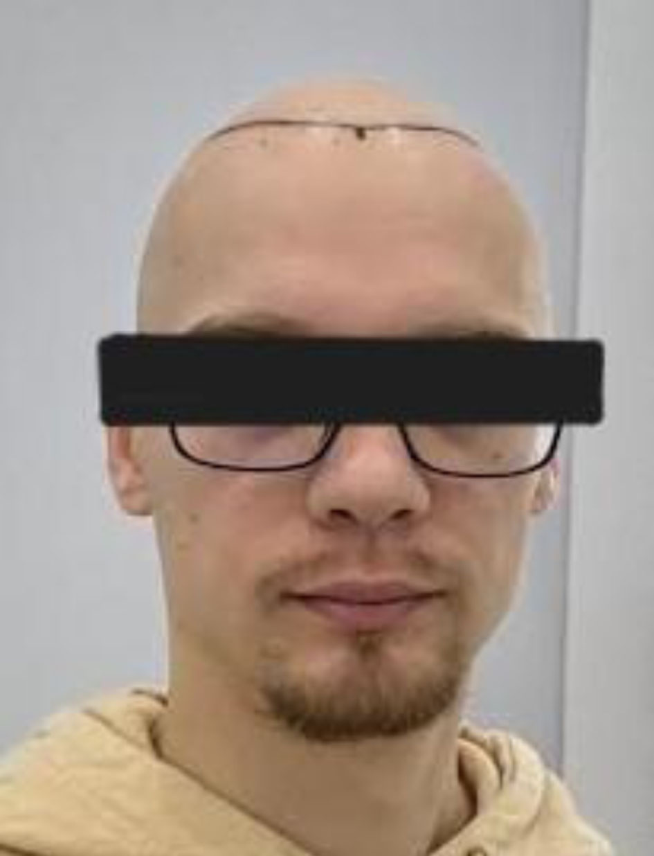 Click for large image | Figure 1. Hair transplantation recipient area immediately after surgery. Clinical photograph showing the scalp immediately after hair transplantation. Approximately one-quarter of the scalp is covered with transplanted follicular grafts in the recipient area. FUE: follicular unit excision. |
Within 48 h after surgery, the patient developed swelling originating from the recipient area, which rapidly extended to the forehead and the left periorbital region, accompanied by pain and a fever reaching 39 °C (Fig. 2). He was brought to the hospital by his family physician and admitted for further evaluation. Despite appropriate emergency care, his condition deteriorated within a few hours. The swelling markedly increased and involved both periorbital regions, with pronounced eyelid edema, erythema, and hemorrhagic-purulent bullae (Fig. 3). Due to pain and swelling, he was unable to open his eyes. Progressive hypotension and unstable hemodynamics developed, requiring transfer to the ICU.
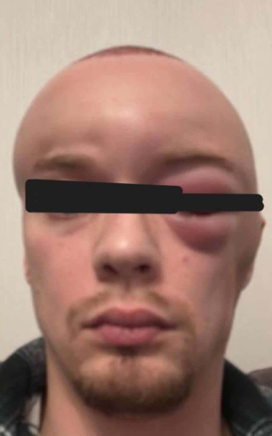 Click for large image | Figure 2. Early postoperative edema (48 h after surgery). Clinical image obtained 48 h after hair transplantation showing progressive swelling originating from the recipient area and extending to the forehead and left periorbital region, with erythema and pain. |
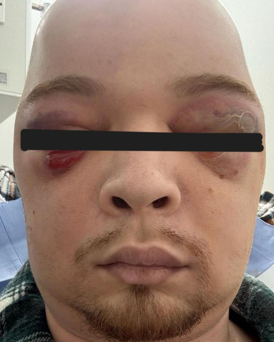 Click for large image | Figure 3. Rapidly progressive bilateral periorbital involvement. Photograph demonstrating extensive bilateral periorbital edema, erythema, and hemorrhagic–purulent bullae. The patient was unable to open his eyes due to severe swelling and pain. This stage was accompanied by hypotension and systemic inflammatory response requiring intensive care unit admission. |
On admission, laboratory findings revealed markedly elevated inflammatory markers: C-reactive protein (CRP) 193 mg/L (reference range < 5 mg/L), procalcitonin (PCT) 51.43 ng/mL (reference range < 0.05 ng/mL), and lactate 9.68 mmol/L (reference range 0.5–2.2 mmol/L). Leukopenia was also present (white blood cell (WBC) 3.41 × 10⁹/L (reference range 4–10 × 109/L)). Blood, urine, and wound cultures were obtained for microbiological testing, and empirical broad-spectrum antibiotic therapy was initiated.
Despite several days of treatment, the patient’s clinical condition worsened. He developed atrial fibrillation with a tachysystolic response, which was successfully controlled with appropriate therapy. Bilateral pleural effusions were detected. The patient was managed under the supervision of a multidisciplinary team, including a cardiologist, a surgeon, an ophthalmologist, and an infectious disease specialist. Laboratory results showed further deterioration with CRP 432 mg/L (reference < 5 mg/L), PCT 51.43 ng/mL (reference < 0.05 ng/mL), and worsening leukopenia. Multiple purulent lesions with seropurulent discharge appeared on the scalp, many of which were covered with crusts. Empirical antibiotic therapy was modified; however, blood, urine, and wound cultures remained negative throughout hospitalization. Liver and renal function tests, as well as other biochemical parameters, remained within normal limits.
By the ninth day of hospitalization, gradual clinical improvement was observed. Hemodynamic parameters stabilized, and the patient became afebrile. Facial edema subsided, although areas of skin necrosis developed in the eyelids, accompanied by severe blepharospasm. The left upper eyelid showed near-complete tissue compromise with a high risk of necrosis and sinus thrombosis. The left eye could not be opened even with assistance, whereas the right eye had conjunctival hyperemia but a transparent cornea (Figs 4 and 5).
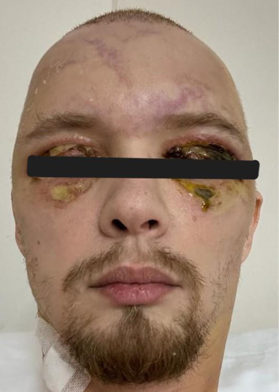 Click for large image | Figure 4. Eyelid necrosis and tissue compromise. Image demonstrating necrotic changes of the upper eyelids, particularly on the left side. Conjunctival hyperemia is visible in the right eye. |
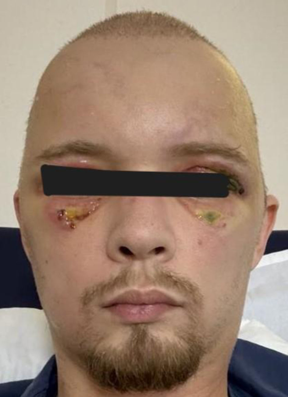 Click for large image | Figure 5. Advanced eyelid necrosis with asymmetric ocular involvement. Clinical image demonstrating advanced necrotic changes of the upper eyelids, more pronounced on the left side, with evident tissue breakdown and loss of skin integrity. The right eye shows conjunctival hyperemia without corneal involvement. These findings reflect progression of severe periorbital soft-tissue injury in the setting of a fulminant inflammatory process. |
After several days, a necrotomy of the left upper eyelid was performed, leaving a skin defect that later required autologous skin grafting after discharge.
By the 23rd day of hospitalization, swelling extended to the cervical soft tissues, predominantly on the left side, associated with limited neck movement and painful, markedly enlarged lymph nodes. A computed tomography (CT) scan of the neck revealed soft-tissue edema with multiple small fluid collections (Fig. 6).
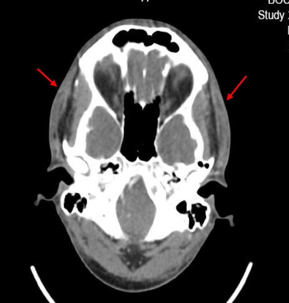 Click for large image | Figure 6. Diffuse cervical soft-tissue swelling. Axial CT image demonstrating diffuse soft-tissue swelling with fat stranding (arrows) consistent with inflammatory edema, without evidence of abscess formation. CT: computed tomography. |
The patient continued treatment with imipenem, moxifloxacin, and dexamethasone, which led to progressive stabilization. Laboratory parameters normalized, and swelling resolved completely. However, residual frontal muscle atony and right-sided facial nerve paresis persisted (Fig. 7).
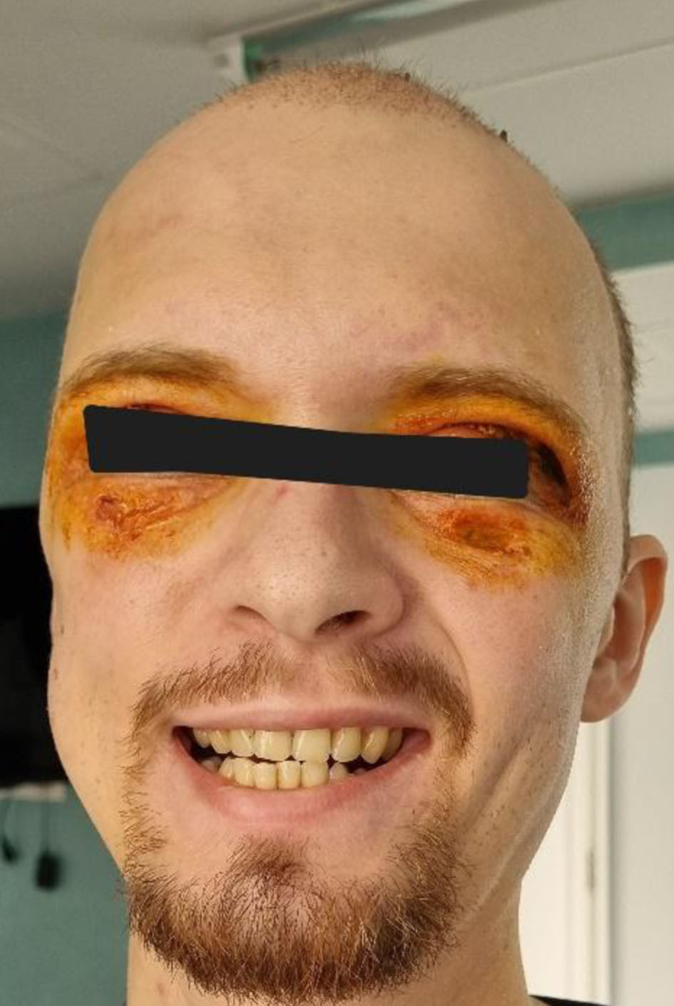 Click for large image | Figure 7. Residual neurological deficits at discharge. Clinical image demonstrating residual frontal muscle akinesia and right-sided facial nerve paresis after resolution of edema and systemic stabilization. These neurological deficits persisted despite overall clinical recovery. |
The patient was discharged home in stable condition with resolution of systemic inflammatory signs, normalization of laboratory parameters, and significant reduction of cervicofacial edema. Several attempts to taper corticosteroids gradually resulted in the recurrence of facial and scalp edema. Approximately 6 months later, he was transitioned to azathioprine therapy, which he continues to this date—nearly 1.5 years post-discharge. His current condition remains satisfactory, with full clinical recovery.
| Discussion | ▴Top |
Hair transplantation is widely regarded as a safe and minimally invasive surgical procedure with a low incidence of severe complications. Most postoperative reactions are mild and transient, including edema, minor pain, pruritus, or folliculitis. Serious complications such as extensive soft-tissue infection, necrosis, or systemic inflammatory response are exceedingly rare but have been described in isolated reports [1, 2].
Postoperative facial edema is among the more common early reactions after hair transplantation. It usually develops between the second and fourth postoperative day, initially affecting the forehead and sometimes extending to the periorbital region. In most cases, this edema resolves spontaneously without clinical consequences. The use of corticosteroids in tumescence solutions or postoperative positioning has been suggested to reduce the severity of swelling, although evidence remains limited [1].
Infectious complications after hair transplantation are uncommon due to the rich vascular supply of the scalp and adherence to sterile surgical techniques. When infections do occur, they typically present as localized folliculitis or cellulitis. However, severe infections involving deeper soft tissues, including cervicofacial regions, have been reported in rare cases and may lead to systemic inflammatory responses and tissue necrosis [2–4].
In the present case, the patient developed rapidly progressive cervicofacial edema accompanied by systemic inflammatory signs, including hypotension and markedly elevated inflammatory markers. Despite extensive microbiological testing, no bacterial growth was identified in cultures. The absence of confirmed microbiological infection together with the favorable clinical response to corticosteroid therapy raises the possibility that an exaggerated inflammatory or immune-mediated response contributed to the clinical presentation.
A few reports in the literature describe severe complications after dermatologic or cosmetic procedures, including necrosis and neurological deficits such as facial nerve involvement. Potential mechanisms include vascular compromise, inflammatory edema causing tissue ischemia, or secondary infection leading to progressive soft-tissue damage [3, 5].
This case highlights the importance of early recognition of atypical postoperative reactions following hair transplantation. Rapid progression of edema, severe pain, systemic inflammatory signs, or periorbital involvement should prompt immediate multidisciplinary evaluation and aggressive management. Early intervention may prevent irreversible tissue injury and reduce the risk of long-term functional deficits.
Advances in FUE and improved surgical techniques have further increased procedural demand [6, 7].
Although hair transplantation is generally considered a safe procedure with a low rate of severe complications, rare cases of rapidly progressive inflammatory or infectious reactions have been reported in the literature. Such reactions may mimic aggressive soft-tissue infections and can lead to extensive cervicofacial edema, tissue necrosis, and systemic inflammatory response. Early differentiation between severe infection, vascular compromise, and immune-mediated inflammatory reactions can be challenging but is crucial for timely management. In the present case, the absence of microbiological growth despite significant systemic inflammation and the favorable response to corticosteroid therapy raise the possibility that immune-mediated mechanisms may have contributed to the clinical course. Similar observations have been described in isolated reports of severe postoperative reactions following dermatologic or cosmetic procedures, emphasizing the importance of multidisciplinary evaluation and individualized therapeutic strategies.
Conclusion
This case highlights that even a technically straightforward and routinely performed procedure such as hair transplantation can precipitate a fulminant, life-threatening inflammatory process with rapid cervicofacial progression, hemodynamic instability, and irreversible soft-tissue damage. While the majority of postoperative reactions are mild and self-limited, our report underscores the importance of maintaining clinical vigilance for early warning signs—disproportionate edema, escalating pain, fever, hypotension, and periorbital involvement—which may indicate an evolving severe infection or inflammatory cascade.
Prompt recognition, early intensive care support, multidisciplinary management, and timely escalation of antimicrobial and immunomodulatory therapy were crucial in preventing catastrophic outcomes for our patient, despite residual neurological deficits. The protracted inflammatory course and responsiveness to long-term immunosuppression also suggest that non-infectious immunological mechanisms may contribute to certain severe postoperative reactions, warranting consideration in future diagnostic and therapeutic approaches.
Increased awareness of such rare but severe complications is essential for clinicians performing or evaluating patients after hair transplantation. Early identification of atypical postoperative courses, rapid intervention, and careful monitoring may significantly improve patient outcomes and reduce the risk of permanent functional or aesthetic impairment. This case contributes valuable insight to the limited global literature and reinforces the need for heightened vigilance, comprehensive assessment, and collaborative management in similar presentations.
The combination of severe systemic inflammation, negative microbiological findings, and favorable response to corticosteroid therapy strongly suggests that an exaggerated immune-mediated inflammatory response may have played a central role in the pathogenesis.
Acknowledgments
The authors would like to thank the multidisciplinary team involved in the management of this patient, including specialists in infectious diseases, cardiology, surgery, ICU doctors, and ophthalmology.
Financial Disclosure
The authors declare that no financial support or funding was received for the preparation of this manuscript.
Conflict of Interest
The authors declare that they have no conflict of interest related to this publication.
Informed Consent
Written informed consent was obtained from the patient for publication of this case report and accompanying images.
Author Contributions
Ekaterine Tcholadze: conceptualization, patient management, manuscript drafting, and final approval of the manuscript. Malvina Javakhadze: literature review and manuscript drafting. Lia Goliadze: clinical data collection and manuscript editing. Marine Kanashvili: patient management and critical revision of the manuscript. Megi Kobakhidze: data interpretation and manuscript review. Aleksandre Tavartkiladze: radiological evaluation and interpretation. Nia Iakobashvili: literature analysis and manuscript preparation. Tinatin Kutubidze: supervision and critical revision of the manuscript. All authors read and approved the final manuscript.
Data Availability
The authors declare that data supporting the findings of this study are available within the article.
| References | ▴Top |
- Garg AK, Garg S. Complications of hair transplant procedures: causes and management. Indian J Plast Surg. 2021;54(4):477-482.
doi pubmed pmc - Chen J, Qu Q, Guo Y, Hu Z, Li X, Fan Z, Yang J, et al. Recipient site scalp necrosis: A rare postoperative complication of hair transplantation. J Cosmet Dermatol. 2024;23(2):622-629.
doi pubmed - Ozturk P, et al. Recipient site necrosis after follicular unit extraction: clinical features and management. Dermatol Surg. 2022;48(9):1068-1074.
- Abraham JR, Singh RP, Srivastava SK, Mammo DA. Hair transplantation complicated by methicillin-resistant Staphylococcus aureus sepsis and endogenous endophthalmitis. Retin Cases Brief Rep. 2024;18(3):301-304.
doi pubmed - Majerowski J, Xu YG. Acute, temporary, unilateral facial nerve palsy after dermatologic surgery: a report of 3 cases. J Cutan Med Surg. 2017;21(5):445-448.
doi pubmed - Vano-Galvan S, Bisanga CN, Bouhanna P, Farjo B, Gambino V, Meyer-Gonzalez T, Silyuk T, et al. An international expert consensus statement focusing on pre and post hair transplantation care. J Dermatolog Treat. 2023;34(1):2232065.
doi pubmed - International Society of Hair Restoration Surgery. Hair restoration surgery statistics and clinical resources. Available at: https://ishrs.org/ (Accessed September 22, 2024).
This article is distributed under the terms of the Creative Commons Attribution 4.0 International License (CC BY 4.0), which permits unrestricted use, distribution, and reproduction in any medium, including commercial use, provided the original work is properly cited.
Journal of Medical Cases is published by Elmer Press Inc.