Figures
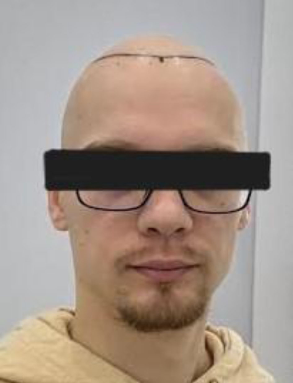
↓ Figure 1. Hair transplantation recipient area immediately after surgery. Clinical photograph showing the scalp immediately after hair transplantation. Approximately one-quarter of the scalp is covered with transplanted follicular grafts in the recipient area. FUE: follicular unit excision.
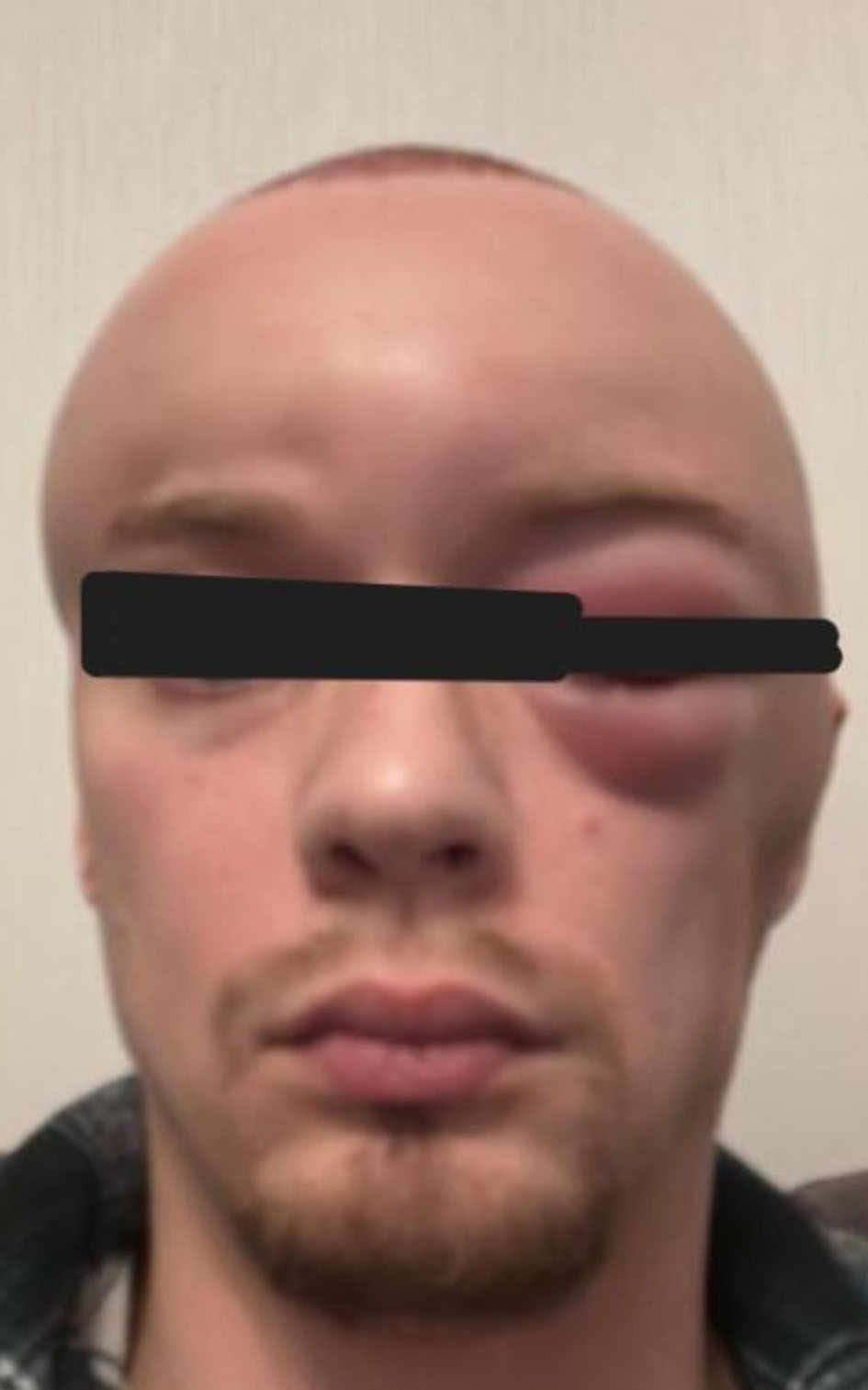
↓ Figure 2. Early postoperative edema (48 h after surgery). Clinical image obtained 48 h after hair transplantation showing progressive swelling originating from the recipient area and extending to the forehead and left periorbital region, with erythema and pain.
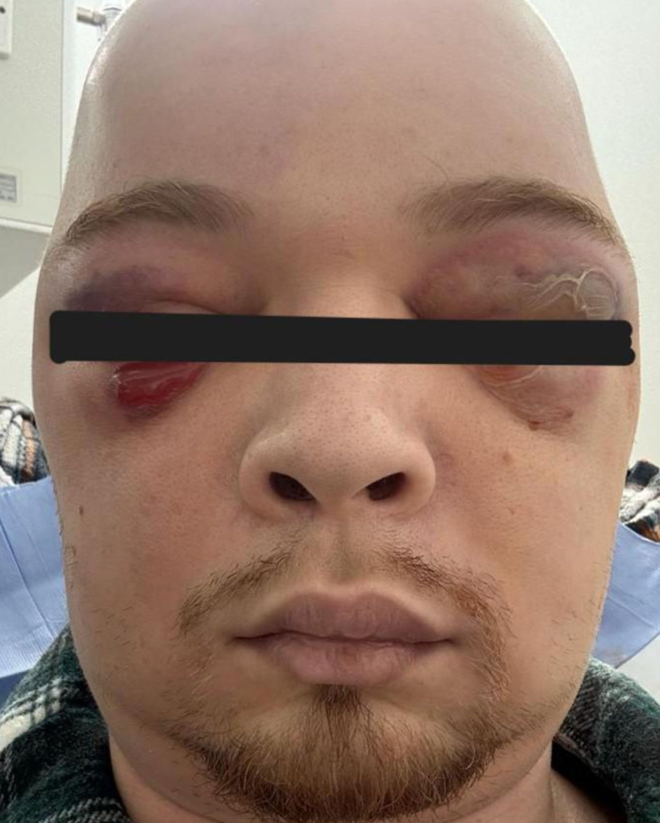
↓ Figure 3. Rapidly progressive bilateral periorbital involvement. Photograph demonstrating extensive bilateral periorbital edema, erythema, and hemorrhagic–purulent bullae. The patient was unable to open his eyes due to severe swelling and pain. This stage was accompanied by hypotension and systemic inflammatory response requiring intensive care unit admission.
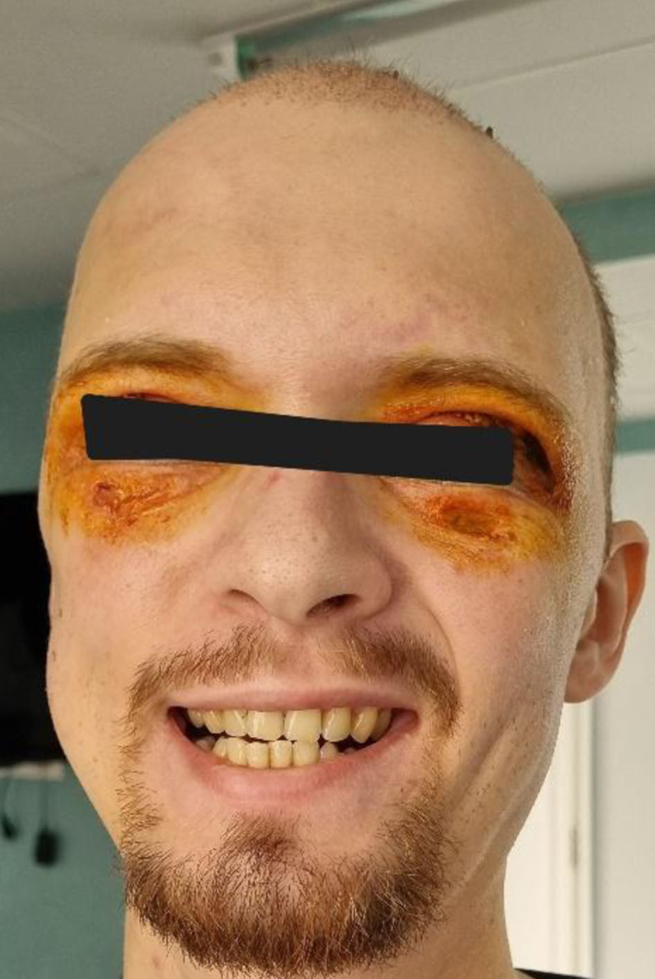
↓ Figure 4. Eyelid necrosis and tissue compromise. Image demonstrating necrotic changes of the upper eyelids, particularly on the left side. Conjunctival hyperemia is visible in the right eye.
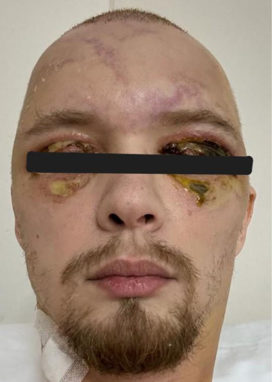
↓ Figure 5. Advanced eyelid necrosis with asymmetric ocular involvement. Clinical image demonstrating advanced necrotic changes of the upper eyelids, more pronounced on the left side, with evident tissue breakdown and loss of skin integrity. The right eye shows conjunctival hyperemia without corneal involvement. These findings reflect progression of severe periorbital soft-tissue injury in the setting of a fulminant inflammatory process.
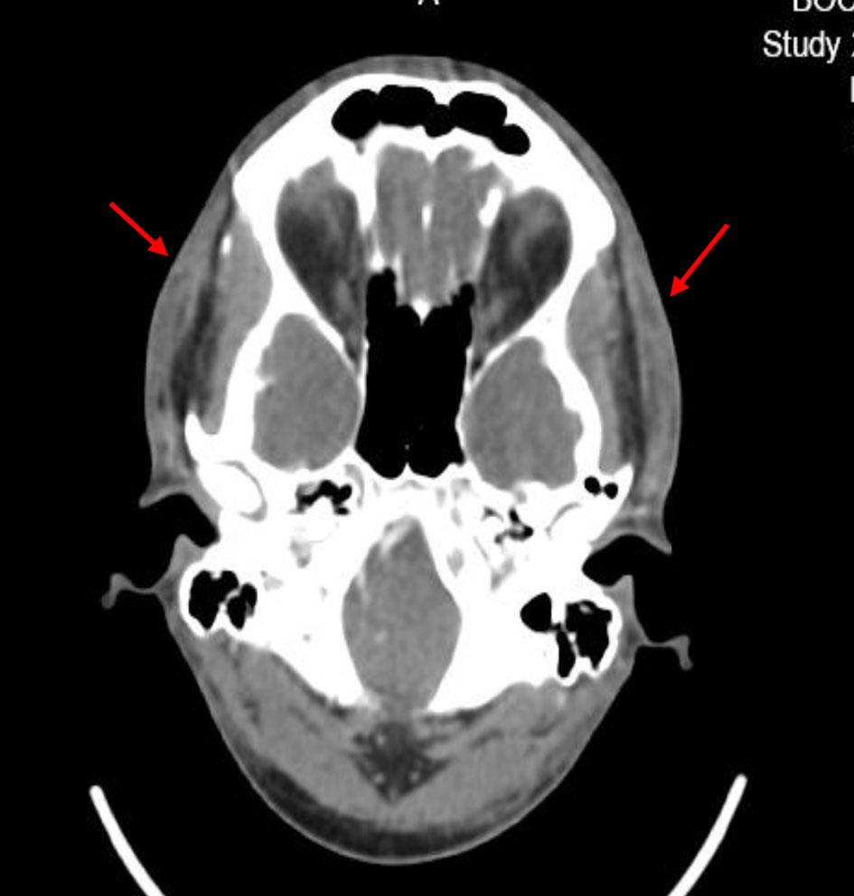
↓ Figure 6. Diffuse cervical soft-tissue swelling. Axial CT image demonstrating diffuse soft-tissue swelling with fat stranding (arrows) consistent with inflammatory edema, without evidence of abscess formation. CT: computed tomography.
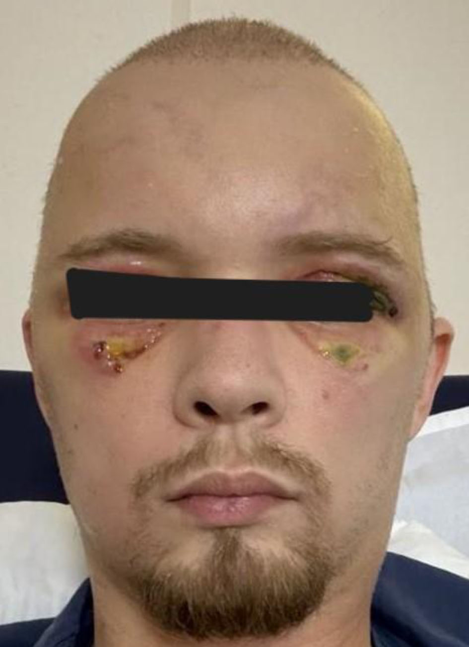
↓ Figure 7. Residual neurological deficits at discharge. Clinical image demonstrating residual frontal muscle akinesia and right-sided facial nerve paresis after resolution of edema and systemic stabilization. These neurological deficits persisted despite overall clinical recovery.