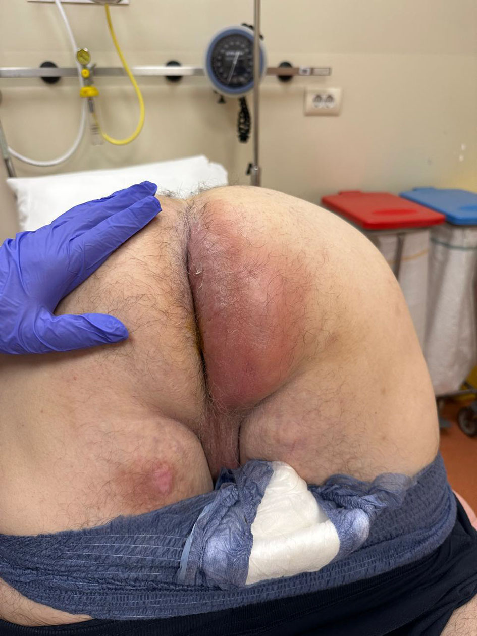
↓ Figure 1. Large perianal abscess.
| Journal of Medical Cases, ISSN 1923-4155 print, 1923-4163 online, Open Access |
| Article copyright, the authors; Journal compilation copyright, J Med Cases and Elmer Press Inc |
| Journal website https://jmc.elmerpub.com |
Case Report
Volume 17, Number 11, November 2026, pages 670-684
Cardiac Magnetic Resonance-Supported Diagnosis of Acute Myocarditis Mimicking Acute Coronary Syndrome in a Patient With Sepsis Due to a Massive Perianal Abscess
Figures
Tables
| Parameter | Patient value | SOFA component | SOFA score |
|---|---|---|---|
| CNS: central nervous system; GCS: Glascow Comma Scale; MAP: mean arterial pressure; SOFA: Sequential Organ Failure Assessment. | |||
| Respiratory rate | 26 breaths/min | Respiratory | - |
| PaO2 | 69 mm Hg | Respiratory | - |
| Oxygen supplementation | 10 L/min Venturi mask | Respiratory | - |
| Estimated FiO2 | ∼0.40 | Respiratory | - |
| PaO2/FiO2 ratio | ∼173 | Respiratory | 2 |
| Platelets | 170 × 103/µL | Coagulation | 0 |
| Bilirubin | 1.2 mg/dL | Liver | 0 |
| Blood pressure | 100/35 mm Hg | Cardiovascular | - |
| Estimated MAP | ∼57 mm Hg | Cardiovascular | - |
| Norepinephrine | 0.05 µg/kg/min | Cardiovascular | 3 |
| Heart rate | 117 beats/min | - | - |
| GCS | 15 | CNS | 0 |
| Mental status | Slightly agitated | CNS | - |
| Creatinine | 1.7 mg/dL | Renal | 1 |
| Lactate | 3.6 mmol/L | - | - |
| SpO2 | 95% | - | - |
| Infection | Massive perianal abscess | Clinical context | - |
| Total SOFA score | 6 | ||
| Parameter | Before treatment | After treatment | Reference range/interpretation |
|---|---|---|---|
| CRP: C-reactive protein. | |||
| Procalcitonin | 2.28 ng/mL | 0.41 ng/mL | 0.5–2 ng/mL: suspected sepsis; > 2 ng/mL: sepsis |
| CRP | 27.49 mg/dL | 0.48 mg/dL | 0–0.5 mg/dL |
| White blood cell count | 25.38 × 10³/µL | 19.45 × 10³/µL | 4.0–10.5 × 10³/µL |
| Surgical-site microbiological cultures | Staphylococcus epidermidis; Enterococcus faecalis; Escherichia coli | ||
| Blood culture | Escherichia coli | ||
| Intravenous antibiotic regimen (10 days) | Piperacillin/tazobactam 4.5 g three times daily; levofloxacin 500 mg once daily; metronidazole 500 mg three times daily | ||
| Step | Diagnostic question/challenge | Examination/finding | Diagnostic conclusion/exclusion |
|---|---|---|---|
| ABG: arterial blood gas; ACS: acute coronary syndrome; CAD: coronary artery disease; CMR: cardiac magnetic resonance; CT: computed tomography; CK-MB: creatine kinase myocardial band; ECG: electrocardiogram; MINOCA: myocardial infarction with non-obstructive coronary arteries; MR: mitral regurgitation; LGE: late gadolinium enhancement; LV: left ventricular; LVEF: left ventricle ejection fraction; NT-proBNP: N-terminal pro-B-type natriuretic peptide; RCA: right coronary artery. | |||
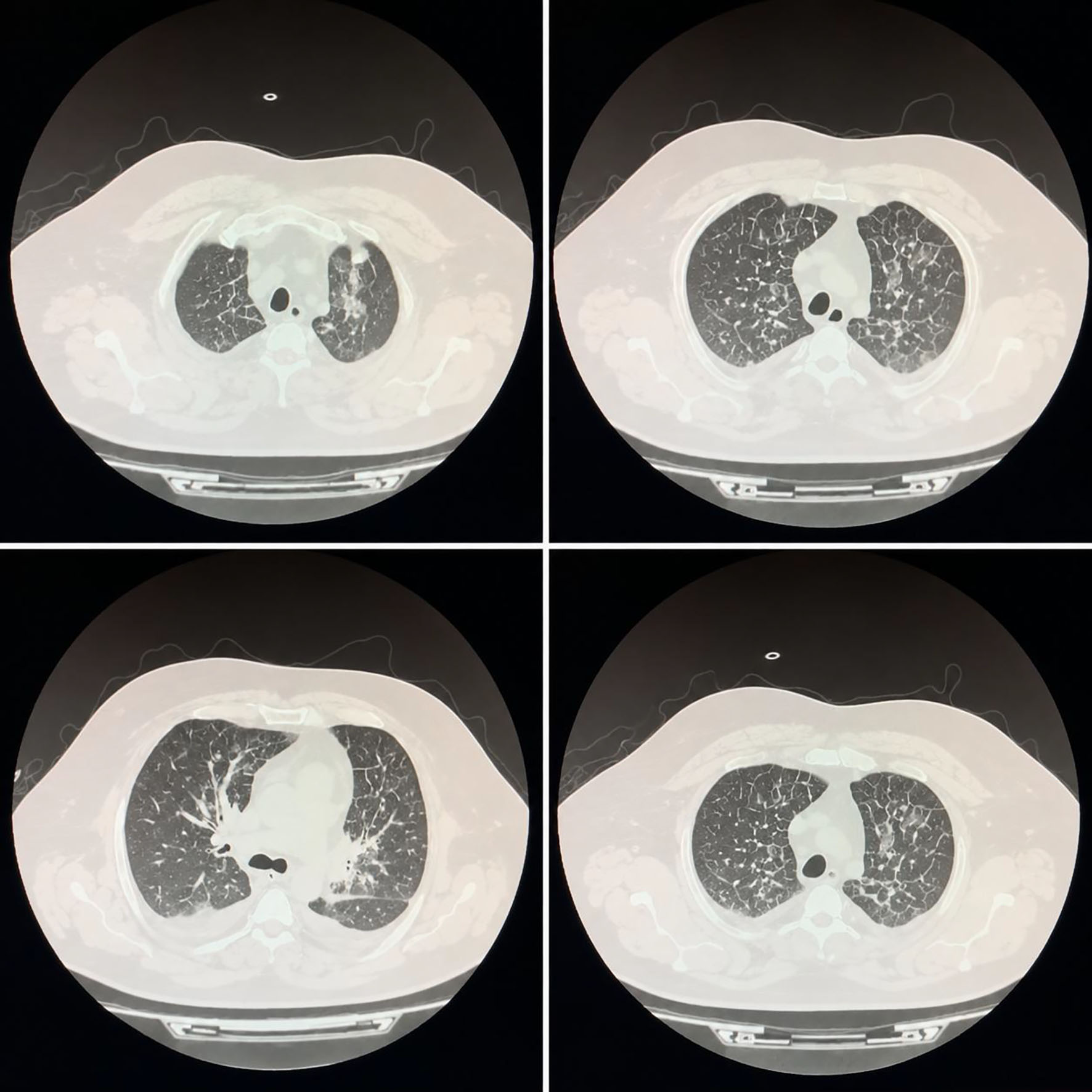
| 1. Respiratory deterioration | What caused the acute postoperative hypoxemia? | ABG showed PaO2 of 51 mm Hg; chest CT showed pulmonary vascular congestion, bilateral pleural effusions, ground-glass opacities, and septal thickening | Pulmonary edema identified |
| 2. Fluid overload | Could pulmonary edema be explained by excessive fluid administration? | 1,100 mL intra-operative fluids; 1,800 mL urine output after furosemide; net fluid balance −700 mL | Simple fluid overload was less likely |
| 3. New cardiac dysfunction | Was there acute deterioration in cardiac function? | Echocardiography: LVEF ∼35%, global hypokinesis, new moderate-to-severe functional MR, without significant regional wall-motion abnormalities | Acute myocardial dysfunction confirmed, with a predominantly global rather than regional pattern |
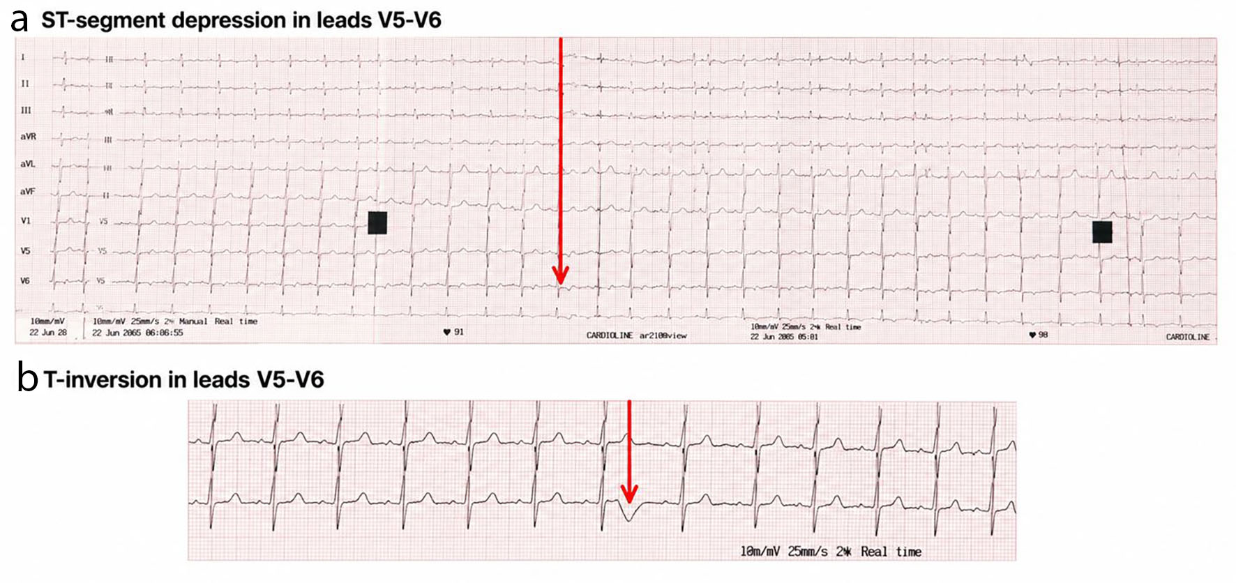
| 4. Acute myocardial injury | Was the cardiac deterioration associated with myocardial injury? | Hs-troponin 1,950 ng/L and CK-MB 350 ng/mL; ECG showed ST depression and T-wave inversion in V5–V6 | ACS was strongly suspected |
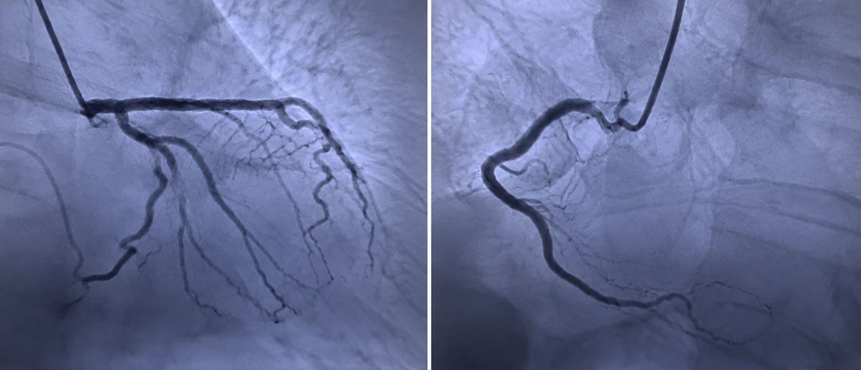
| 5. Obstructive coronary disease | Was this an acute myocardial infarction due to coronary obstruction? | Urgent coronary angiography: only 40% non-obstructive RCA stenosis; normal left coronary system | Obstructive ACS excluded; MINOCA considered |
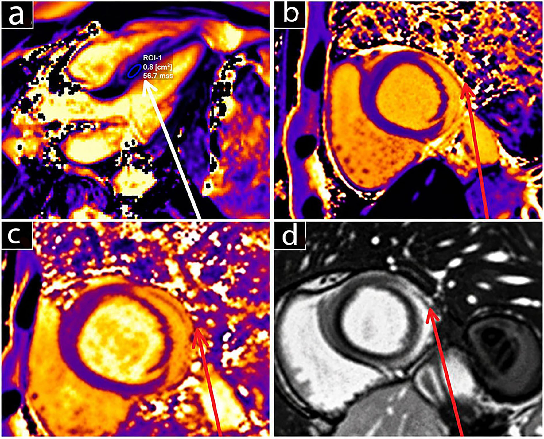
| 6. Myocarditis vs. septic cardiomyopathy | What explained the myocardial injury in the setting of severe sepsis? | CMR: elevated T2 values indicating myocardial edema and LGE involving the interventricular septum and LV lateral wall | Active myocardial inflammation demonstrated, supporting acute myocarditis |
| 7. Histological confirmation | Could myocarditis be confirmed by biopsy? | Endomyocardial biopsy was declined by the patient | Diagnosis was established non-invasively by clinical context, exclusion of obstructive CAD, and CMR findings |
| 8. Diagnostic confirmation by outcome | Was the myocardial dysfunction reversible? | LVEF recovered to 55%, MR resolved, NT-proBNP decreased from 3,846 to 95 pg/mL, and cardiac enzymes normalized | Reversible inflammatory myocardial injury consistent with acute myocarditis |
| Time point | Clinical course/symptoms | Investigations and findings | Treatment/management | Outcome / diagnostic interpretation |
|---|---|---|---|---|
| ABG: arterial blood gas; ACE: angiotensin converting enzyme; BMI: body mass index; CT: computed tomography; CMR: cardiac magnetic resonance; CXR: chest X-ray; CK-MB: creatine kinase myocardial band; ECG: electrocardiogram; EMB: endomyocardial biopsy; hs-Tn: hs-troponin; ICU: intensive care unit; IV: intravenous; LGE: late gadolinium enhancement; LV: left ventricular; LVEF: left ventricle ejection fraction; MR: mitral regurgitation; RCA: right coronary artery; SOFA: Sequential Organ Failure Assessment. | ||||
| Day 10 | Fever (40 °C), right perianal pain/swelling, mild cough, and fatigue | Large perianal abscess identified | - | Severe local infection with systemic inflammatory response. |
| Day 5 | Persistent symptoms with temporary improvement | - | Outpatient IV antibiotics | Subsequent clinical deterioration. |
| Day 0 – hospital admission | Fever 40 °C, tachypnea, diaphoresis; BMI 42.1 kg/m2 | SOFA score 6; large perianal abscess | Broad-spectrum antibiotics; surgical consultation | Severe sepsis/septic shock secondary to perianal abscess. |
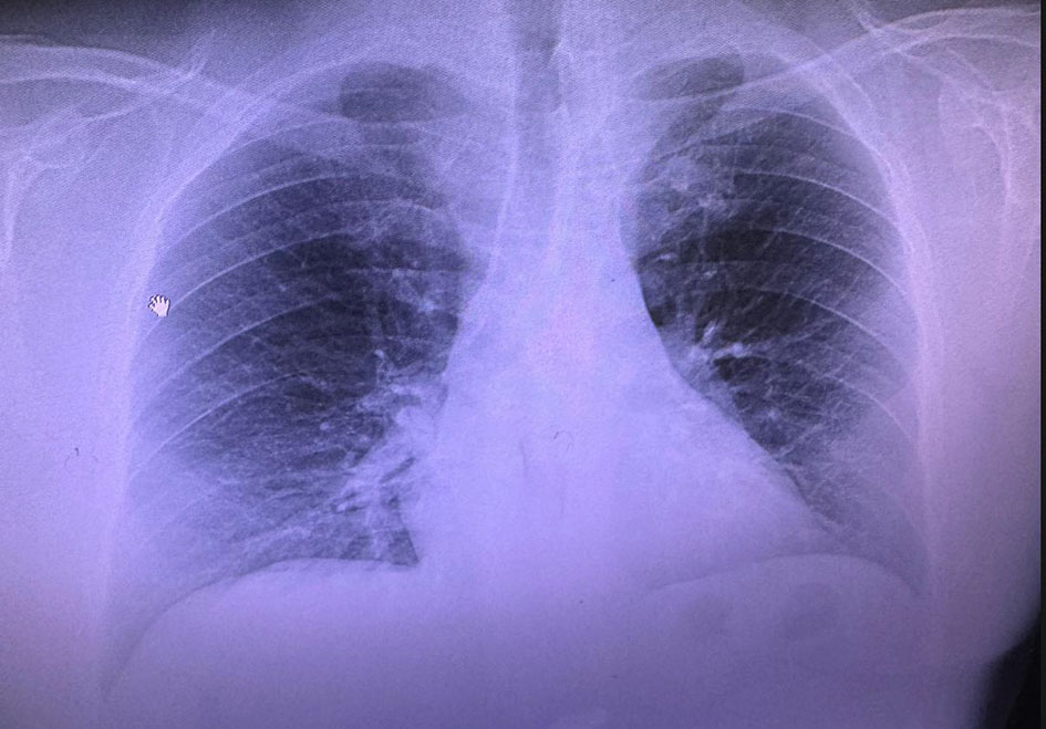
| Day 0 – pre-operative assessment | Large right perianal abscess extending into the gluteal region | CXR: hilar enlargement/resolving pneumonia; SpO2 95–96%. Echo: preserved LV function, mild-to-moderate MR | Emergency surgical drainage planned | Normal LV systolic baseline. |
| Day 0 – surgery | Emergency abscess drainage | Radial arterial monitoring; uncomplicated intubation | General anesthesia; propofol, fentanyl, vecuronium, sevoflurane, remifentanil | Surgery completed in ∼1 h. |
| Day 0 – emergence | Severe hypertension (201/77 mm Hg) and hypoxemia (SpO2 88–92%) | ABG: pH 7.33, PaO2 51, PaCO2 39 mm Hg | Furosemide 60 mg IV; net fluid balance −700 mL | Pulmonary edema initially suspected. |
| Early postoperative period | Persistent oxygen requirement, chest discomfort, and basal crackles | CT: bilateral pleural effusions, vascular congestion, ground-glass opacities, septal thickening | Cardiopulmonary monitoring and cardiac evaluation | Cardiac dysfunction increasingly suspected. |
| Early postoperative period – cardiac evaluation | New cardiac dysfunction | ECG: sinus tachycardia 119 bpm, ST depression/T-wave inversion V5–V6. Echo: LVEF 35%, moderate to severe MR. Hs-Tn 1,950 ng/L; CK-MB 350 ng/mL | Cardiology consultation | ACS/acute MI initially suspected. |
| Early postoperative ICU period | Hemodynamic instability requiring intensive monitoring | Adequate tissue perfusion maintained | Dobutamine 7.5 µg/kg/min + norepinephrine 0.05–0.1 µg/kg/min | Hemodynamic stabilization. |
| Early ICU period – coronary evaluation | Ongoing myocardial injury evaluation | 40% non-obstructive RCA stenosis; normal left coronary tree | No coronary intervention | Obstructive ACS excluded; MINOCA/non-ischemic injury considered. |
| After hemodynamic stabilization – CMR | Persistent concern for myocarditis | CMR: elevated septal T2 values and LGE involving the interventricular septum/LV lateral wall | EMB declined | Findings supported acute myocarditis. |
| ICU days 1–6 | Progressive cardiac and respiratory improvement | Stable tissue perfusion | Antibiotics, diuretics, oxygen, vasoactive/inotropic support | Transferred to cardiology after 6 days. |
| Day 6 – transfer from ICU | Continued improvement | - | Diuretics, ACE inhibitor, digitalis, oxygen, antibiotics, wound care | Continued recovery. |
| Day 10 | Ongoing recovery | Echo: LVEF 45%, moderate residual MR | Conservative heart failure management | Improved LV function. |
| Three-month follow-up | Clinically recovered | Complete recovery of cardiac function; MR resolved | Follow-up | Complete healing of perianal wound/fistula and favorable outcome. |
| Feature | Sepsis-induced cardiomyopathy | Myocarditis |
|---|---|---|
| BNP: B-type natriuretic peptide; CRP: C-reactive protein; ECG: electrocardiogram; ESR: erythrocyte sedimentation rate; GLS: global longitudinal strain; LGE: late gadolinium enhancement; NT-proBNP: N-terminal pro-B-type natriuretic peptide. | ||
| Underlying cause | Systemic inflammatory response to sepsis causing reversible myocardial dysfunction | Inflammatory injury of the myocardium, most commonly viral, autoimmune, toxic, or immune-mediated |
| Clinical context | Occurs during severe sepsis or septic shock | May occur following a viral prodrome, autoimmune disease, drug exposure, or independently of sepsis |
| Typical onset | Concurrent with septic illness | Acute, subacute, or chronic; may precede or occur without systemic infection |
| Symptoms | Signs of sepsis with circulatory failure; heart failure symptoms may be masked | Chest pain, dyspnea, palpitations, fatigue, syncope, heart failure |
| Fever | Common due to sepsis | May be present if infectious; absent in autoimmune forms |
| Hemodynamics | Vasodilatory shock with reduced systemic vascular resistance; myocardial depression contributes to hypotension | Variable; may range from stable hemodynamics to cardiogenic shock (fulminant myocarditis) |
| ECG findings | Sinus tachycardia; non-specific ST-T changes; occasional arrhythmias | ST-segment and T-wave abnormalities, PR depression, conduction disturbances, ventricular arrhythmias |
| Cardiac biomarkers | Troponin mildly to moderately elevated; BNP/NT-proBNP frequently elevated | Troponin often markedly elevated; BNP/NT-proBNP elevated with ventricular dysfunction |
| Inflammatory markers | Markedly elevated (CRP, procalcitonin, leukocytosis) reflecting sepsis | Elevated CRP and ESR; procalcitonin usually normal unless concomitant bacterial infection |
| Echocardiography | Global biventricular systolic dysfunction, ventricular dilatation, reduced ejection fraction, usually without regional wall motion abnormalities | Regional or global ventricular dysfunction; increased wall thickness (edema), pericardial effusion may be present |
| Speckle-tracking echocardiography | Reduced global longitudinal strain, usually diffuse | Reduced GLS with regional abnormalities corresponding to inflamed myocardium |
| Cardiac magnetic resonance | Usually non-specific; myocardial edema may occur, but late gadolinium enhancement is uncommon | Diagnostic modality of choice; demonstrates myocardial edema and non-ischemic LGE according to updated Lake Louise criteria |
| Coronary angiography | Usually normal if performed | Usually normal unless concomitant coronary artery disease |
| Endomyocardial biopsy | Not indicated | Gold standard for definitive diagnosis in selected patients, particularly fulminant or unexplained cases |
| Treatment | Source control, antibiotics, hemodynamic support, vasopressors/inotropes, management of septic shock | Guideline-directed heart failure therapy, treatment of underlying cause, immunosuppression in selected cases, mechanical circulatory support if required |
| Recovery | Usually reversible within 7–10 days after resolution of sepsis | Variable; complete recovery, persistent ventricular dysfunction, dilated cardiomyopathy, or death |
| Prognosis | Primarily determined by severity of sepsis rather than cardiac dysfunction alone | Depends on etiology and severity; fulminant myocarditis has high early mortality, but survivors often recover well |
| Diagnostic modality | Key findings | Diagnostic value |
|---|---|---|
| AV: atrioventricular; BNP: B-type natriuretic peptide; CK-MB: creatine kinase myocardial band; CMR: cardiac magnetic resonance; CT: computed tomography; EF: ejection fraction; EMB: endomyocardial biopsy; NT-proBNP: N-terminal pro-B-type natriuretic peptide; PCR: polymerase chain reaction. | ||
| Clinical presentation | Chest pain, dyspnea, palpitations, syncope, fatigue, acute or chronic heart failure; recent viral illness or autoimmune disease may be present | Raises clinical suspicion but is not diagnostic |
| Electrocardiogram | Sinus tachycardia, ST-segment elevation or depression, T-wave inversion, PR depression, AV block, bundle branch block, ventricular arrhythmias | Non-specific; useful for identifying conduction abnormalities and arrhythmias |
| Cardiac biomarkers | Elevated cardiac troponin (I or T); elevated CK-MB; elevated BNP or NT-proBNP if ventricular dysfunction is present | Indicates myocardial injury but does not establish the cause |
| Inflammatory markers | Elevated C-reactive protein, erythrocyte sedimentation rate, leukocytosis; procalcitonin may help identify concomitant bacterial infection | Supports inflammatory process but lacks specificity |
| Transthoracic echocardiography | Regional or global left ventricular dysfunction, reduced ejection fraction, increased wall thickness due to edema, diastolic dysfunction, pericardial effusion | First-line imaging to assess cardiac function and exclude alternative diagnoses |
| Speckle-tracking echocardiography | Reduced global longitudinal strain, often with regional impairment | Detects subtle myocardial dysfunction before EF declines |
| Cardiac magnetic resonance | Myocardial edema (T2-weighted imaging or T2 mapping), non-ischemic late gadolinium enhancement, increased native T1 and extracellular volume | Preferred non-invasive diagnostic test; diagnosis based on the updated Lake Louise Criteria |
| Coronary angiography or CT coronary angiography | Normal coronary arteries | Excludes acute coronary syndrome in appropriate patients |
| Endomyocardial biopsy | Histological evidence of inflammatory infiltrates with myocyte necrosis; immunohistochemistry and molecular testing can identify inflammatory cell type and viral genome | Gold standard for definitive diagnosis; recommended in selected patients (e.g., fulminant, unexplained, or treatment-resistant myocarditis) |
| Virological and autoimmune testing | Viral PCR, serology (limited role), autoimmune antibodies, eosinophil count when clinically indicated | Identifies underlying etiology and guides management in selected cases |
| Diagnostic criteria | Combination of compatible clinical presentation with CMR findings and/or histological confirmation by EMB | Diagnosis should integrate clinical, imaging, laboratory, and histopathological findings |
| Reference | Focus of the cited study/recommendation | Relevance to the present case | Present case: corresponding finding | Contribution to diagnostic reasoning |
|---|---|---|---|---|
| ACS: acute coronary syndrome; CAD: coronary artery disease; CMR: cardiac magnetic resonance; ECG: electrocardiogram; LGE: late gadolinium enhancement; LV: left ventricular; LVEF: left ventricle ejection fraction; SICM: sepsis-induced cardiomyopathy. | ||||
| Beesley et al [4] | Sepsis-induced cardiomyopathy and its clinical recognition | Establishes SICM as an important alternative explanation for myocardial dysfunction during sepsis | New global LV dysfunction developed during severe bacterial sepsis | SICM had to be considered before attributing the dysfunction to myocarditis |
| Romero-Bermejo et al [7] | Sepsis-induced cardiomyopathy | Describes reversible myocardial dysfunction associated with sepsis | LVEF decreased from preserved pre-operatively to 35% postoperatively | Demonstrates why septic cardiomyopathy was an important differential diagnosis |
| Borkowski et al [8] | Clinical complexity of sepsis-induced cardiomyopathy | Highlights diagnostic overlap between sepsis and cardiac dysfunction | Marked troponin elevation, LV dysfunction and hemodynamic instability occurred in the setting of sepsis | Supports the need for systematic cardiac evaluation rather than assuming SICM |
| Aissaoui et al [9] | Contemporary understanding, diagnosis, and assessment of SICM | Provides the framework for evaluating myocardial dysfunction in sepsis | Echocardiography demonstrated new global LV dysfunction without significant regional abnormalities | Global dysfunction supported SICM as a differential but did not establish it |
| Sato et al [12] | SICM as a heterogeneous syndrome and role of echocardiography | Emphasizes multimodal assessment of septic cardiac dysfunction | Serial echocardiography demonstrated marked deterioration followed by recovery | Serial cardiac imaging helped establish the evolution and reversibility of dysfunction |
| Martin et al [15] | Mechanisms and clinical implications of the septic heart | Explains inflammatory myocardial injury during severe sepsis | Severe bacterial sepsis coincided with marked myocardial injury | Provides biological plausibility for sepsis-associated inflammatory myocardial injury |
| Lampejo et al [18] | Diagnosis and management of acute myocarditis | Defines the clinical and diagnostic spectrum of myocarditis | Troponin elevation, ECG abnormalities, and new LV dysfunction | Demonstrates that the patient’s presentation was compatible with acute myocarditis but non-specific |
| Ammirati and Moslehi [19] | Diagnosis and treatment of acute myocarditis | Supports multimodal diagnostic assessment | CMR was performed after coronary angiography excluded obstructive CAD | Supports the stepwise transition from suspected ACS to myocarditis |
| Domínguez et al [20] | Consensus approach to myocarditis and inflammatory cardiomyopathy | Supports integration of clinical, laboratory, and imaging findings | Clinical context + biomarkers + echocardiography + coronary angiography + CMR | Reinforces the multimodal diagnostic approach used in this case |
| Ferreira et al [26] | CMR evaluation of non-ischemic myocardial inflammation | Directly relevant to the decisive investigation | Elevated T2 values and non-ischemic LGE in the septum/LV lateral wall | CMR provided the principal non-invasive evidence supporting acute myocarditis |