Figures
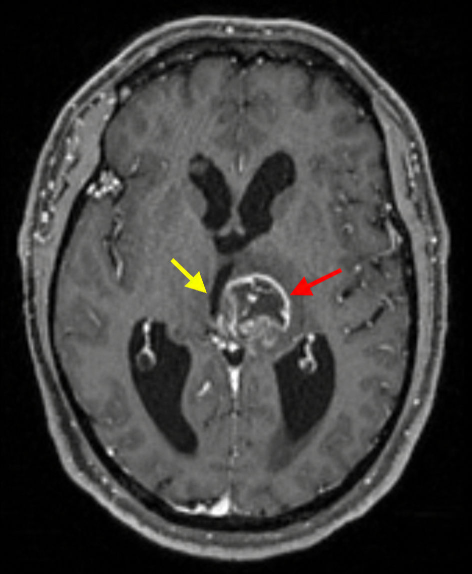
↓ Figure 1. Initial MRI brain, T1-weighted sequence (axial view), demonstrating a predominantly T1-hypointense intra-axial mass centered within the left thalamus with extension into the left midbrain (red arrow). Significant mass effect is present with compression and rightward deviation of the third ventricle (yellow arrow). MRI: magnetic resonance imaging.
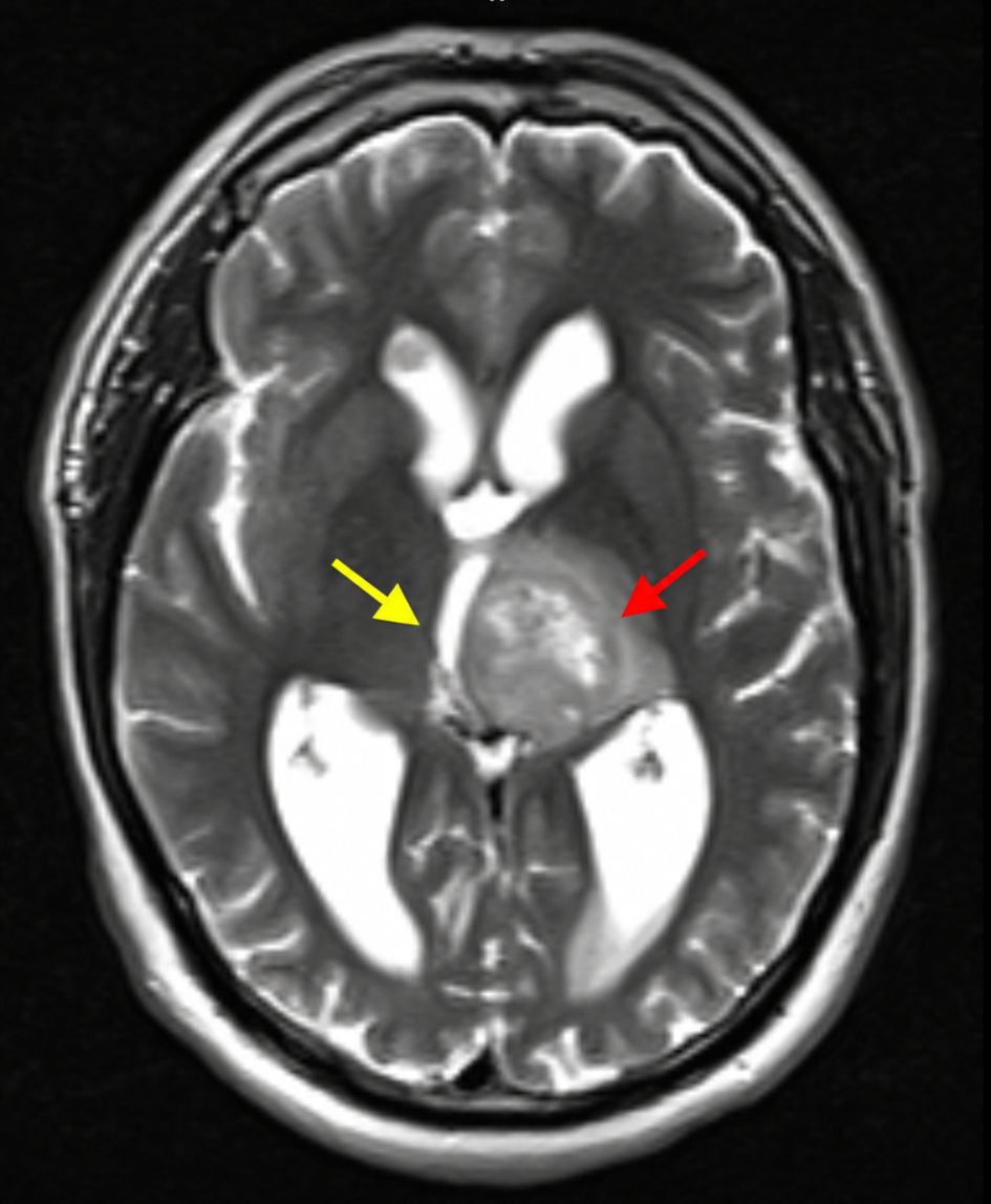
↓ Figure 2. Initial MRI brain, T2-weighted sequence (axial view), demonstrating a heterogeneous left thalamic mass with mixed T2 hypo- and hyperintense signal characteristics (red arrow). Extensive surrounding vasogenic edema is evident, extending into adjacent deep white matter structures (yellow arrow). MRI: magnetic resonance imaging.
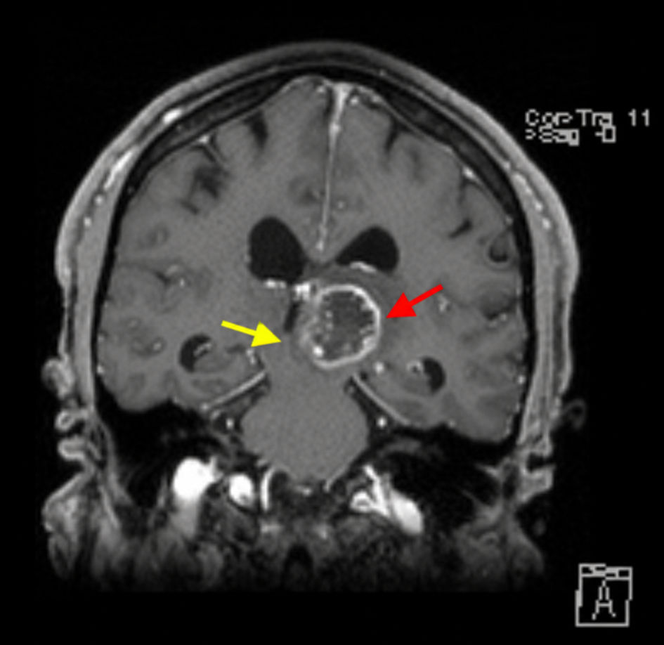
↓ Figure 3. Initial MRI brain, post-contrast T1-weighted sequence (coronal view), showing a heterogeneously enhancing lesion centered within the left thalamus with extension into the midbrain (red arrow). Associated mass effect and third-ventricular compression are demonstrated (yellow arrow). MRI: magnetic resonance imaging.
↓ Figure 4. Follow-up MRI brain obtained after completion of radiotherapy, post-contrast T1-weighted sequence (axial view), demonstrating interval reduction in the size of the heterogeneously enhancing left thalamic lesion, consistent with treatment response (yellow arrow). MRI: magnetic resonance imaging.
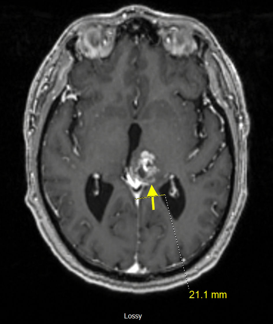
↓ Figure 5. Follow-up MRI brain obtained after completion of radiotherapy, T1-weighted sequence (coronal view), demonstrating interval reduction in the size of the heterogeneously enhancing left thalamic lesion, consistent with treatment response (yellow arrow). MRI: magnetic resonance imaging.
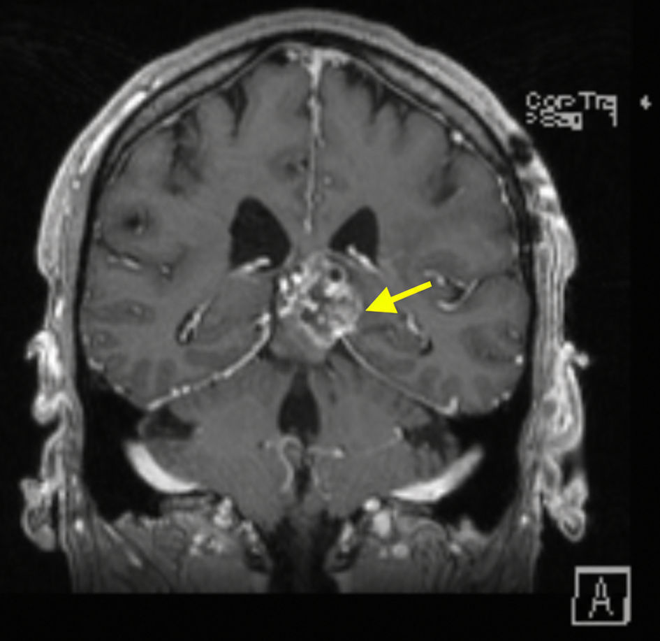
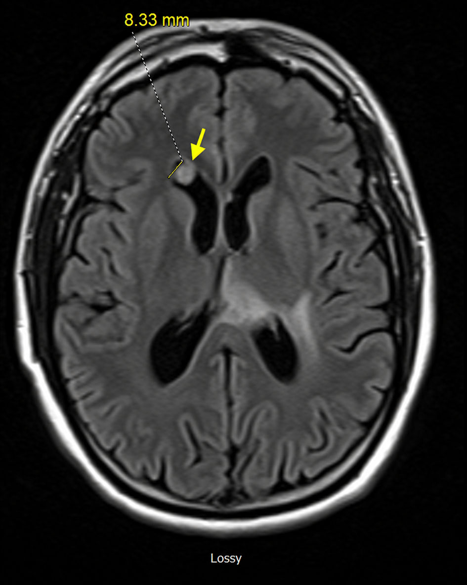
↓ Figure 6. Follow-up MRI brain, post-contrast T1-weighted sequence (axial view), demonstrating a small enhancing subependymal nodule along the floor of the anterior right lateral ventricle near the frontal horn, concerning for possible early subependymal dissemination (yellow arrow). MRI: magnetic resonance imaging.