Journal of Medical Cases, ISSN 1923-4155 print, 1923-4163 online, Open Access
Article copyright, the authors; Journal compilation copyright, J Med Cases and Elmer Press Inc
Journal website https://jmc.elmerpub.com

Case Report

Volume 17, Number 11, November 2026, pages 637-641


Pregnancy-Associated Vulval Lymphangiectasia With a Non-Vesicular Wart-Like Appearance

Figures

↓  Figure 1. Serial vulval examinations showing multiple skin-colored papules on both labia before delivery (a) and 1 day postpartum (b). The lesions began to regress by 2 days postpartum (c) and had almost completely regressed by 4 weeks postpartum (d).
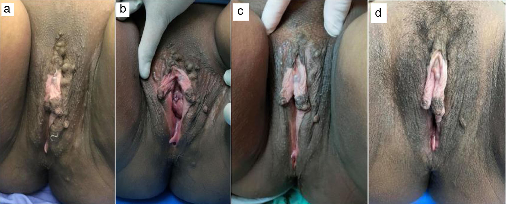
Figure 1.
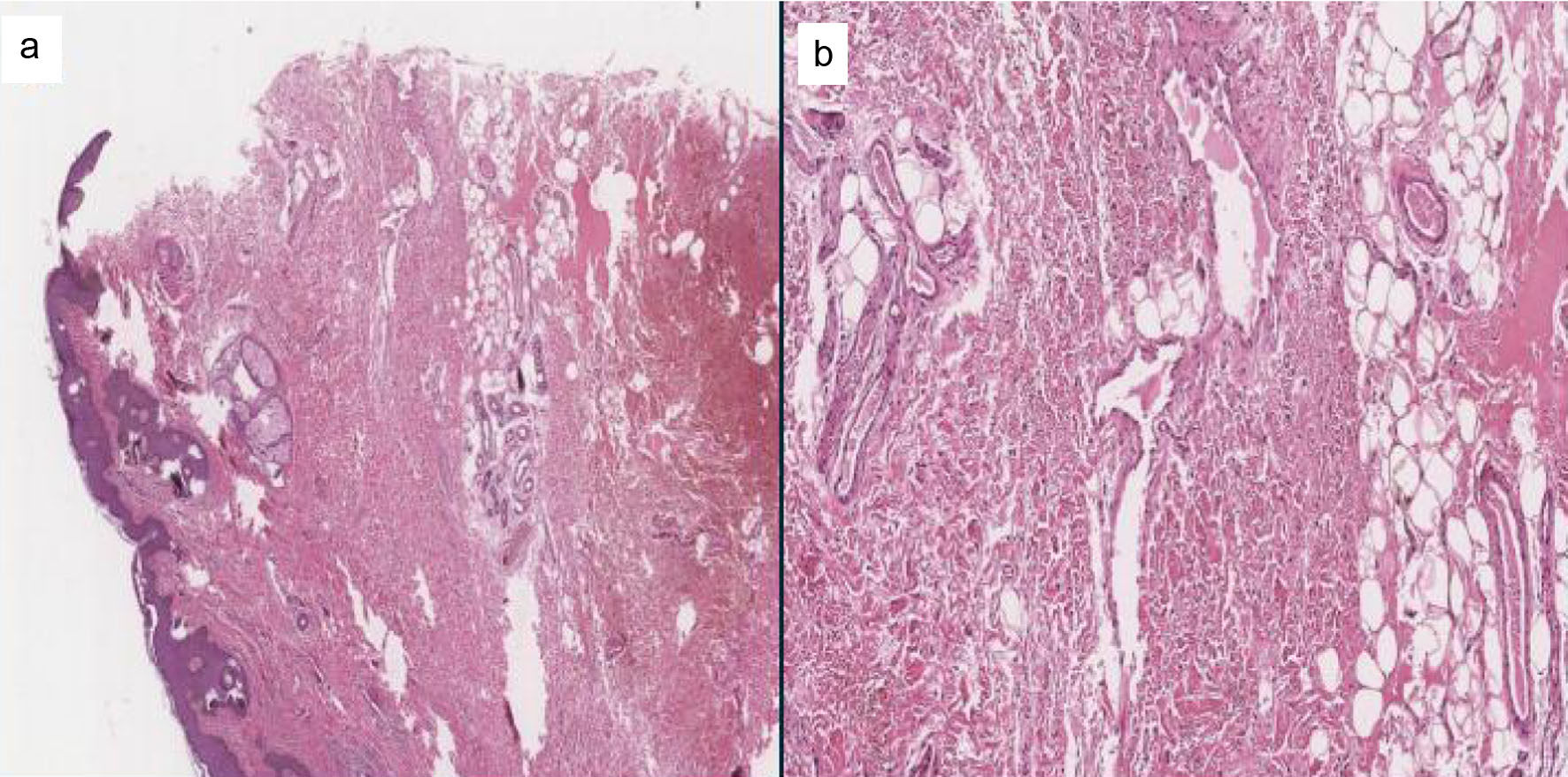
↓  Figure 2. Histopathological findings of the vulval lesion. (a) Hematoxylin and eosin staining showing dilated, thin-walled vascular space within the superficial dermis (original magnification, × 20). (b) Higher-magnification view showing the dilated, thin-walled vascular space lined by a single layer of flattened endothelial cells (original magnification, × 40).
Figure 2.