Figures
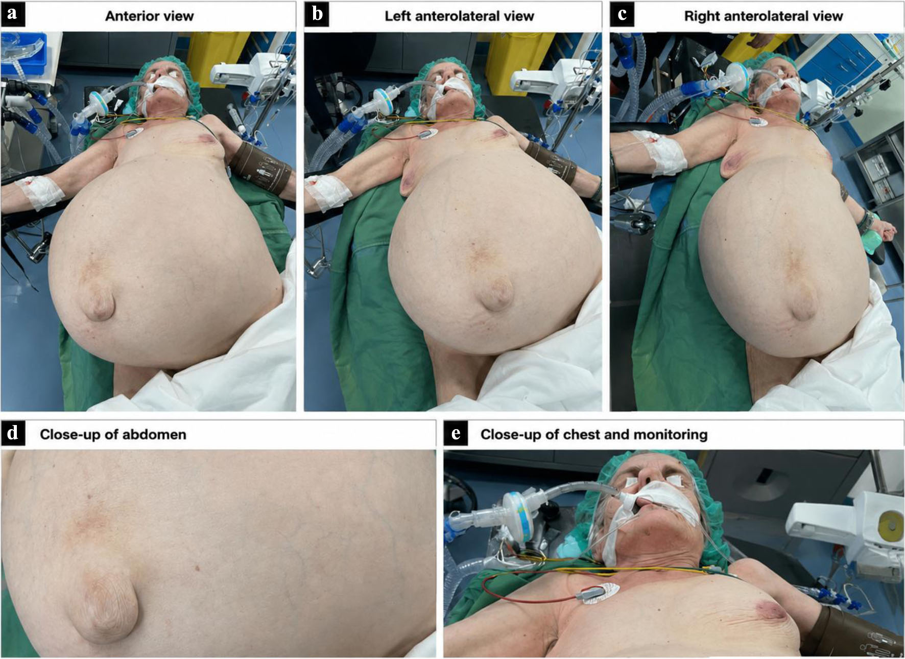
↓ Figure 1. A giant tumor caused marked abdominal distension and increased intra-abdominal pressure.
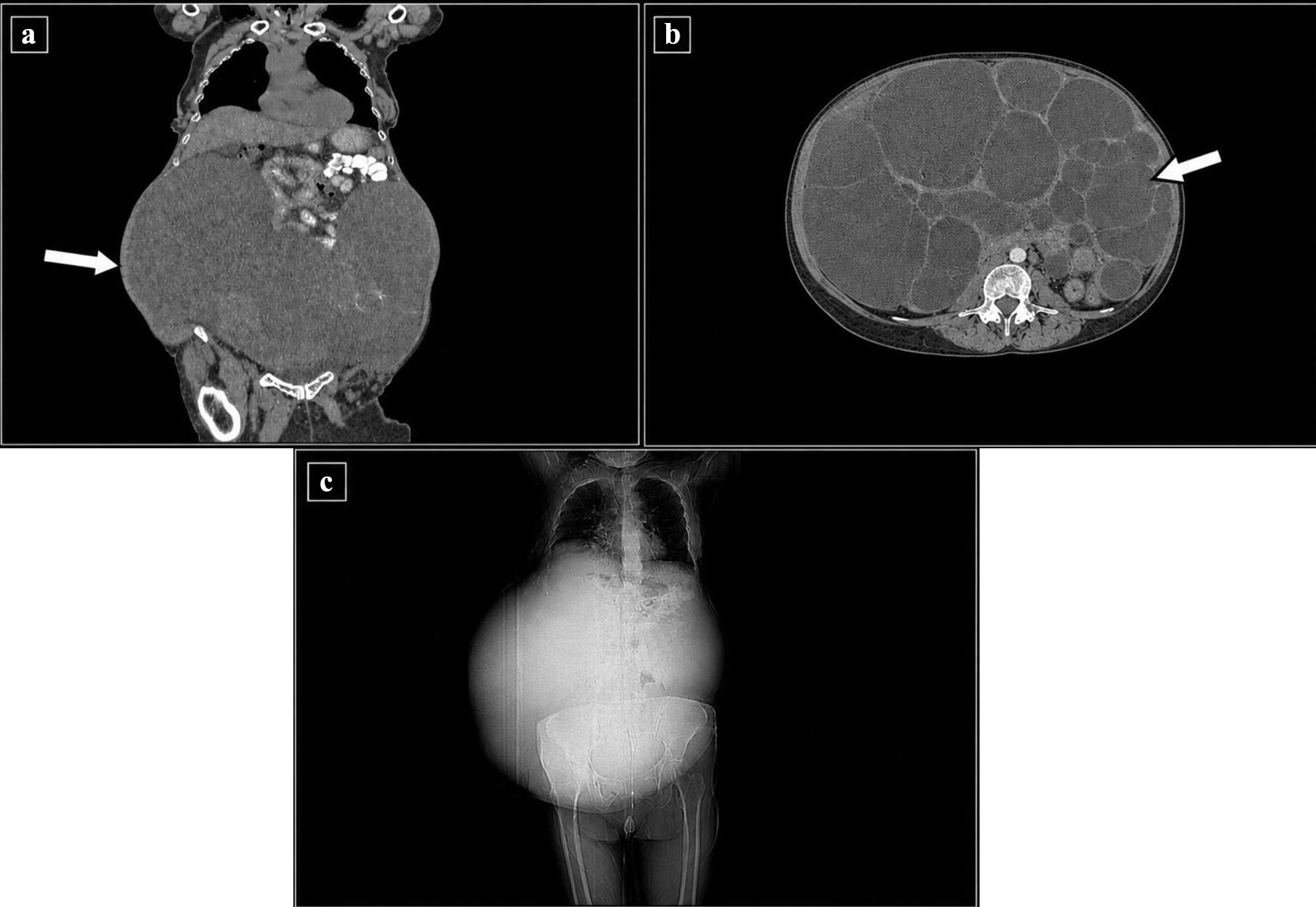
↓ Figure 2. Computed tomography (CT) scan examination revealed large intra-abdominal tumor. (a) Coronal contrast-enhanced CT image showing a large intra-abdominal tumor occupying the lower abdomen. Arrow indicates giant tumor. (b) Axial contrast-enhanced CT image demonstrating the multi-cystic nature of the tumor. Arrow indicates cysts. (c) CT scout image demonstrating the marked abdominal distention caused by the large intra-abdominal mass.
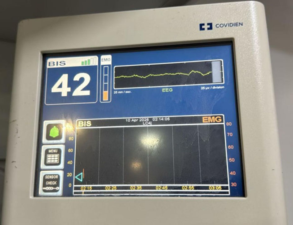
↓ Figure 3. Bispectral Index (BIS) monitoring showed a value of 42, indicating an adequate depth of anesthesia during the procedure and helping guide anesthetic titration despite significant intraoperative hemodynamic fluctuations.
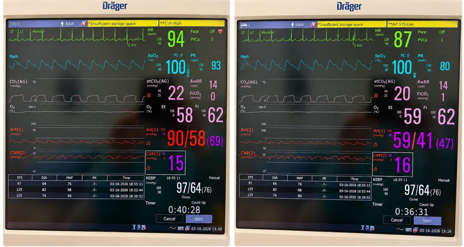
↓ Figure 4. Hemodynamic parameters during different intraoperative phases (a) and hypotension (ART 1 red wave) occurred only by patient’s abdominal repositioning (b). In both monitoring views central venous pressure (CVP, red wave) is high, indicating intra-abdominal pressure.
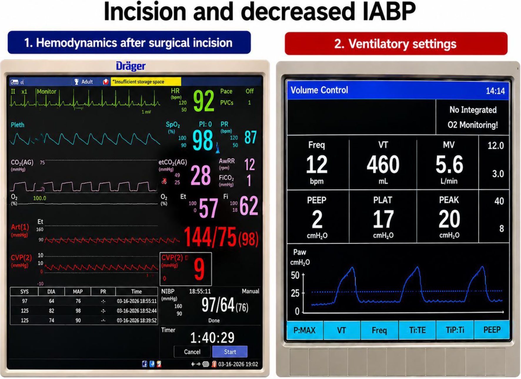
↓ Figure 5. Hemodynamic and ventilatory parameters improved following surgical incision and relief of intra-abdominal pressure. HR: heart rate; SpO2: pulse oximetry; ETCO2: end-tidal CO2; AwRR: airway respiratory rate; Art: arterial pressure; CVP: central venous pressure; NIBP: non-invasive blood pressure; PI: Perfusion Index; PR: pulse rate; PVCs: premature ventricular contractions; VT: tidal volume; MV: minute ventilation; PEEP: positive end-expiratory pressure; PLAT: plateau pressure; PEAK: peak inspiratory pressure; Paw: airway pressure; Ti: inspiratory time; TE: expiratory time; TiP: pause time.
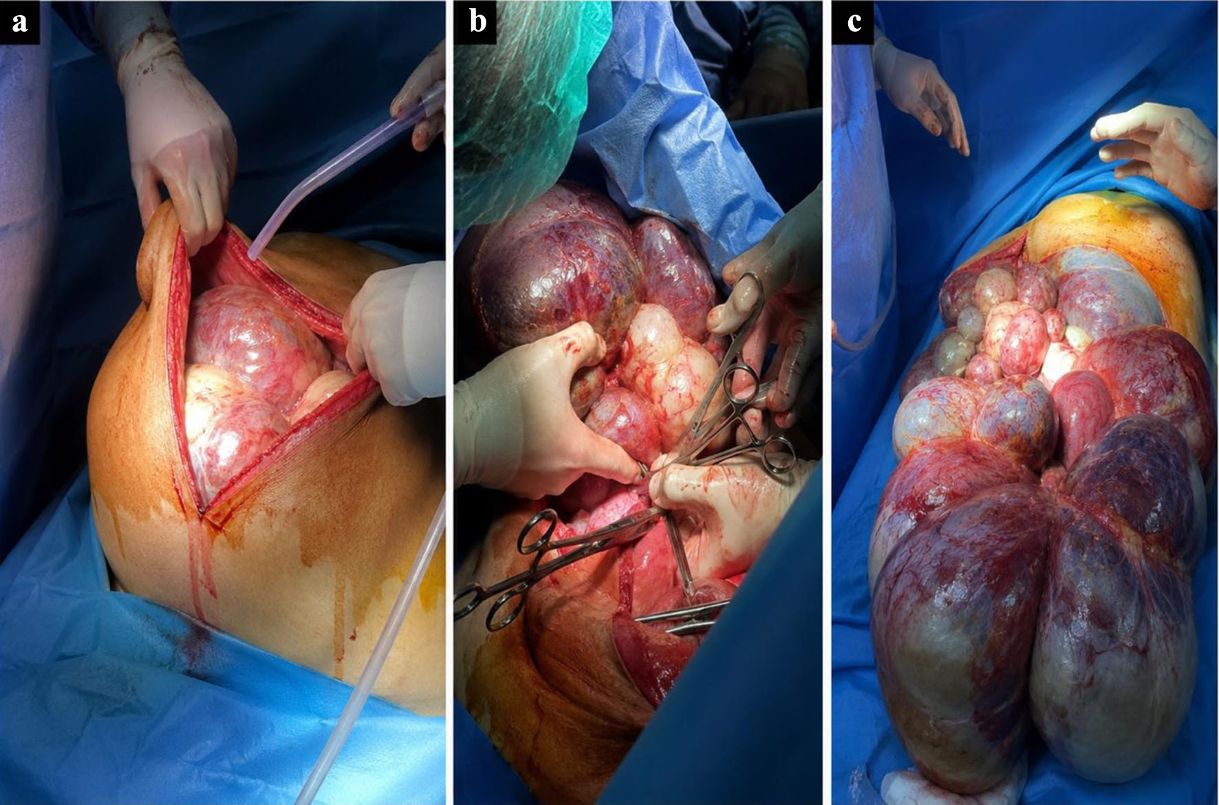
↓ Figure 6. Different surgical stages, from the surgical incision (a) and preparation of the anatomical resection plan (b) to complete removal of the giant tumor (c).
Table
↓ Table 1. Perioperative Hemodynamic Changes and Intraoperative Clinical Data Associated With Giant Intra-Abdominal Tumor Resection
| Variable | Before mass removal | After mass removal |
|---|
| aHemodynamic stability was achieved with crystalloid administration and noradrenaline infusion under invasive monitoring. CVP: central venous pressure. |
| Arterial blood pressure (mm Hg) | 59/41 | Improved with vasopressor and fluid therapya |
| CVP (mm Hg) | 16 | 1 |
| Peak inspiratory pressure (cm H2O) | 34 | 20 |
| Plateau pressure (cm H2O) | 29 | 17 |
| Urinary output | 2,000 mL |
| Estimated blood loss | 200 mL |
| Ringer’s lactate administered | 3,500 mL |
| Hemoglobin | 9.5 g/dL |
| Lactate | 1.3 mmol/L |
| Time to extubating | 2 h after ICU admission |