Figures
↓ Figure 1. Diagnosis. (a) Preoperative clinical images: occlusal and buccal views, showing white temporary filling material on the occlusal surface. (b) Periapical radiographs of tooth #26 taken before and after root canal treatment (prior to apical surgery). The red arrow indicates the separated endodontic instrument. (c) Preoperative left maxillary CBCT scan, sagittal and axial slices. CBCT: cone-beam computed tomography.

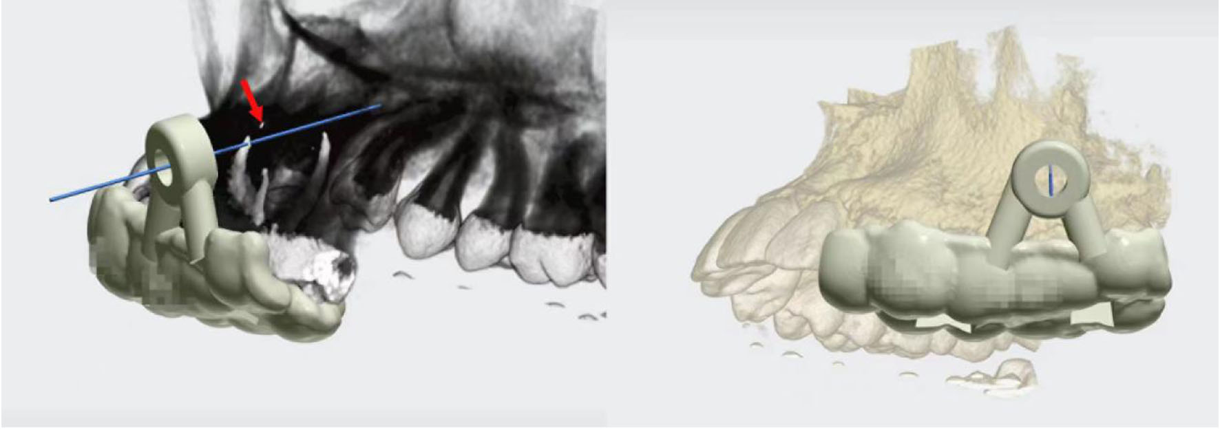
↓ Figure 2. Digital surgical guide designed with 3Shape Unite software; the red arrow points to the separated endodontic instrument.
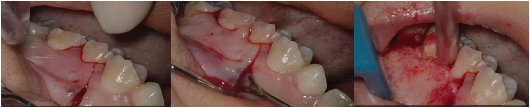
↓ Figure 3. Angular flap (#25–#27 buccal, intrasulcular), full-thickness vertical/horizontal incisions, flap elevation, bone exposure.
↓ Figure 4. Digital surgical guide seated, trephine windowing, buccal bone plate removal, and apical localization.
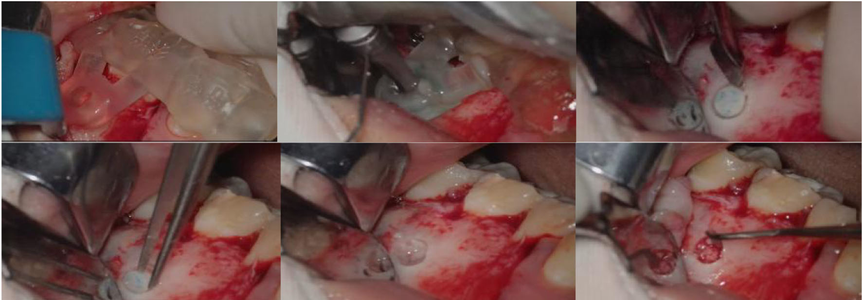
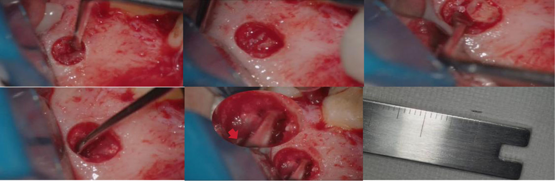
↓ Figure 5. Apicoectomy of tooth #26, debridement of the periapical cyst, and removal of the separated instrument, the red arrow points to the separated endodontic instrument.
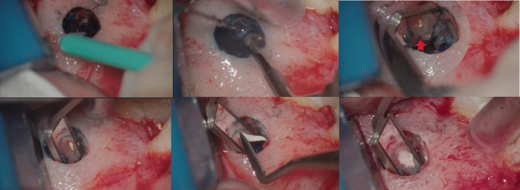
↓ Figure 6. Methylene blue staining, retrograde preparation, and retrograde filling. Developmental malformation of the mesiobuccal root (red arrow).
↓ Figure 7. Postoperative periapical radiographs. (a) Immediate postoperative; (b) 4-month follow-up; (c) 9-month follow-up; (d) 1-year follow-up; (e) 2-year follow-up.

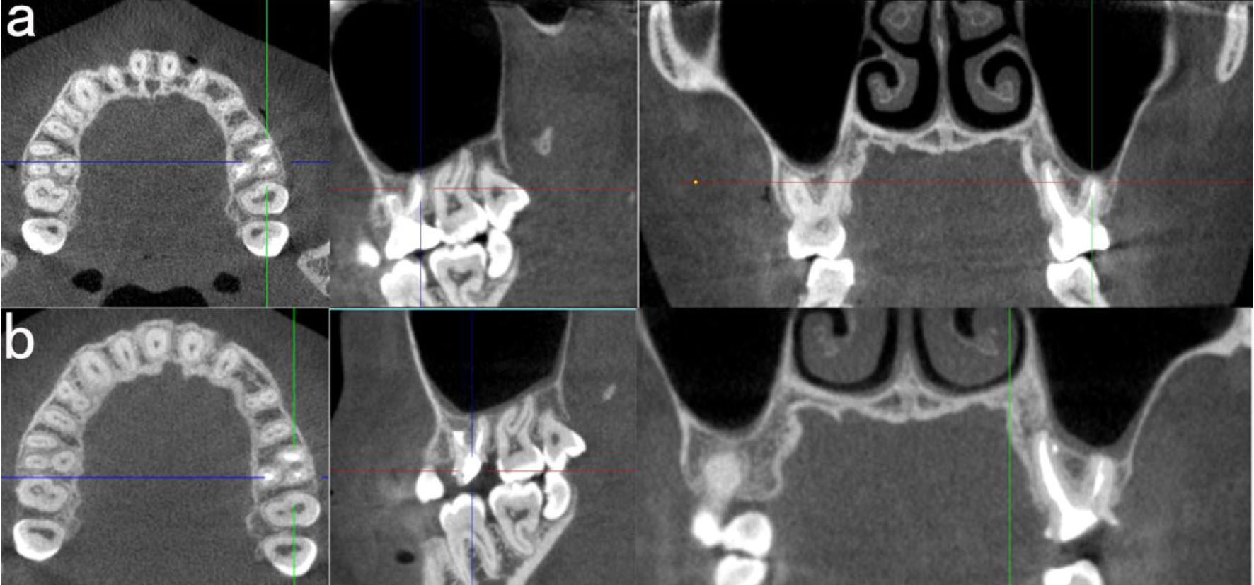
↓ Figure 8. Postoperative CBCT. (a) 1-year follow-up; (b) 2-year follow-up. CBCT: cone-beam computed tomography.
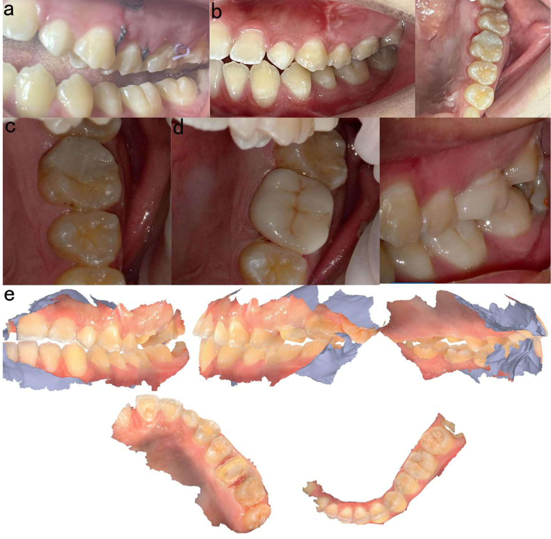
↓ Figure 9. Intraoral photographs. (a) Before suture removal (10 days postoperatively); (b) 1-year follow-up; (c) 2-year follow-up; (d) Intraoral view after crown restoration (2 years); (e) Intraoral scan of the crown restoration.