Figures
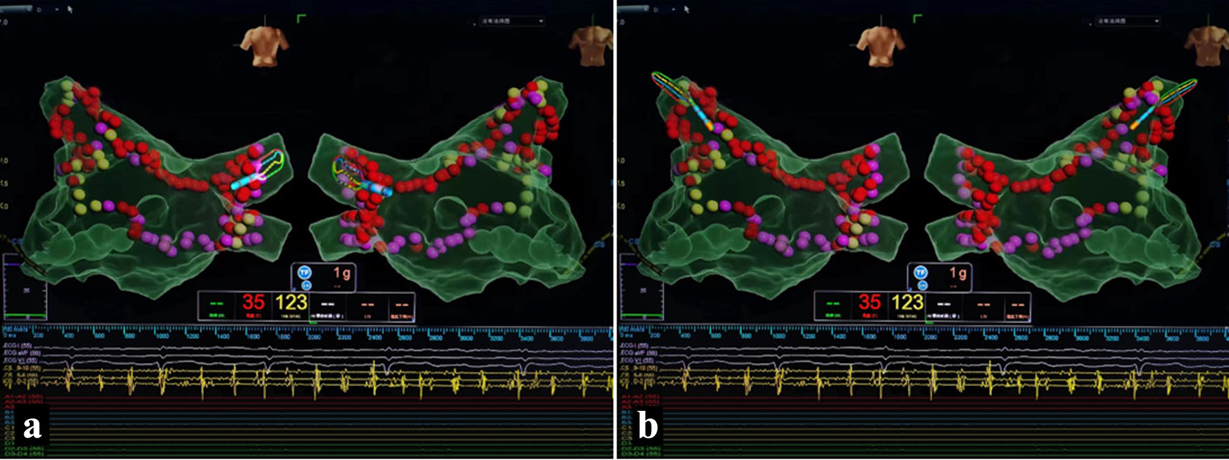
↓ Figure 1. Pulmonary vein isolation. (a) Right pulmonary vein isolation. (b) Left pulmonary vein isolation.
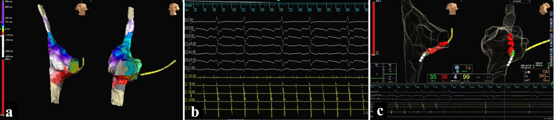
↓ Figure 2. Electrophysiological examination and ablation of atrial flutter. (a) Early activation on CS9-0, indicating right atrial origin of flutter. (b) Mapping of the right atrium using an HD-Grid mapping catheter revealed counterclockwise cavo-tricuspid isthmus-dependent atrial flutter. (c) Pressure-sensing saline-irrigated catheter was used as the ablation catheter to create a line across the tricuspid isthmus. The power was set at 35 W, the temperature at 43 °C, and the saline irrigation rate at 17 mL/min. The sinus rhythm was restored during the ablation procedure.
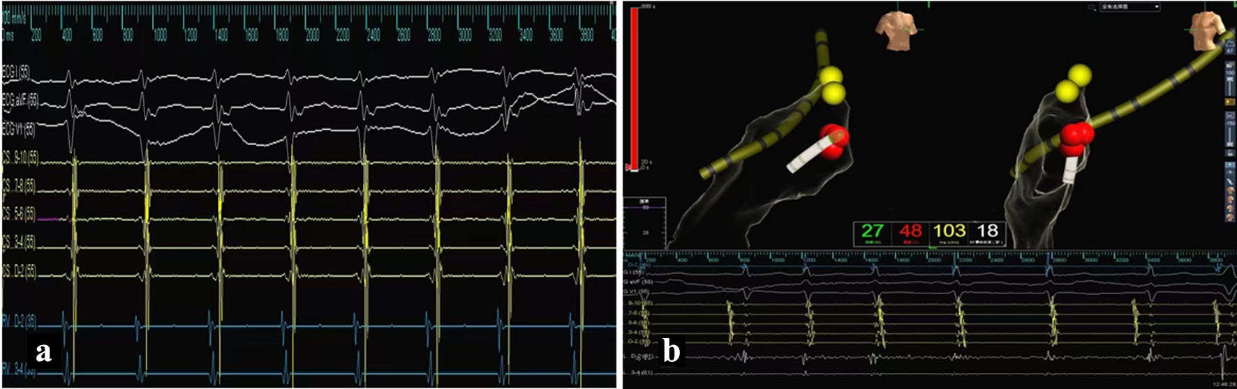
↓ Figure 3. Electrophysiological examination and ablation of supraventricular tachycardia. (a) Dual-pathway supraventricular tachycardia (SVT) was induced. (b) Target points for ablation were identified in the slow-pathway region. Using a 50 W, 55 °C temperature-controlled mode, effective ablation was indicated by the appearance of slow junctional rhythm, consolidated for 120 s, and confirmed by the lack of recurrence after the procedure.
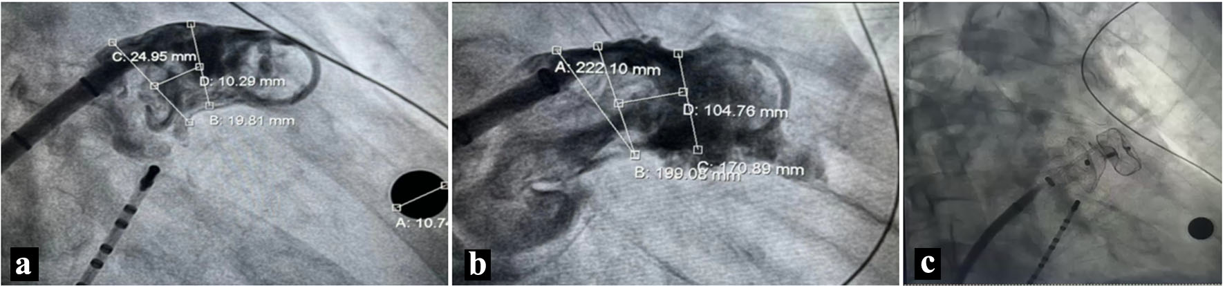
↓ Figure 4. Left atrial appendage (LAA) occlusion. (a) The asymmetry of the LAA’s upper and lower edges was noted, with a longer upper ridge, measuring a fixation zone of 17–19 mm, and an LAA orifice of 20 mm. (b) With a clear landing zone, a 20–26 mm LACBES occluder was selected for occlusion. (c) The successful deployment of the occluder, which indicated it can be released according to PAST principles.