Journal of Medical Cases, ISSN 1923-4155 print, 1923-4163 online, Open Access
Article copyright, the authors; Journal compilation copyright, J Med Cases and Elmer Press Inc
Journal website https://jmc.elmerpub.com

Case Report

Volume 17, Number 10, October 2026, pages 558-562


Well-Differentiated Liposarcoma of the Spermatic Cord Involving Testicular Parenchyma

Figures

↓  Figure 1. Coronal contrast-enhanced CT of the abdomen and pelvis demonstrating bilateral fat-containing inguinal masses (red arrows). The larger right-sided lesion extends through the inguinal canal into the right hemiscrotum and was initially interpreted as an inguinal hernia containing mesenteric fat. Retrospective review demonstrated internal soft tissue nodularity and mass effect concerning liposarcoma. CT: computed tomography.
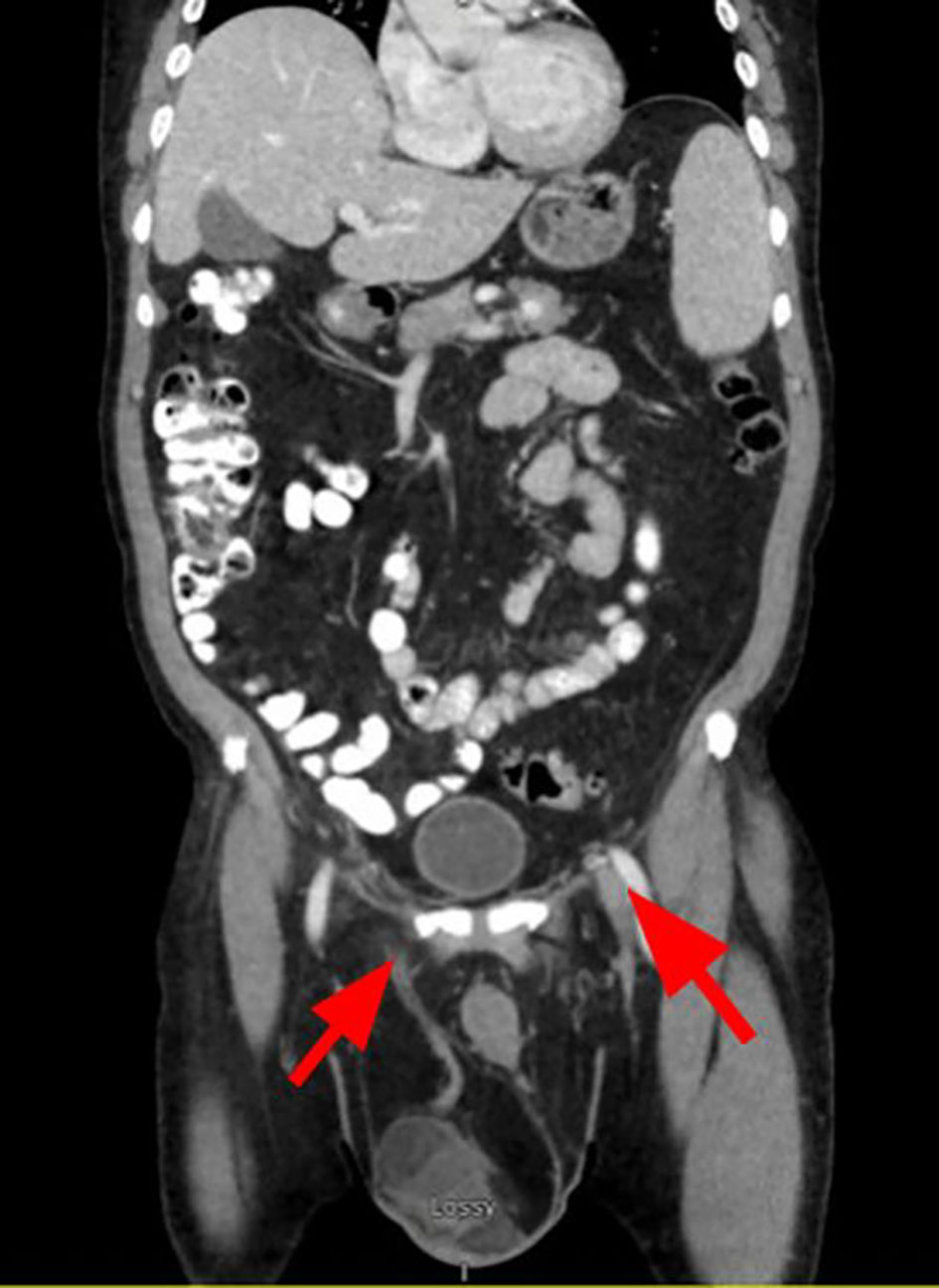
Figure 1.
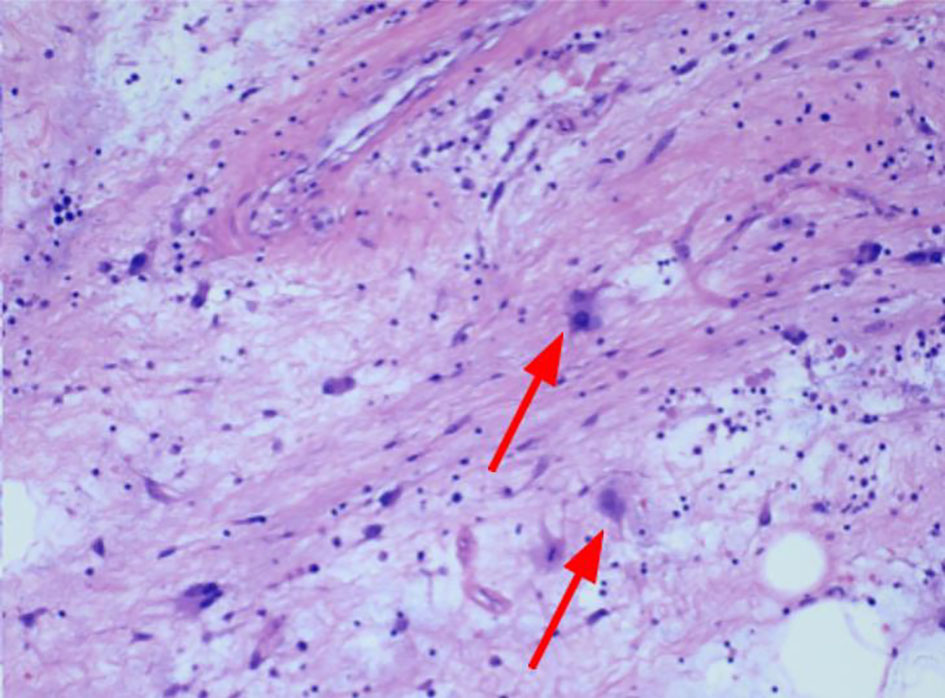
↓  Figure 2. Hematoxylin and eosin stain slides of excised scrotal mass. The tumor is composed of adipocytes with bands of fibrotic stroma showing markedly atypical cells with enlarged, hyperchromatic nuclei (red arrows).
Figure 2.