Figures
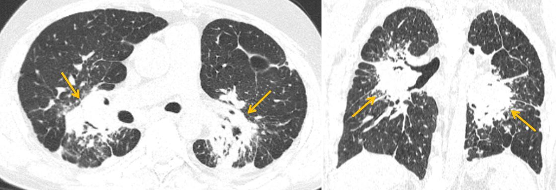
↓ Figure 1. Axial and coronal HRCT images (lung window) demonstrating heterogeneous perihilar consolidation with airless conglomerates (yellow arrows), preserved bronchial patency, and linear pleural adhesions. HRCT: high-resolution computed tomography.
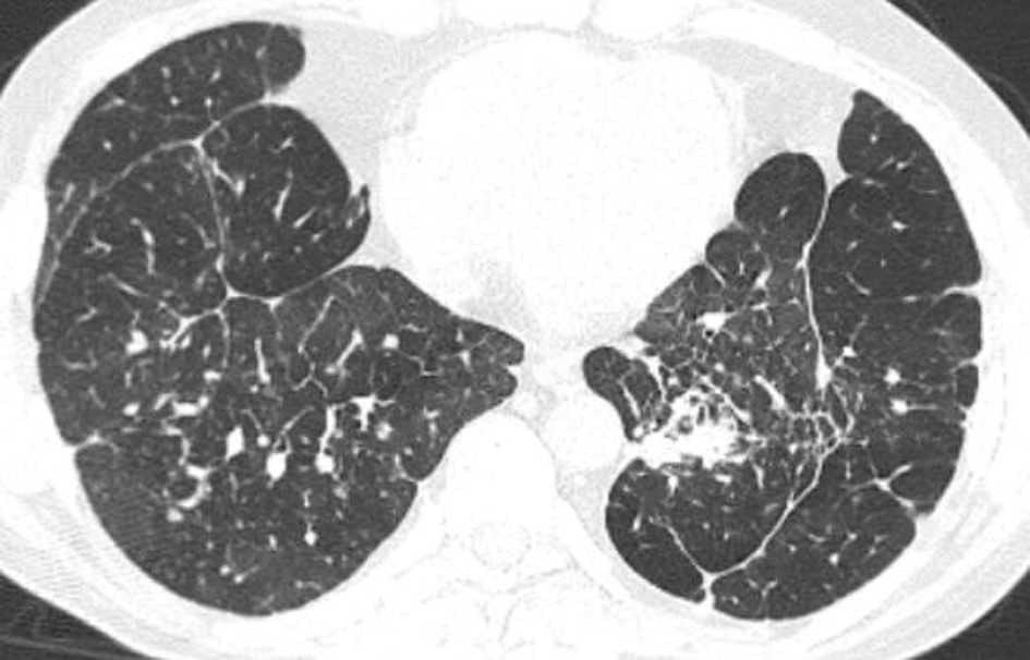
↓ Figure 2. Axial HRCT image (lung window) demonstrating diffuse interstitial reticular changes with interlobular septal thickening, nodular thickening of bronchovascular bundles, and ill-defined ground-glass opacities with preserved bronchial patency. HRCT: high-resolution computed tomography.
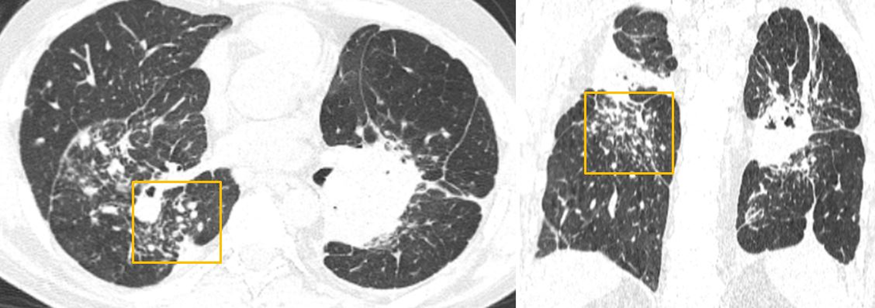
↓ Figure 3. Axial and coronal HRCT images (lung window) demonstrating multiple polymorphic nodules (yellow box) distributed along the bronchovascular bundles and interlobar pleura, without evidence of coalescence. HRCT: high-resolution computed tomography.
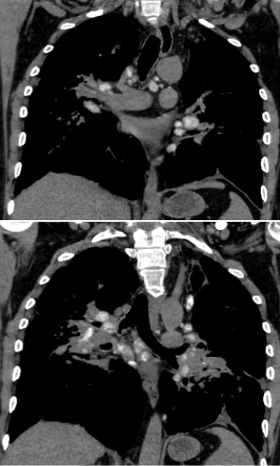
↓ Figure 4. Coronal HRCT image (mediastinal window) demonstrating multiple enlarged mediastinal and hilar lymph nodes involving the upper and lower paratracheal, subaortic, pulmonary, subcarinal, and paraaortic stations. HRCT: high-resolution computed tomography.
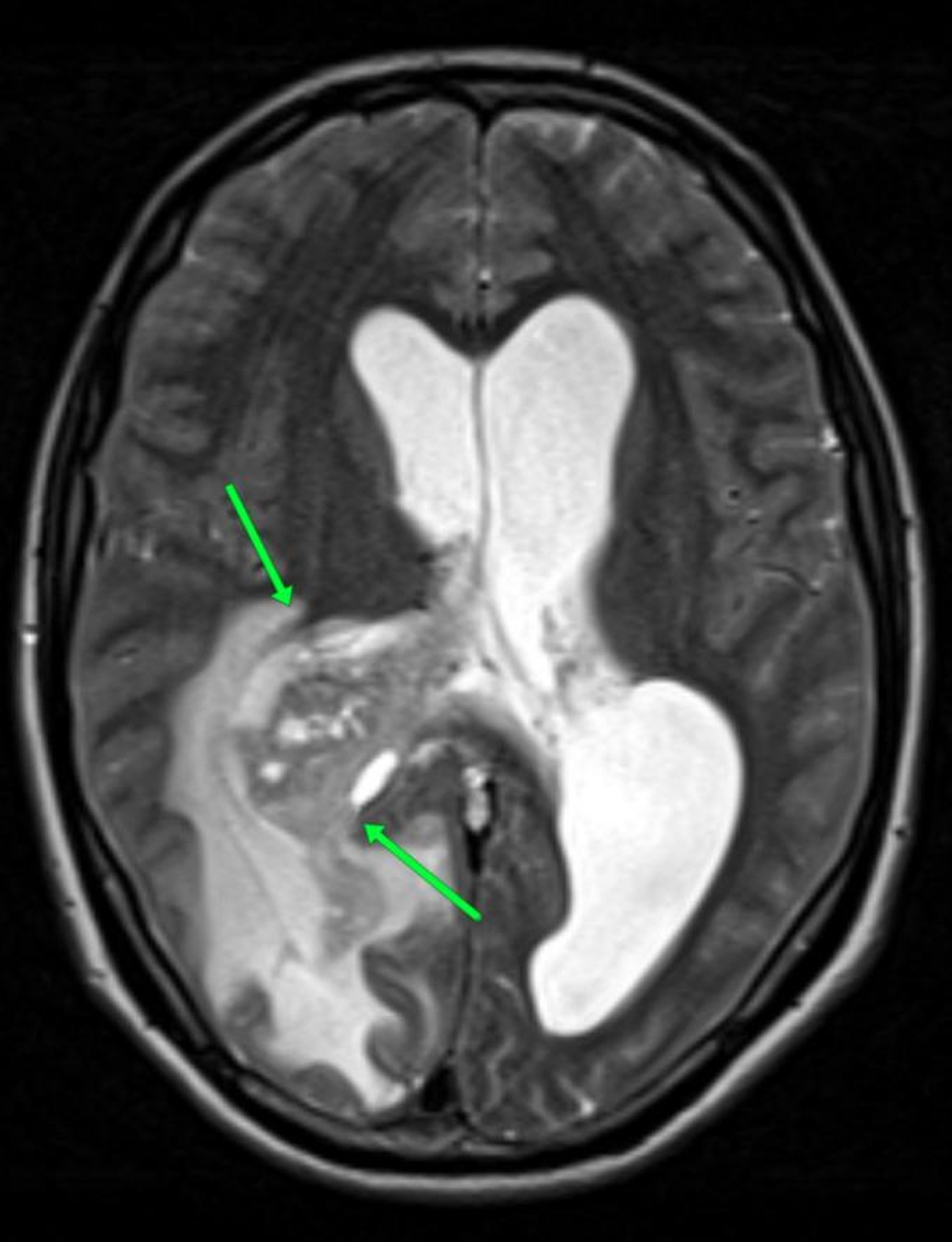
↓ Figure 5. Axial T2-weighted MRI demonstrating a large intraventricular mass (green arrows) arising from the choroid plexus in the atrium of the right lateral ventricle, measuring approximately 6–7 cm in maximal dimension and greater than 3 cm in thickness. MRI: magnetic resonance imaging.
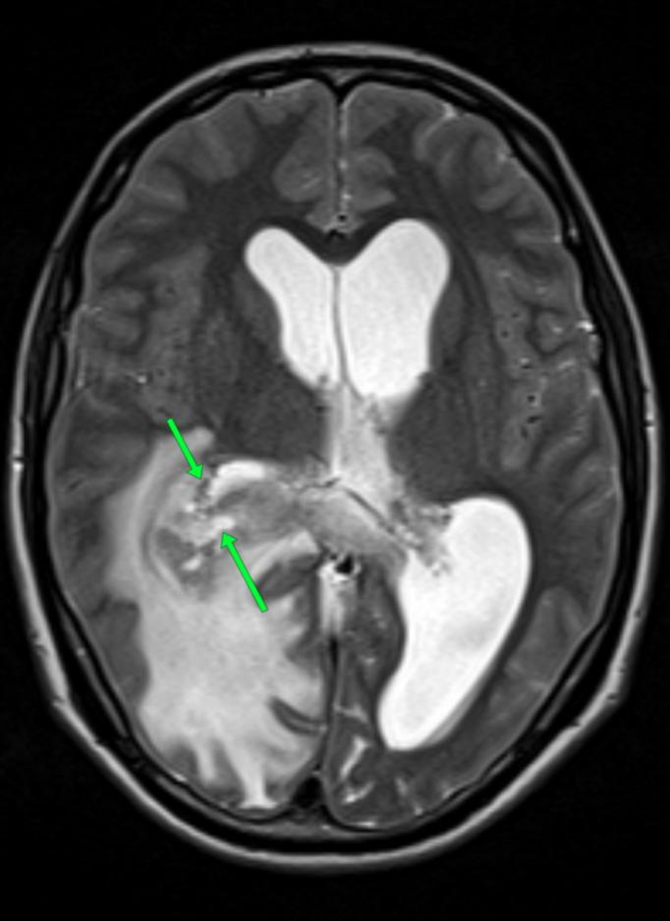
↓ Figure 6. Axial T2-weighted MRI demonstrating a heterogeneous lesion with multiple septations and mixed cystic and solid components, without diffusion restriction on DWI. DWI: diffusion-weighted imaging; MRI: magnetic resonance imaging.
↓ Figure 7. Post-contrast axial T1-weighted MRI demonstrating intense, heterogeneous enhancement of the lesion (green arrows). MRI: magnetic resonance imaging.
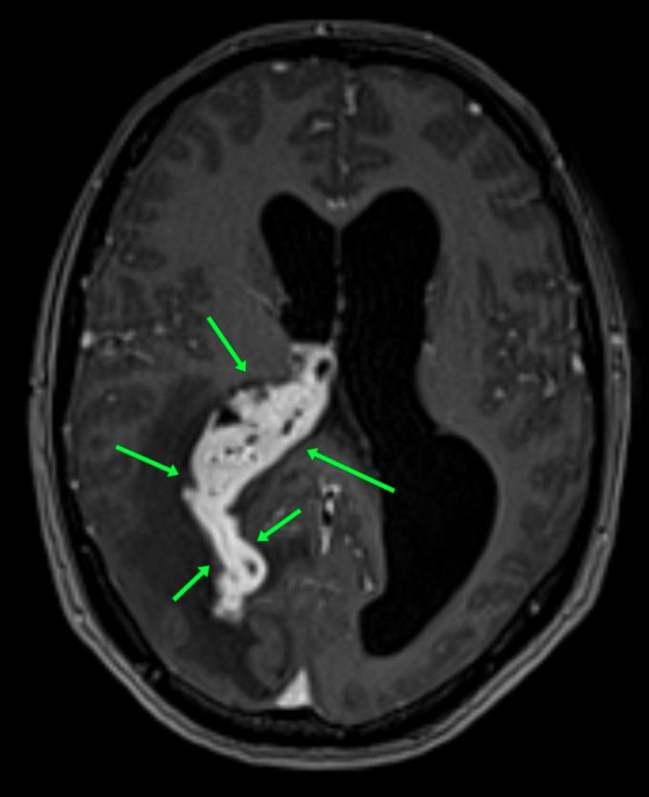
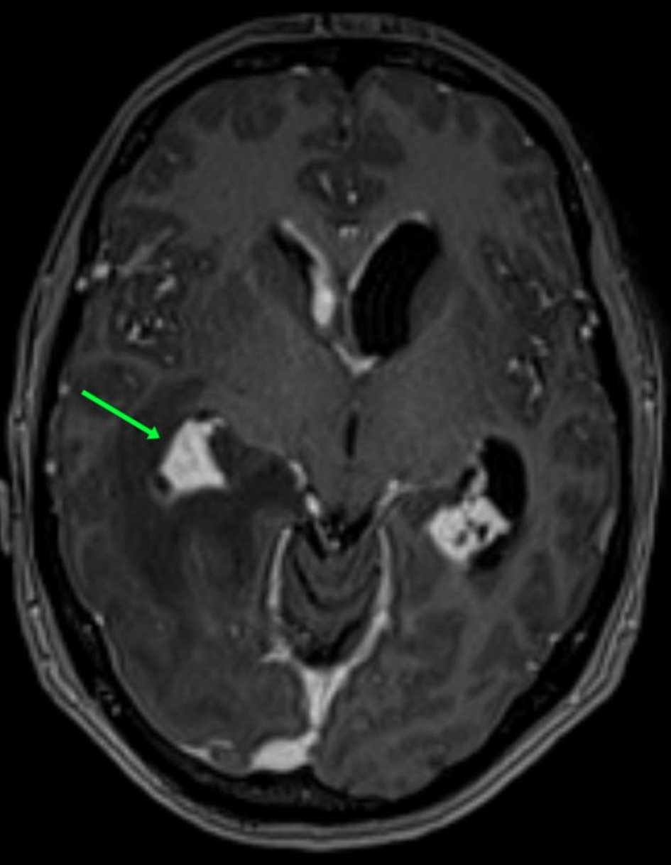
↓ Figure 8. Post-contrast axial T1-weighted MRI demonstrating extension of the lesion into the posterior and temporal horns of the right lateral ventricle, with near-complete obliteration and distortion of the ventricular lumen (green arrows). MRI: magnetic resonance imaging.
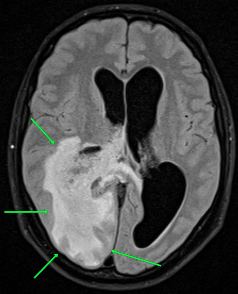
↓ Figure 9. Axial T2-FLAIR MRI demonstrating marked periventricular white matter edema involving the adjacent temporal and occipital lobes (green arrows). FLAIR: fluid-attenuated inversion recovery; MRI: magnetic resonance imaging.
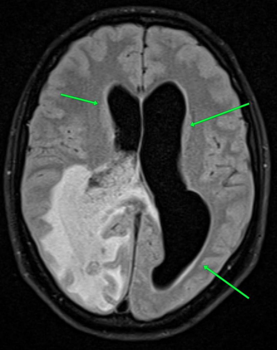
↓ Figure 10. Axial T2-FLAIR MRI demonstrating moderate dilation of the lateral ventricles, more pronounced on the contralateral side (green arrows), consistent with obstructive hydrocephalus. FLAIR: fluid-attenuated inversion recovery; MRI: magnetic resonance imaging.
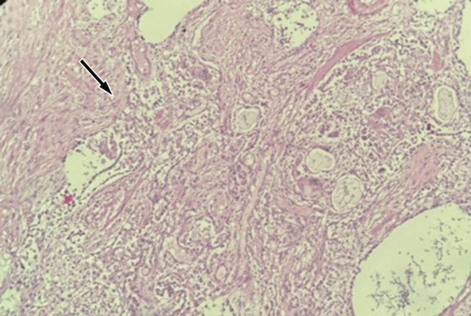
↓ Figure 11. Histological section of the lung demonstrating marked interstitial fibrosis with distortion of the surrounding pulmonary architecture (arrow) and associated chronic inflammatory changes (hematoxylin and eosin stain, × 200).
↓ Figure 12. Histological section obtained from a grossly abnormal central nervous system lesion, demonstrating epithelioid granulomatous inflammation (arrow) with a peripheral lymphocytic infiltrate (arrowhead) (hematoxylin and eosin stain, × 200).
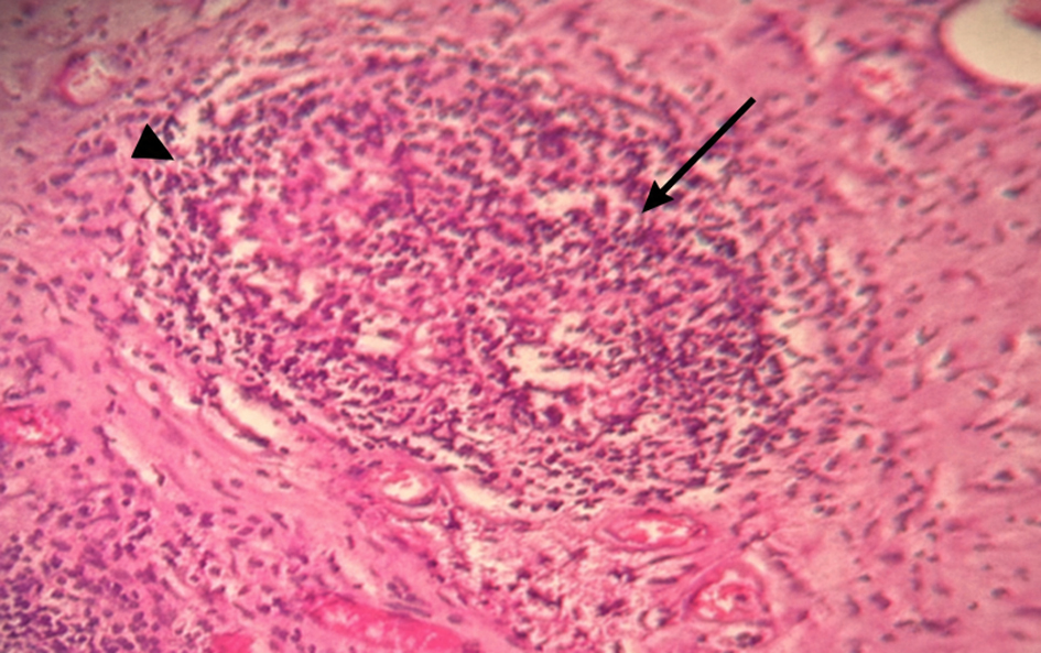
↓ Figure 13. Histological section obtained from the central nervous system lesion demonstrating non-necrotizing granulomatous inflammation (arrow) with a peripheral lymphocytic infiltrate (arrowhead) (hematoxylin and eosin stain, ×200).
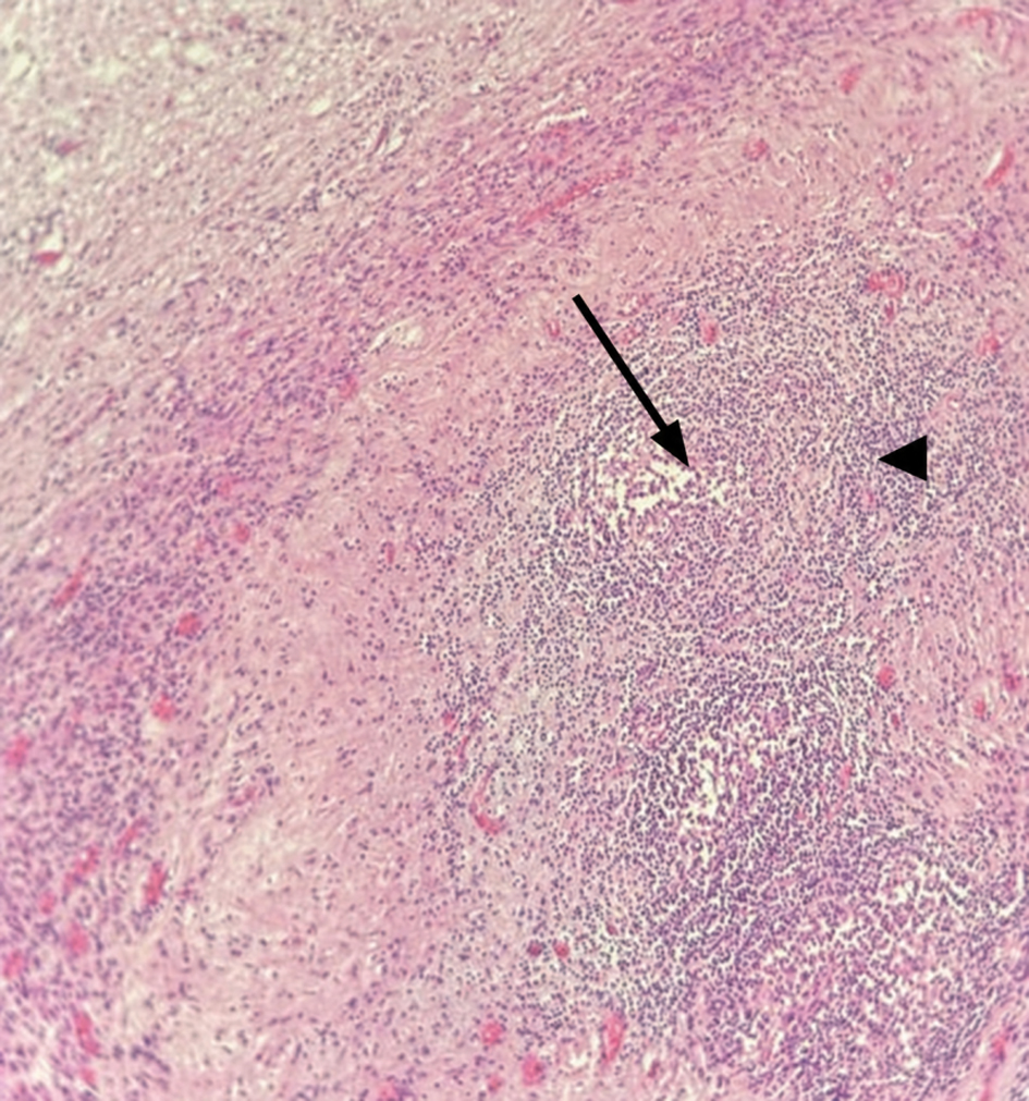
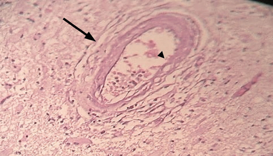
↓ Figure 14. Histological section of a central nervous system vessel demonstrating concentric fibrotic thickening of the vessel wall (arrow) and marked luminal narrowing (arrowhead) (hematoxylin and eosin stain, ×200).