Figures
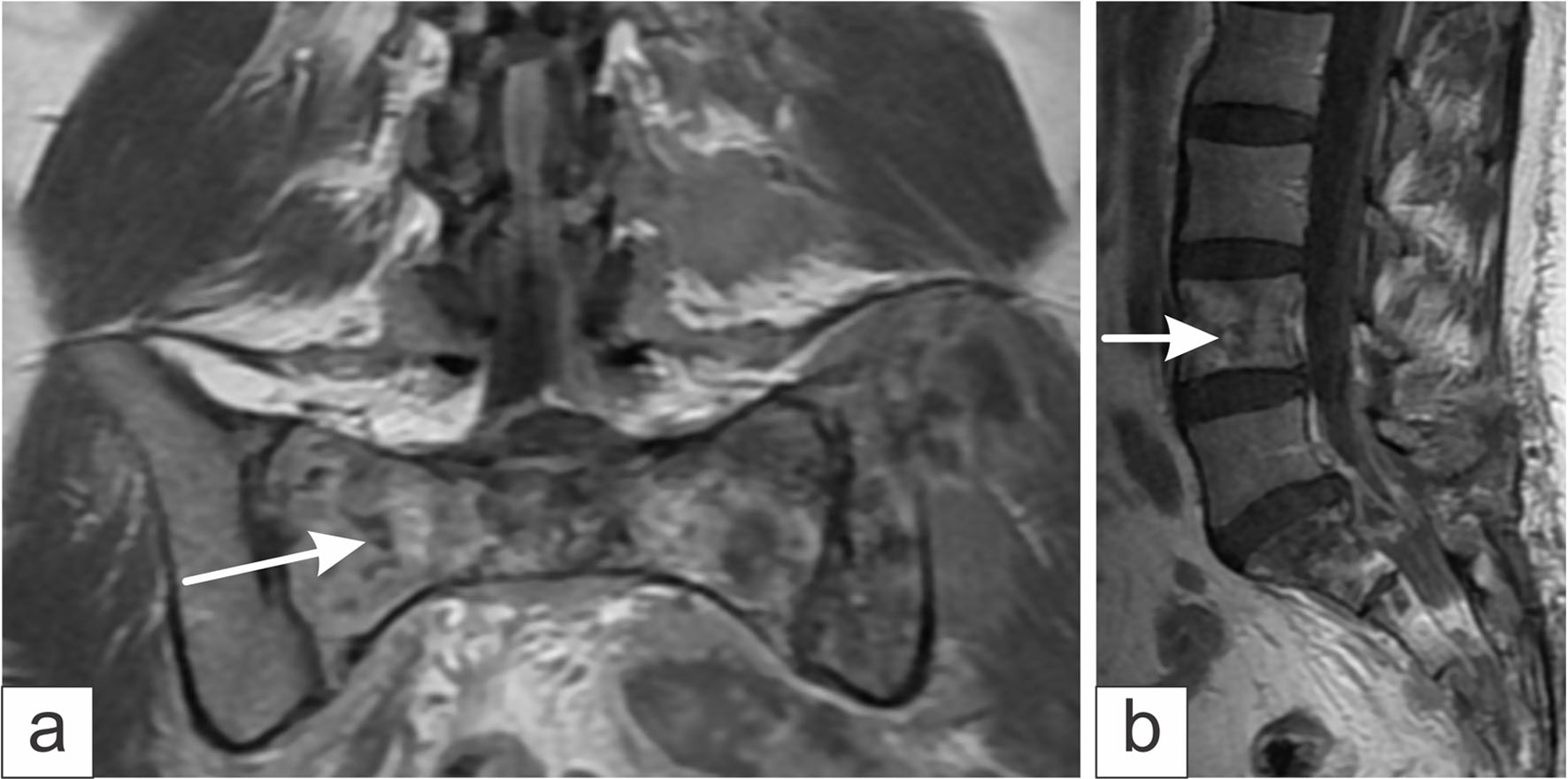
↓ Figure 1. Magnetic resonance imaging obtained using the T2-weighted sequence. (a) Coronal section shows infiltration with multiple lytic and infiltrative lesions at the level of the sacrum (white arrow) and left iliac crest. (b) Sagittal section showing lytic lesions at the level of L4 (white arrow) and the sacrum.
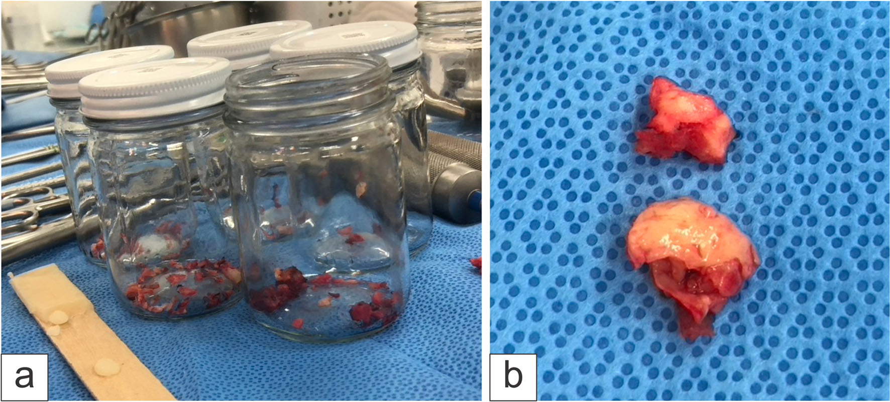
↓ Figure 2. Photographs of (a) sacral bone biopsies and L5 laminectomy, and (b) tissue compressing the extradural S1 region, yellowish in color and soft in consistency.
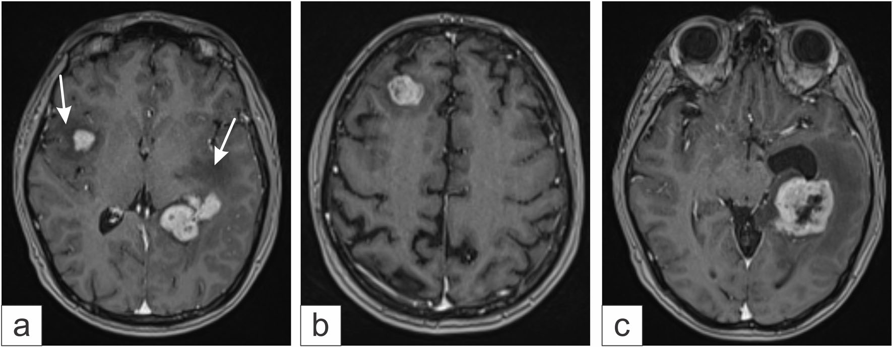
↓ Figure 3. Gadolinium-enhanced brain magnetic resonance imaging. (a) Multiple metastases with perilesional edema (white arrows) are observed. (b) Right frontal metastases. (c) Metastases in the left temporal region.
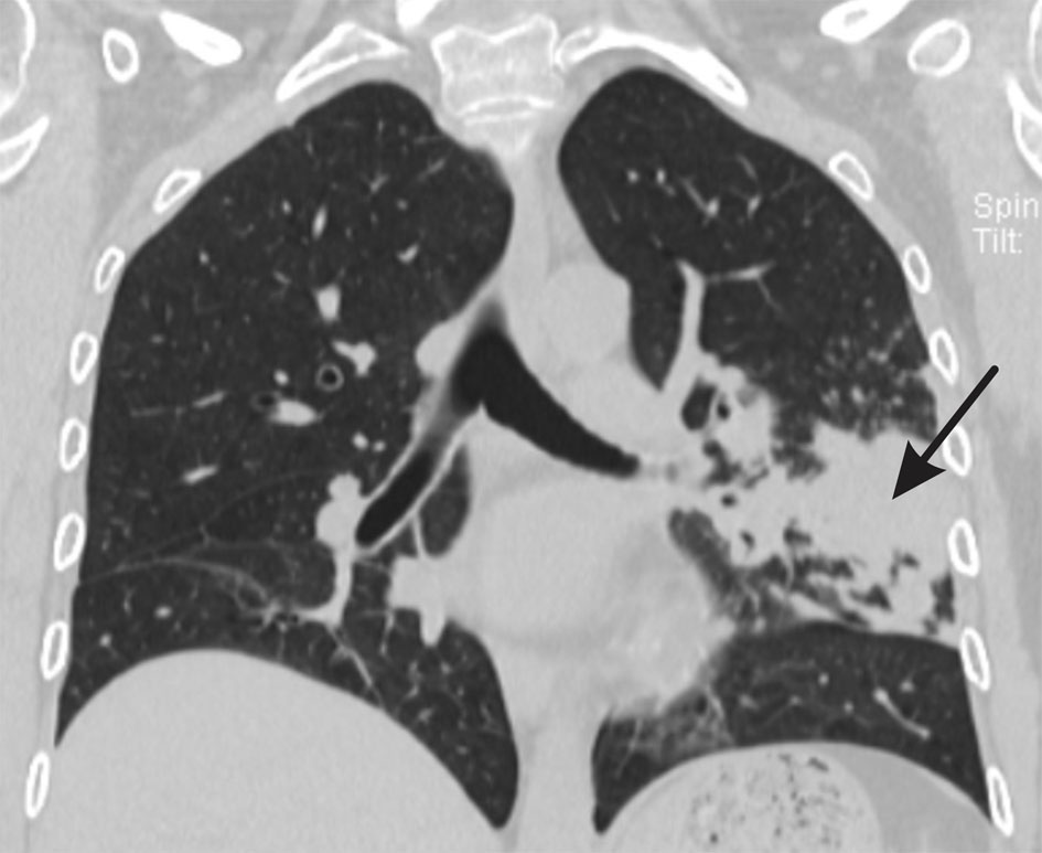
↓ Figure 4. Coronal chest CT scan with lung window showing an infiltrate in the left lower lobe (black arrow), consistent with probable pulmonary metastasis from non-Hodgkin lymphoma.
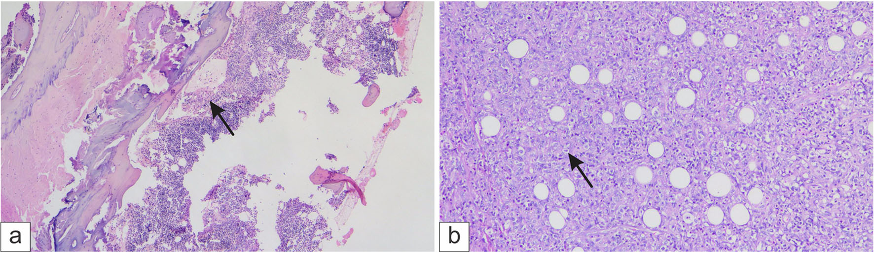
↓ Figure 5. Histological analysis of the trabecular bone. (a) Infiltration by neoplastic lymphoma cells (black arrow) and (b) Diffuse infiltration of large cells (black arrow).
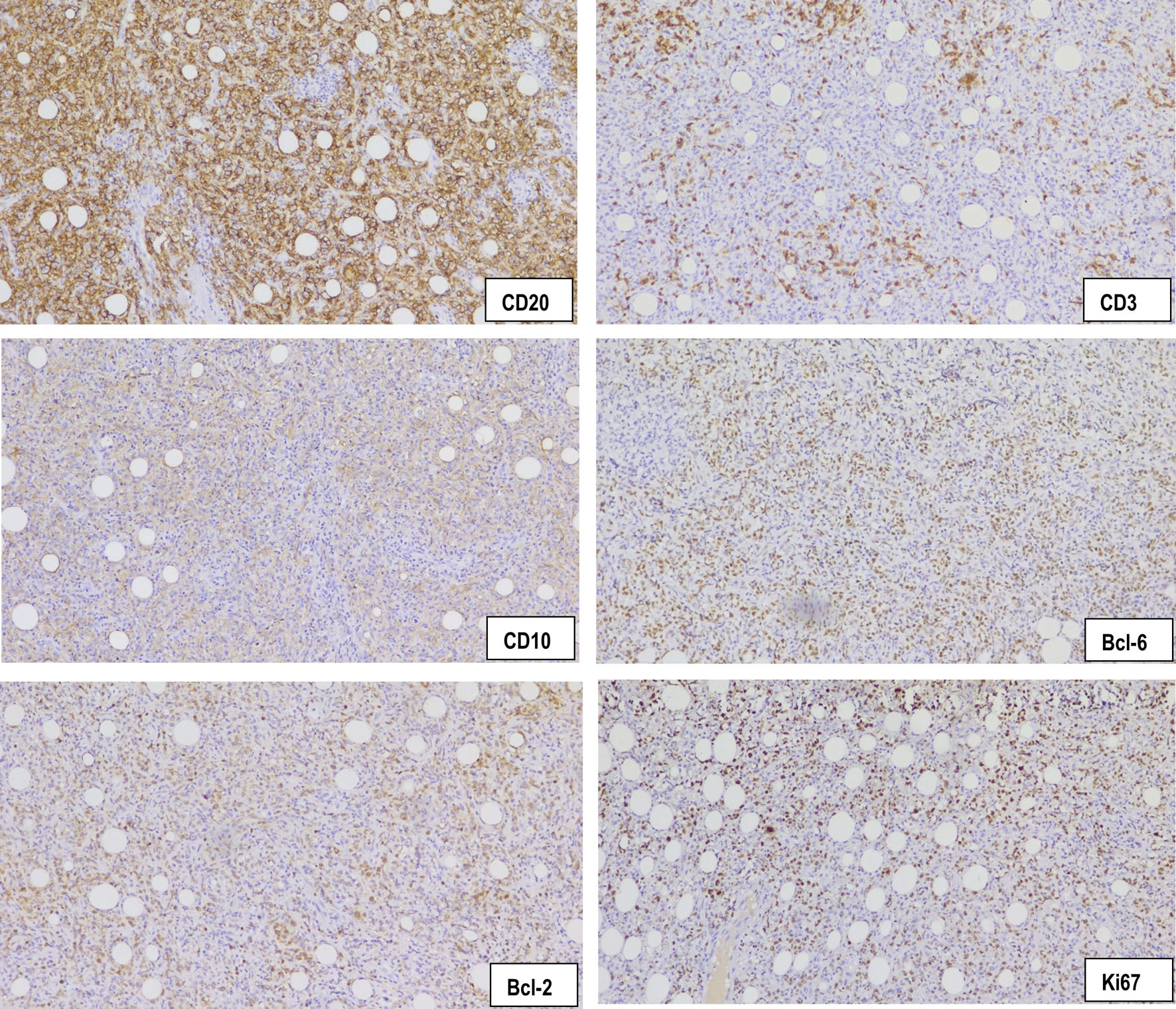
↓ Figure 6. Immunohistochemical analysis of the trabecular bone: CD20, × 40, membranous staining in neoplastic lymphocytes; CD3, × 40, negative in neoplastic lymphocytes expressed in residual T lymphocytes; CD10, 40X, membranous staining in neoplastic lymphocytes; Bcl-6, × 40, nuclear staining in neoplastic lymphocytes; Bcl-2, × 40, membranous staining in neoplastic lymphocytes; Ki67, × 40, nuclear staining in 90% of the neoplastic lymphocytes.
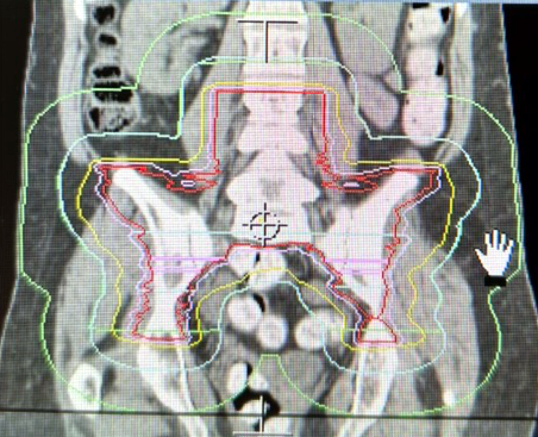
↓ Figure 7. External beam radiotherapy treatment plan using IMRT to the lumbosacral region and iliac crests. A dose of 45 Gy to the target volume, delineated by the red isodose curves was delivered.