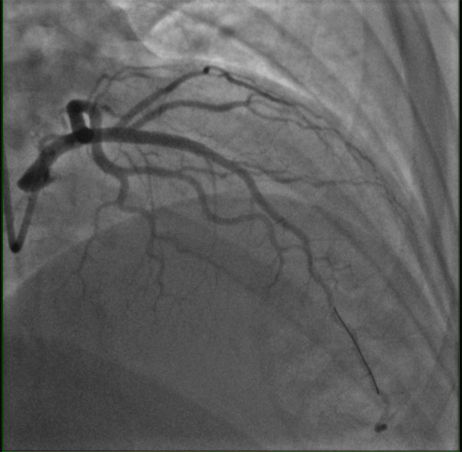
↓ Figure 1. Coronary angiography demonstrating a severe, smooth 95% stenosis of the mid segment of the left anterior descending coronary artery (LAD). The lesion has a concentric, non-calcified appearance without angiographic evidence of dissection, intraluminal thrombus, or ulceration. Preserved distal vessel filling is noted, consistent with high-grade fixed stenosis without complete occlusion. This degree of luminal compromise exceeded the critical threshold for flow-limiting obstruction, creating the substrate for episodic platelet-mediated cyclic flow reductions as described in the Folts model.
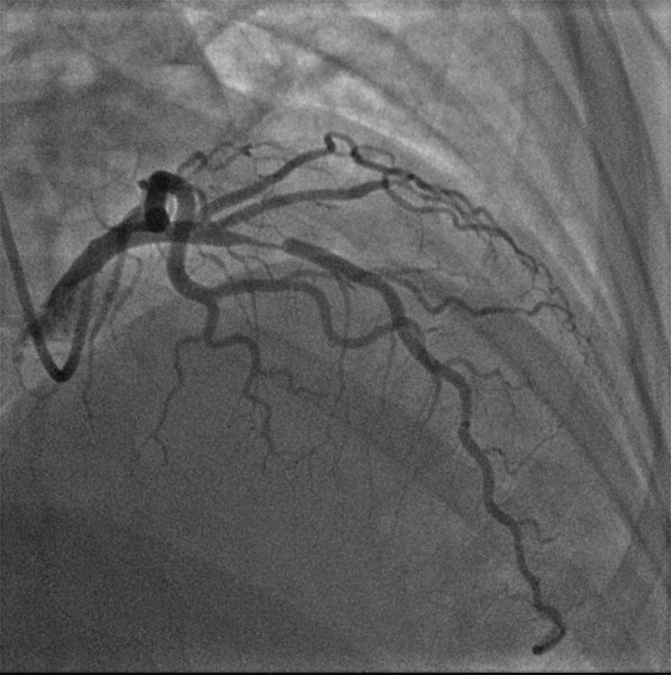
↓ Figure 2. Post-intervention coronary angiography following intravascular ultrasound (IVUS)-guided percutaneous coronary intervention (PCI) with placement of a single drug-eluting stent in the mid left anterior descending coronary artery (LAD). The stent demonstrates complete expansion without geographic miss, residual stenosis, or edge dissection. TIMI-3 anterograde flow is restored throughout the mid and distal LAD and its diagonal branches, with no evidence of distal embolization or no-reflow phenomenon. The post-dilation result, achieved with a 4.0 × 15 mm non-compliant balloon at 14 atmospheres, confirms adequate luminal gain and stent apposition.