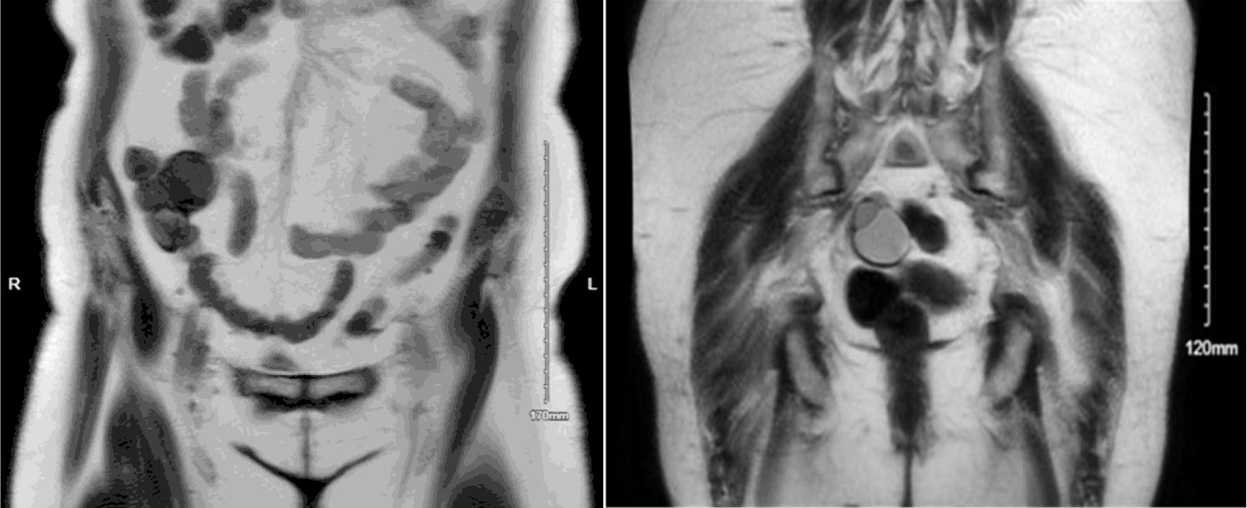
↓ Figure 1. Coronal MRI images demonstrate an omental cystadenofibroma and an ovarian cystadenofibroma. The omental lesion appears as a well-circumscribed cystic mass within the anterior abdominal cavity, consistent with a benign omental implant, while the ovarian cystadenofibroma is located lower in the pelvis adjacent to the uterus, showing mixed cystic and solid components typical of epithelial origin. MRI: magnetic resonance imaging.