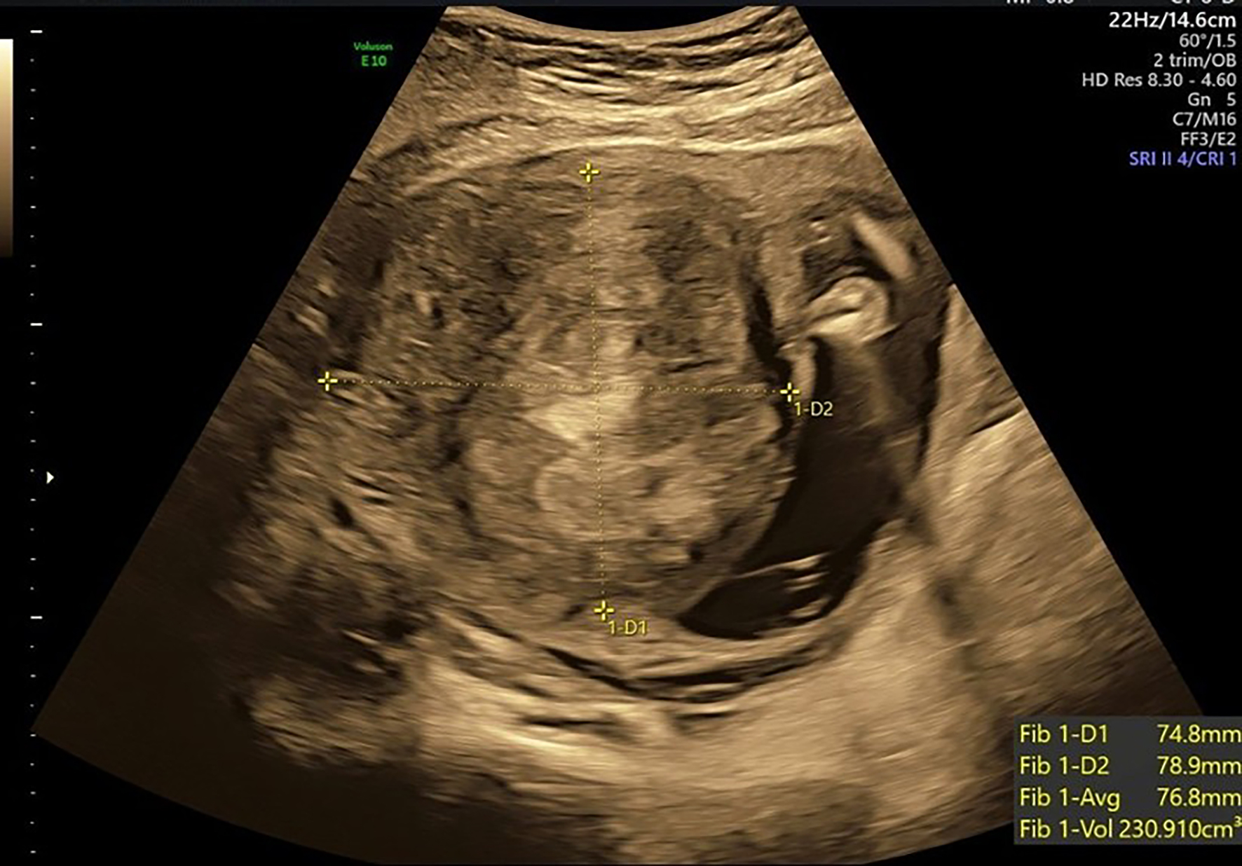
↓ Figure 1. Prenatal ultrasound at 20 weeks’ gestation showing an anterior-wall intramural fibroid (yellow cross-marks) measuring approximately 7.5 × 7.9 cm. The lesion was in the lower anterior uterine segment.
| Journal of Medical Cases, ISSN 1923-4155 print, 1923-4163 online, Open Access |
| Article copyright, the authors; Journal compilation copyright, J Med Cases and Elmer Press Inc |
| Journal website https://jmc.elmerpub.com |
Case Report
Volume 17, Number 6, June 2026, pages 275-279
Trans-Endometrial Cesarean Myomectomy for a Large Anterior Lower-Segment Intramural Fibroid Preventing Hysterotomy Closure
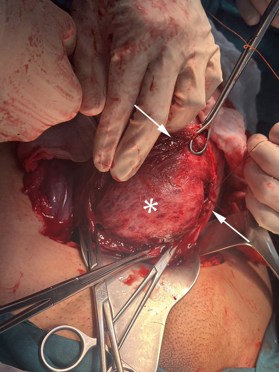
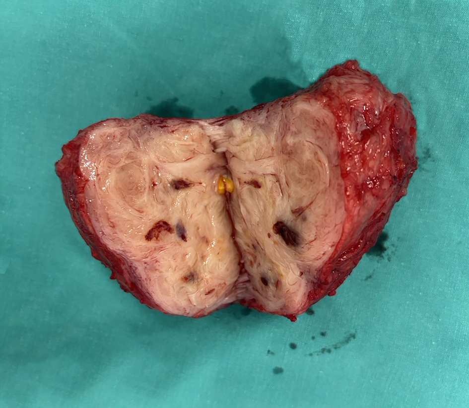
Figures