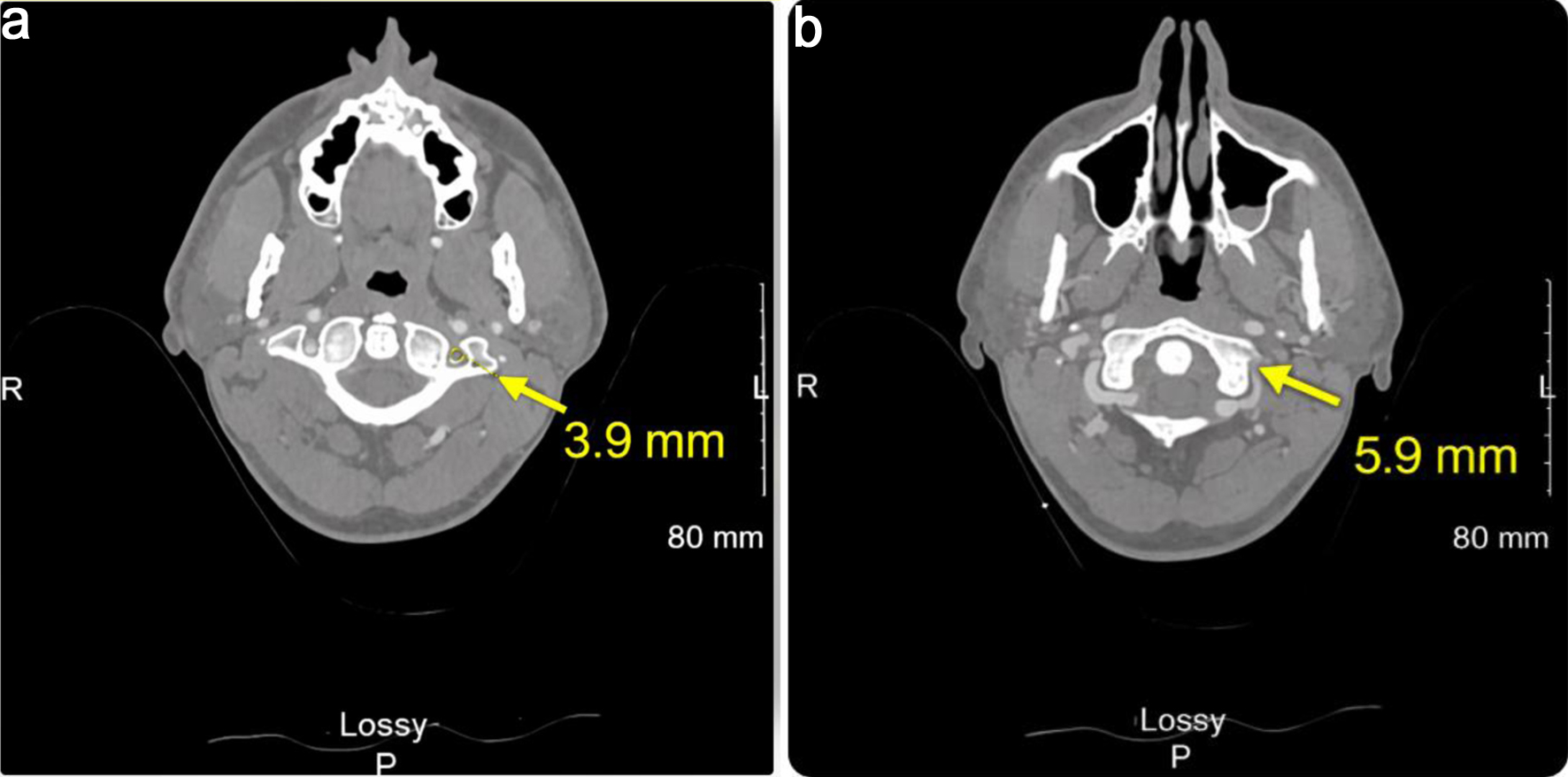
↓ Figure 1. Axial computed tomography angiography image of the head and neck acquired in early 2025 (series 2, image 156 (a) and image 162 (b); slice thickness: 1.25 mm; location: –58.25 mm). Impressions indicate acute lacunar infarction in the left cerebellar hemisphere and small venous angioma in the left parietal lobe.
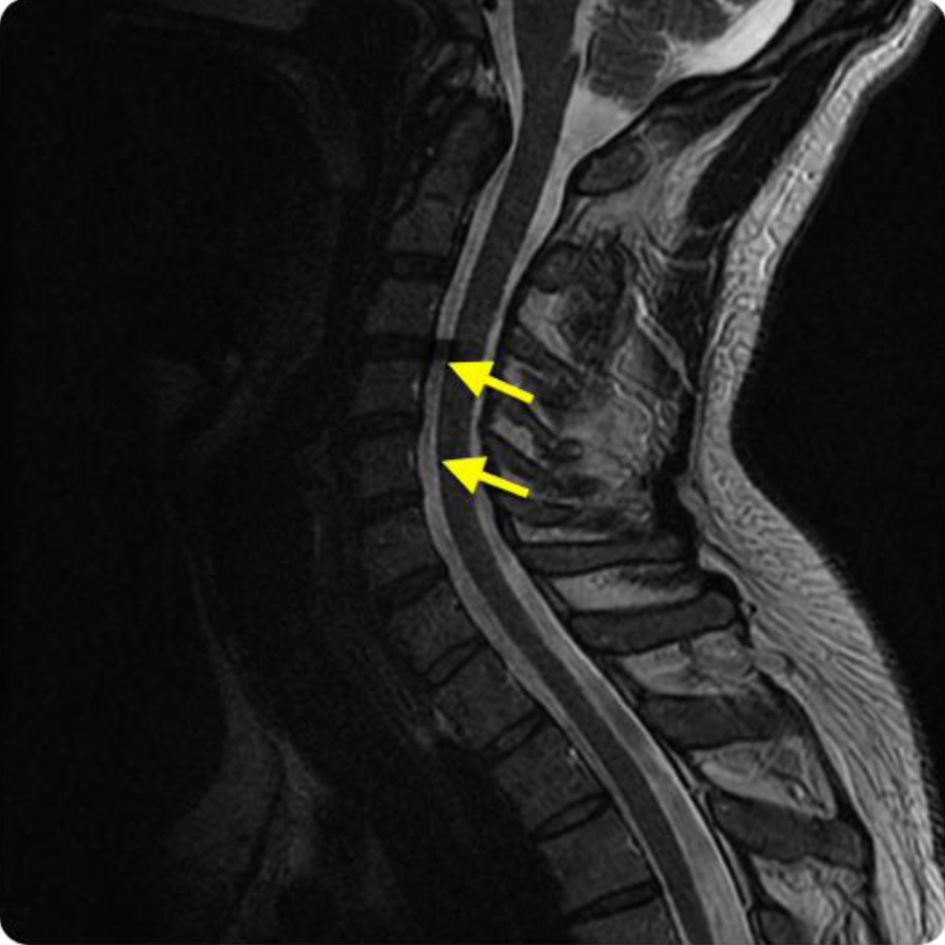
↓ Figure 2. Sagittal magnetic resonance angiography of cervical spine without contrast acquired early to mid-2025 (image 11, slice thickness: 3.0 mm; location: 15.96). Impressions indicate moderate neural foraminal narrowing at C3-C4 and C4-C5 with possible central punctate T2 signal abnormality within the central cord at C6-C7.