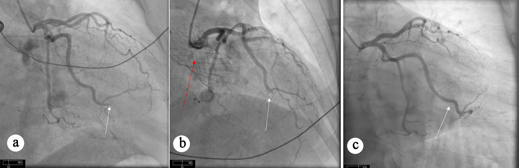
↓ Figure 1. Angiographic views showing coronary arteries. (a) Diagnostic angiogram before TAVR procedure showing angiographically normal coronary arteries without any evidence of obstructive CAD. (b) Occlusion of LAD and diagonal branch artery. (c) Occlusion of OM artery. (d, e) Restoration of flow can be seen in LAD (white arrow), diagonal branch (red arrow), and OM artery (blue arrow) after aspiration thrombectomy. CAD: coronary artery disease; LAD: left anterior descending; OM: obtuse marginal; TAVR: transcatheter aortic valve replacement.
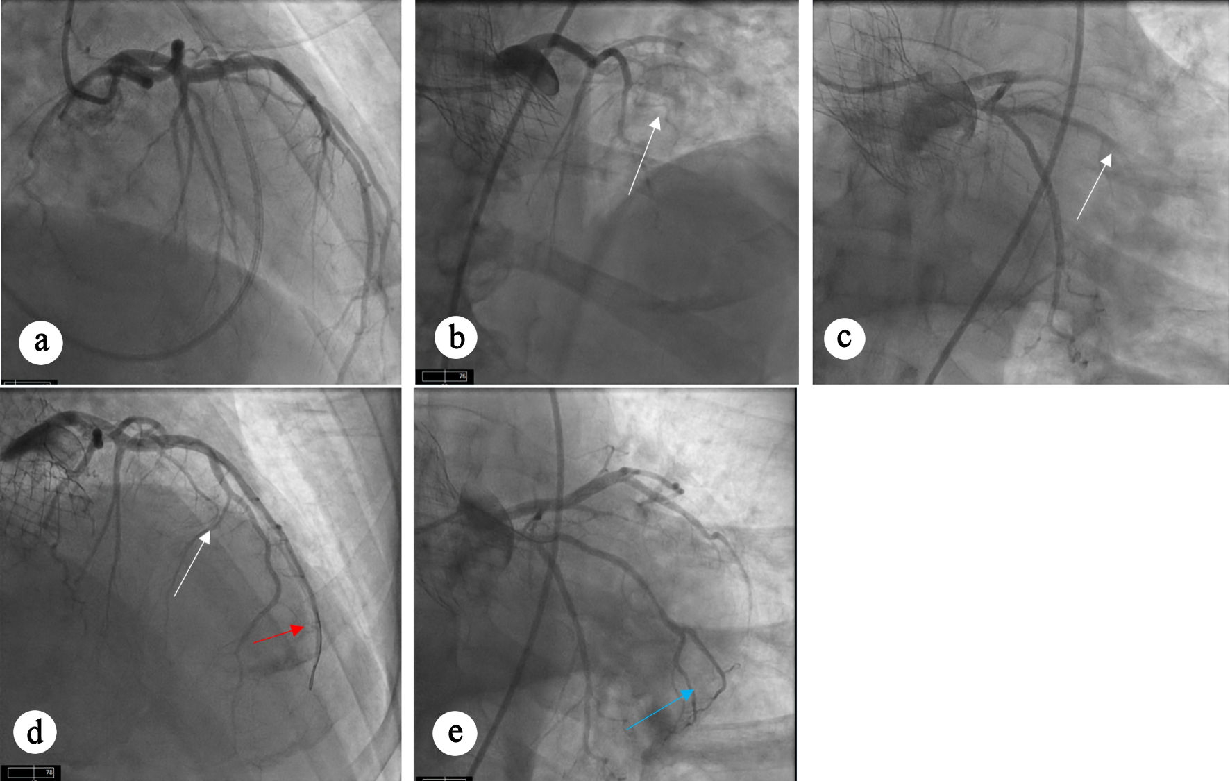
↓ Figure 2. Angiographic views showing occlusion and restoration of flow after intervention. (a) Occlusion of OM branches (white arrows) can be seen. (b) Restoration of flow can be seen in OM branches (white arrow) after aspiration thrombectomy. TAVR valve (red arrow) can also be appreciated. (c) Diagnostic angiogram before TAVR procedure showing patent OM branches. OM: obtuse marginal; TAVR: transcatheter aortic valve replacement.
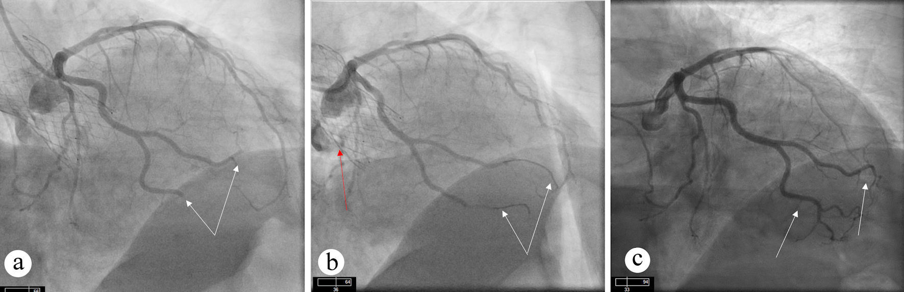
↓ Figure 3. Angiographic views showing persistent occlusion of OM branch. (a) Occlusion of OM artery (white arrow) can be seen. (b) Persistent occlusion of OM artery (white arrow) despite aspiration thrombectomy. TAVR valve (red arrow) can also be seen. (c) Diagnostic angiogram before TAVR procedure showing patent OM artery without obstructive CAD. CAD: coronary artery disease; OM: obtuse marginal; TAVR: transcatheter aortic valve replacement.