Journal of Medical Cases, ISSN 1923-4155 print, 1923-4163 online, Open Access
Article copyright, the authors; Journal compilation copyright, J Med Cases and Elmer Press Inc
Journal website https://jmc.elmerpub.com

Case Report

Volume 17, Number 6, June 2026, pages 258-262


An Atypical Case of Superior Vena Cava Syndrome Due to Sarcomatoid Lung Cancer With Pericardial Metastasis

Figures

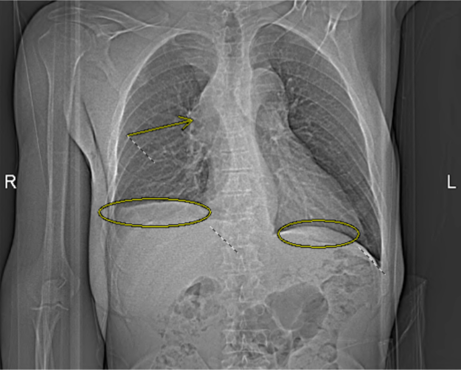
↓  Figure 1. Computed tomography angiography (CTA) pulmonary chest X-ray obtained on the day of admission demonstrates bilateral pleural effusions (circled) and a mediastinal mass causing severe superior vena cava (SVC) compression and right upper lobe pulmonary artery obstruction (arrow).
Figure 1.
↓  Figure 2. Computed tomography (CT) scan of the chest obtained on hospital day 2 shows a 13 × 16 mm mass in the lateral right upper lobe with irregular margins and extension to the pleura, consistent with findings during early hospitalization for suspected superior vena cava (SVC) syndrome.
Figure 2.
↓  Figure 3. Computed tomography (CT) ion scan of the chest obtained on hospital day 4, depicting pericardial effusion.
Figure 3.
↓  Figure 4. Chest X-ray obtained on hospital day 14, depicting an increase of right pleural effusion. Trace left pleural effusion and bibasilar areas of consolidation that may reflect atelectasis.
Figure 4.