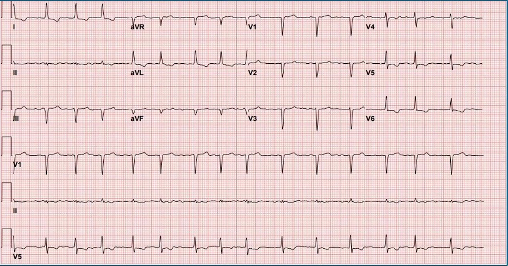
↓ Figure 1. Electrocardiogram on presentation showing atrial fibrillation with rapid ventricular response, inferior Q waves, and probable anteroseptal Q waves.
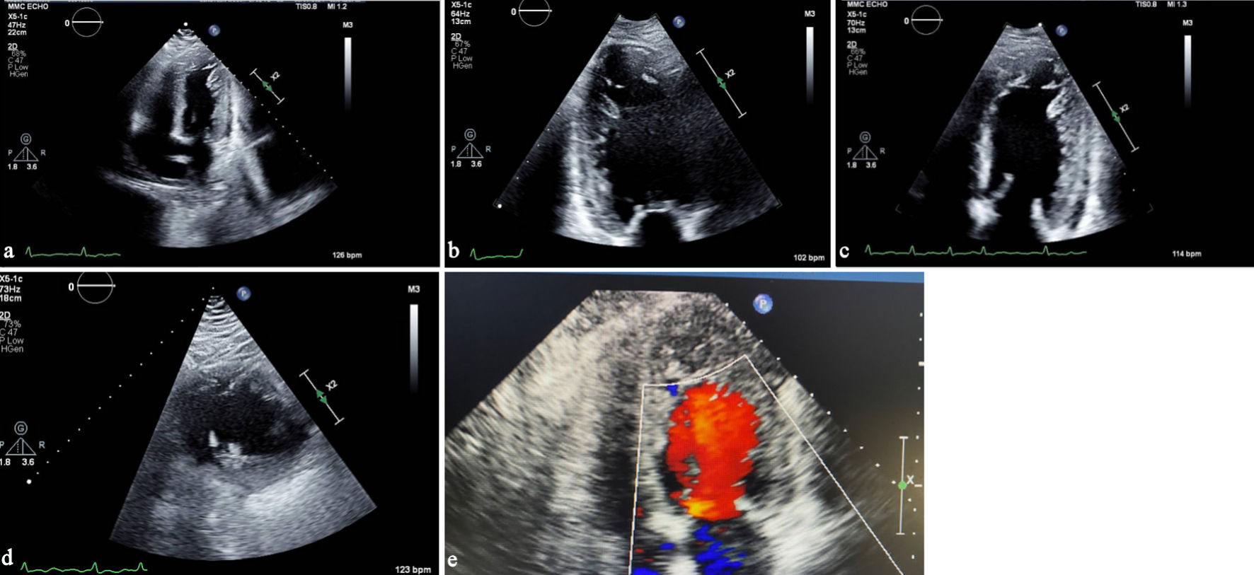
↓ Figure 2. (a) Transthoracic echocardiography, apical four-chamber view, demonstrating prominent trabeculations in the left ventricular apex. (b) Apical two-chamber view showing a two-layered myocardium with deep intertrabecular recesses. (c) Transthoracic echocardiography showing a left ventricular-focused apical four-chamber view illustrating a noncompacted-to-compacted myocardial ratio exceeding 2.0. (d) Parasternal short-axis view illustrating the noncompacted-to-compacted myocardial ratio exceeding 2.0 in systole. (e) Apical four-chamber view focused on the apex showing color-flow Doppler into recesses of apical trabeculations.
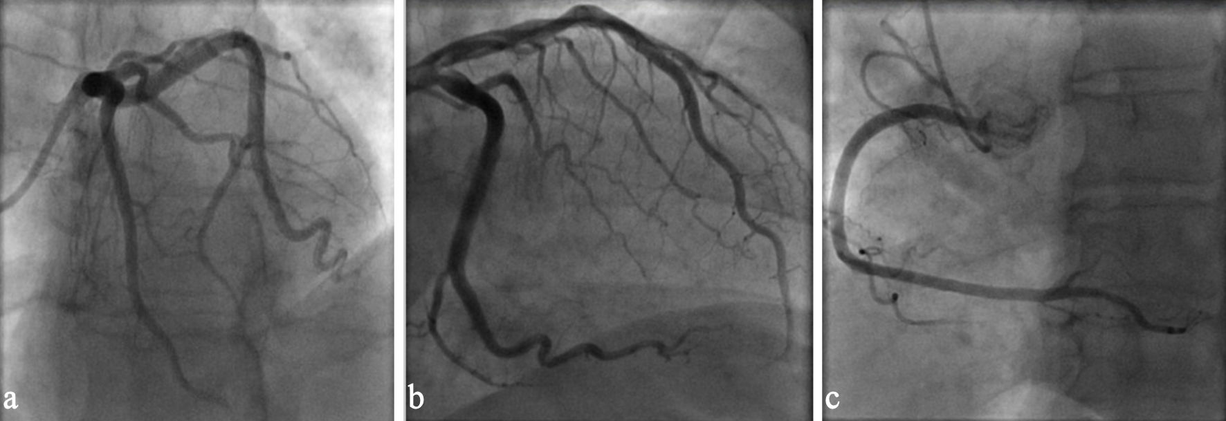
↓ Figure 3. (a) Coronary angiography left anterior oblique view demonstrating patent left main and left anterior descending coronary arteries. (b) Coronary angiography showing unobstructed left circumflex coronary artery. (c) Coronary angiography right anterior oblique view demonstrating patent right coronary artery.